

Abstract
There are numerous contributions to the development of ophthalmology from Switzerland, a country that holds a very special place in the history of medicine from the age of Paracelsus and Vesal to the current time. This review gives an overview over these contributions and the pioneers, among them Johann Friedrich Horner, Hans Goldmann, Jules Gonin, and Walter Rudolf Hess, one of only two ophthalmologists ever awarded the Nobel Prize for Medicine. A leading role in this evolution of modern ophthalmology has been played by physicians from Basel, home of Switzerland's oldest university.
Keywords: Cataract, Flammer syndrome, Glaucoma, History of ophthalmology, Laser, Minimally invasive strabismus surgery, Perimetry, Retinoblastoma, Retinal detachment, Slit lamp, Swiss medicine
Rankings are − whether we like it or not − part of our life. Some are enlightening, a few are annoying. Medicine seems to have, reluctantly though at the beginning, embraced rankings. There are now “The Best Doctors,” “The Best Hospital,” “The Best Medical Schools” lists in many western countries. Those with a reputation for a fair and (more or less) objective assessment usually reflect expertise, skill, and standing within a specialization or subspecialty. In the field of ophthalmology, the annual “Power List” by the online publication The Ophthalmologist is widely noted and shared on social media by many eye care providers. While many of the 2018 Power Listers work in the United States, when it came to the number of ophthalmologists honored by this ranking per capita, the most successful country was Switzerland, together with Singapore and the UK [1].
This result from a non-peer reviewed publication might be regarded as entertaining though by far not thoroughly scientific − it did not, however, happen by accident either. It serves as an indication that this rather small country in the middle of the European continent has contributed to ophthalmology and other fields of medicine closely associated with the functioning of the human visual system (like circulation and blood vessels) in a way that seems disproportionate to its geographical and demographic size. Analyzing these contributions − historical and current ones − and assessing their role in the advancement of ophthalmology is an overdue endeavor, probably in part due to the Swiss' strong sense of personal modesty and their aversion to putting themselves into the limelight. It therefore seems appropriate that an outsider unsuspicious of Helvetic patriotism researches these contributions, which are the subject of this review in which one particular Swiss academic institution, the University of Basel, figures prominently.
The Dawn of Modern Medicine: Vesalius, Ruf, Paracelsus − and a Petty Criminal with an Enduring Role in Medical Education
It is in Basel, in the Anatomical Museum of that Swiss city, where a human skeleton is preserved (though usually not on display to the general public) that is not only most likely the oldest specimen of its kind − a skeleton exclusively destined for medical education − anywhere in the world but is also a relic from the dawning age of modern medicine (Fig. 1). It is marked with the words: “This skeleton was given by the master of anatomy, Andreas Vesalius of Brussels, to this University, when he stayed in Basel in 1543 in order to attend to the publication of his great anatomical work.” It is no historical accident that Vesal was in Basel that year. The city was the foremost printing center in the world in that age, based not only on the expertise of its many printers but also on a political climate that fostered science and knowledge. Basel and other cities in Switzerland − nominally still part of the Holy Roman Empire of the German nation − were much more liberal and tolerant (at least for the standards of the time) than most other European countries. Scholars and authors who wanted their works to be published flocked to Basel, out of reach for the inquisition and other forces of dogmatism. Particularly banned almost everywhere else was research on the human body, its functions, and its ailments. Anatomical knowledge had only modestly advanced since antiquity, i.e. since the age of Galen whose teachings were usually repetitioned. If dissections were allowed, it was mostly on the bodies of criminals who had been executed. One of these involuntary donors to science was the convicted felon Jakob Karrer whom Andreas Vesalius dissected and whose bones have proved so durable. Vesalius moved from Padua to Basel in 1543 and published that year in cooperation with the printer Johannes Oporinus − who not only had learned the still young printing trade but also studied medicine − his great anatomical work, “De Humani Corporis Fabrica” [2, 3] (Fig. 2). Oporinus in his younger days had worked with the other great figure and innovator of 16th century medicine, Paracelsus [4]. A number of this pioneer's works were also first published in Basel.
Fig. 1.

The Basel skeleton, prepared by Vesal.
Fig. 2.

Frontispiece of Andreas Vesalius' “De Humani Corporis Fabrica.”
It was during this age of (relatively) free and uncensored publishing in Basel and other places in Switzerland that the first known genuinely Swiss treatise on ophthalmology was written. Jakob Ruf was born around 1505 in Konstanz on the modern Swiss-German border. Ruf became the official city surgeon of Zurich and left behind an extensive corpus of works in Latin and German which has been edited and published just a few years ago by Hildegard Elisabeth Keller [5]. One of his works, Practica copiosa de arte ophthalmica was written around 1545; two of its four volumes have survived. Ruf died in Zurich in 1558 [6].
On the Way to an Independent Medical Specialty: The First Eye Clinics in Basel and Zurich
These historical roots should be remembered when assessing the development of medicine in Switzerland and particularly in Basel, which today provides its citizens (and quite a number of visitors from abroad) with health care second to none in quality. Especially in ophthalmology, the number of contributors − past and still living − and innovations is by far disproportionate to its modest demographic with a population of about 8.4 million. Ophthalmology, it has to be remembered, used to be a subspecialty of surgery in most countries up to about the beginning of the 19th century. Surgery itself, however, during the ages before had been considered less a part of academic medicine; medical doctors with a university degree did not operate and were by their professional self-estimation averse to come into contact with human blood. Operating on the human eye before the 1800s in most cases meant treating cataract by “couching” the lens, tearing it out of its zonules with a needle and sinking it into the vitreous cavity which a modern eye surgeon shudders to imagine because of the multitudes of potential complications. This intervention was performed by people known as barbers, barber-surgeons, Wundärzte who generally also performed other surgical procedures among which “cutting” the bladder stone seems to be if not the most frequent but at least the one defining their profession. It is no coincidence that the surgeon who published the first German-language textbook in ophthalmology is also the surgeon who published the first treatise in that language on cutting the stone, Georg Bartisch in the capital of Saxony, Dresden, in the late 16th century [7].
Some of these early eye care professionals, usually called oculists, were itinerant, i.e. while they might have had a home base, they were traveling and offering their service for a limited time in cities along their route. One of the most famous was Johann Heinrich Jung-Stilling who gained fame as a pietistic author and who performed eye surgery in Basel 1802/03 [8]. Two centuries after Bartisch, the road to independence for ophthalmology became clearly visible. More and more academically trained physicians specialized in eye care, though often not exclusively. Many still did general surgery: In Basel, J.R. Burckhardt became a cataract surgeon of high repute although he was a professor of internal medicine, in the early 19th century not a completely conservative specialty anymore. His son, August Burckhardt, followed in his father's footsteps and performed eye surgery between 1833 and 1860 [8], just about the time the city got its first real ophthalmologist, Johann Schiess. The first eye hospitals were finally founded. The first private eye clinic in central Europe was most likely the one opened by the Italian oculist Giovanni Casaamata in 1782 − again, in Dresden, a pioneering place in the history of ophthalmology. In 1813, at the height of the Napoleonic Wars, the world's first University Eye Hospital opened its gates in Vienna with Georg Joseph Beer becoming the first chair of ophthalmology in the Austrian capital 5 years later. In Switzerland, the first University Eye clinics were founded in the early 1860s, in Basel and Zurich. Special lectures in ophthalmology for medical students were given in Basel as early as 1823.
Von Graefe's Disciples Take Over: Johann Friedrich Horner
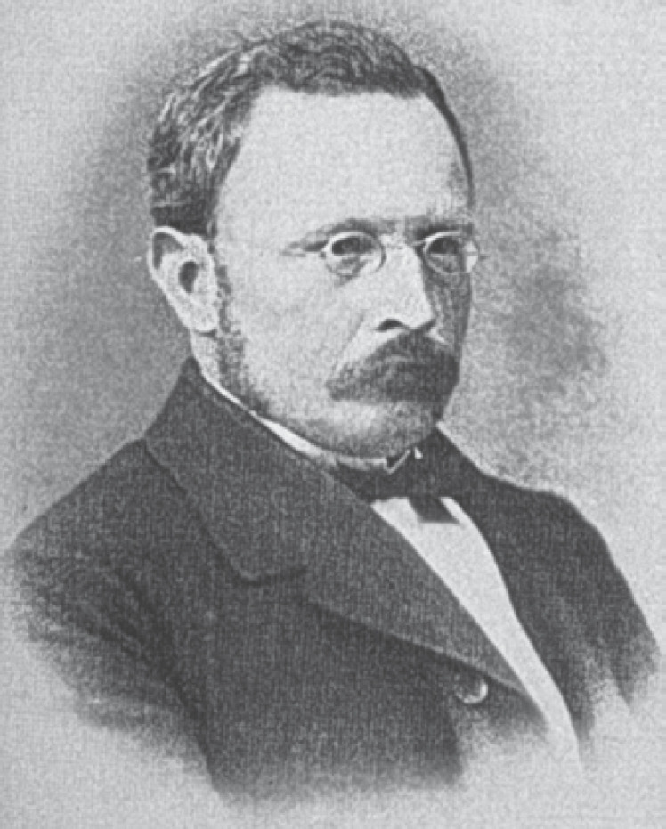
Some countries have something of a “founder” of their nation's ophthalmology. In Germany, for instance, this undoubtedly is Albrecht von Graefe (1828–1870). His contemporary and disciple Johann Friedrich Horner is widely considered the founder of modern scientific Swiss ophthalmology (Fig. 3). Just 3 years younger than von Graefe, Horner was born in 1831 into a family of distinguished physicians in Zurich. After studying and being an assistant to von Graefe, Horner opened his private practice in his hometown first as a general physician; later, he established his own eye clinic called “Hottinghof” after having considered first to open an ophthalmological practice in Basel. The famous surgeon Theodor Billroth (1829–1894), at that time chair of clinical surgery in Zurich (he was appointed to the University in Vienna in 1867 where he gained worldwide fame particularly for his methods of gastrectomy that still bear his name), was instrumental in creating a chair of, and a clinic for, ophthalmology which traditionally had been just a subdivision of surgery. It was Horner who was appointed the first director of the eye clinic at the Cantonal Hospital and thus of the first Swiss University Eye Clinic in 1862; in 1873, he became a full professor of ophthalmology. He was not only a skilled technician but also an outstanding researcher and educator: the number of 28 doctoral students who gained their medical degrees under his guidance and in a just recently established specialty is a pioneering achievement in itself. Horner made a number of scientific discoveries of which the Horner's muscle, the palpebral part of the orbicularis oculi muscle, and Horner's syndrome are the most famous; the latter describing the classical triad of ptosis, miosis and enophthalmus, sometimes combined with other signs like anhidrosis (no sweating) on the affected side of the face as a consequence mostly of some damage to the sympathetic nervous system.
Fig. 3.

Johann Friedrich Horner (1831–1886).
Johann Friedrich Horner seems to have been one of those unfortunate though not entirely rare physicians who work themselves to death for the (presumed) benefit of their patients: due to heart problems, he had to resign from his chair in 1885 and died just one year later at the age of only 55 years [9, 10, 11].
Establishing Academic Ophthalmology in Basel: Johann Schiess
Almost the same age as Horner, Johann Schiess was born in 1833 in Heiden, the oldest of 14 children of the local priest and his wife (Fig. 4). Schiess became an early example of what seems to be a special approach to ophthalmology in Basel, an approach that combines clinical ophthalmology with pathology and the general health of the patient. Schiess began studying medicine at the University of Basel in 1852; a later semester in Würzburg made him a pupil of Rudolf Virchow, the founder of modern pathology. After graduating in 1856, he pursued further studies in Munich and Vienna. The latter one being the most renowned place in medicine of that era, he focused on ophthalmology and was instructed by Eduard Jäger how to use the latest innovation − the ophthalmoscope which for the first time made a thorough examination of the eye's posterior segment possible.
Fig. 4.
Heinrich Schiess (1833–1914).
Schiess at first seemed to follow the usual path for doctors at that time: he opened his own practice as a general practitioner in a small town in Eastern Switzerland where his father was working as a pastor. But 4 years later, in 1861, he decided to dedicate the rest of his professional life to ophthalmology, moved to Basel and opened his office there as the city's first specialized eye doctor. A factor in this decision was the influence Albrecht von Graefe had on him. The by now already famous ophthalmic surgeon from Berlin once a year traveled to Heiden (where Schiess was born) to seek relief in the clear Alpine air for his respiratory ailment that in 1870 led to Graefe's untimely death at age 42: tuberculosis. Instead of relaxing in Heiden, it became kind of a clinic away from the home clinic for von Graefe who saw hosts of patients there and discussed their status with local colleagues − like the young Doctor Schiess. Schiess followed him to Berlin and did an internship at von Graefe's clinic. After that experience with state-of-the-art ophthalmology, there was no turning back for Schiess. Soon after settling in Basel, he decided to open its first eye clinic. In 1864, this precursor of Basel's University Eye Clinic (Fig. 5) opened on Missionsstrasse 45 with 6 beds for patients; in 1877, the clinic moved to its current location on Mittlere Strasse. Schiess was holding lectures at the University since 1863; in addition to 3 h a week on clinical ophthalmology, he offered a 1-h course in histopathology of the eye. The number of his disciples must have been limited since Basel was a distinguished medical school but not an overcrowded one: in 1864, it counted 39 medical students while Vienna boasted more than 1,400. Schiess published quite a number of papers and coped with an ever-increasing workload; in 1871, a special ward for children with ocular ailments was opened. One of his specialties was myopia. He developed strategies to prevent children from becoming myopic (he was shortsighted himself) and saw correcting glasses as a measure of (almost) the last resort. In 1878, Schiess became the first eye specialist to serve as dean of the medical faculty in Basel. Among the illustrious patients who travelled to Heinrich Schiess' Basel was the philosopher Friedrich Nietzsche. The fact that the international congress of ophthalmology (there was just one at that time, unlike the scores in modern times) in 1880 convened in Interlaken with Horner and Schiess as part of the organizational team gives testimony to the reputation of Swiss ophthalmology already at that time. Schiess, who became almost blind in old age, was a somewhat quirky character. He was not what would today be called social and he had his prejudices. A wine connoisseur who was proud of his subterranean collection, women were not allowed to descend into his wine cellar. Schiess was convinced that their presence would turn the wine sour [8].
Fig. 5.

The first Basel Eye Clinic, around 1865.
Otto Haab: Most Likely the First to Describe What Today Is a Leading Cause of Blindness
Horner's successor as director of the University Eye Clinic Zurich was a man whose two major scientific achievements seem to represent the changing focus of ophthalmological practice. The one invention he was primarily praised for by his contemporaries seems to be emblematic for the eye care routine in the industrial age in the late 19th and early 20th century, another of his publications − certainly underrated at that time − points to a major global health care issue in the 21st century with its ageing demographics. Otto Haab (1850–1931) was born in a village that today is part of the city of Winterthur and travelled widely during his years as a medical student and a young doctor − journeys that led him to become a student of some of the great personalities of medicine like Donders and Snellen in the Netherlands, von Arlt in Vienna and even Joseph Lister in Edinburgh, a giant in the history of medicine as the pioneer of antisepsis. Following Horner in 1885, he served for almost 34 years as the director of the Zurich clinic. In his obituary, his perfecting the giant magnet for the extraction of intraocular foreign bodies − a device that was named after Haab − was described as “his most important achievement” [12] (Fig. 6). Injuries at the workplace were common in that age − reaching the ninth or tenth decade of one's life was not. Therefore, the epidemiological importance of clinical observations that Haab published in 1865 (as probably the first ophthalmologist) could not be anticipated: it was a clinical description of a retinal disease afflicting the elderly, an entity that would later be known as AMD − age-related macular degeneration. Yet officially unnamed, he called it “Disease of the Macula Lutea of the Retina due to Old Age (Senile Macular Disease).” Haab was realistic about the prognosis: “The disease, which is incurable, usually attacks both eyes and leads to grave visual disturbance, though the changes in the eye-ground amount to no more than a light or dark mottling, consisting of yellowish-red or yellowish spots, or a slight increase in the pigment, the changes being always most marked at the center of the cornea” [13]. Haab's name also lives on in medical terminology in “Haab's Striae,” breaks in the Descemet membrane of eyes with congenital glaucoma [14] (Fig. 7).
Fig. 6.

Otto Haab (1850–1931) with his giant magnet.
Fig. 7.

The Zurich University Eye Clinic in the late 19th century.
Pioneer with the Slit Lamp, Author of a Classical (and Heavy) Treatise: Alfred Vogt
Calling him a difficult character would also be true of Alfred Vogt − and it would be a quite diplomatic definition of his combative personality. It is probably Vogt who paved the way for the triumph of, and the introduction into every ophthalmological practice, of the one instrument that is quintessential − maybe even more so than the ophthalmoscope − in everyday diagnostics: the slit lamp. It was during his time in Basel that the first edition of his monumental Lehrbuch und Atlas der Spaltlampenbiomikroskopie was published. A one-volume work, it was extended in later editions to consist of three volumes and a record-breaking more than 2,000 illustrations over 1,100 pages. One of his successors in the chairmanship both in Basel and in Zurich, Balder Gloor, has called it a still valid, unsurpassed standard book that even today every physician aspiring to become an ophthalmologist should know [15].
Alfred Vogt was born October 31, 1879, in the village of Menziken in canton Aarau, and began studying medicine in 1899 in Zurich; after the first semester, however, he transferred to Basel University. While Vogt was originally planning to become a surgeon, the director of the Basel University Eye Clinic, Carl Mellinger, sparked Vogt's interest in ophthalmology. Under Mellinger's guidance, Vogt worked on, and published his dissertation about, the detrimental effects of artificial aniline dyes on the eye [16]. It certainly was a fitting subject in a city whose industrial landscape was − and is − dominated by chemical and pharmaceutical factories (Fig. 8).
Fig. 8.

Alfred Vogt (1879–1943).
Leaving the Basel University Eye Clinic in April 1906, Alfred Vogt spent a 2-month internship with Carlo Reymond, a well-regarded ophthalmologist in Turin, and after returning to Switzerland married his sweetheart Marie Bossardt. In August 1906, Vogt opened his own practice as an ophthalmologist in the city of Aarau. Just 3 years later, in addition to that task he became the chief of the eye clinic at the Aargau Cantonal Hospital (not to be confused and differing only in one letter, Aargau is the canton, located in the north of Switzerland and to the east of Basel, while Aarau is the canton's major town). Under Vogt's leadership, the number of patients attending the eye clinic increased sharply as did its reputation. Vogt began his career as a prolific researcher and author, which resulted in − according to a recent count − 246 publications (and a further 234, including 34 dissertations, written by his students and residents) during his lifetime [17]. He investigated the effect of infrared and ultraviolet light on the eye and conducted experiments in cooperation with the physics lab of a local high school [18]. Cataract, its pathogenesis and its treatment, started to become a focal point in Vogt's scientific and clinical endeavors and remained so for the rest of his life [19]. Surprisingly enough, the young ophthalmologist was already on the trajectory course to have his name immortalized in medical terminology: his publication of a case study describing a patient whose eyelashes suddenly turned to gray while at the same time a uveitis was active [20] was later supported by publications of Japanese ophthalmologists. Today, this clinical entity is worldwide known as the Vogt-Koyanagi-Harada disease [21, 22].
A Brilliant Researcher, a Demanding Superior, a Combative Personality
To turn his career as increasingly desired by him from private practice to teaching and researching in the setting of a university clinic, Vogt contacted the University of Zurich to submit a “Habilitation” − a peculiarity of the German-speaking academic world where aspiring scientists will write a thesis-like publication and then − if approved by the university's faculty − will be granted the venia legendi, the permission to teach. Vogt's application in Zurich was rejected; his subsequent distinguished academic career is the more astounding since he had to overcome the obstacle of not having this postdoctoral qualification [23].
When his former boss Mellinger died in 1917, Vogt applied for the position of head of the Basel eye clinic. He was one of several candidates, and while the faculty supported someone else, the university's governing body decided on December 26, 1917, to appoint Vogt director of the clinic and at the same time named him extraordinary professor of ophthalmology [24]. His way of dealing with the patients who started to come to Basel from farther away than any time before the clinic has been founded was utterly direct, sometimes with a touch of insensitivity. An artist, who happened to be a friend of Vogt and who had retinal hemorrhages, for instance, was frankly told: “Well, old pal, with eyes like these you are not going to paint much longer!” [24].
Vogt acted as President of the Swiss Ophthalmological Society from 1920 through 1922. In the latter year, Vogt hosted the first international symposium on slit lamp biomicroscopy, attended by more than 30 ophthalmologists from European countries and even one doctor from Japan − his expertise with that instrument which had resulted one year earlier in the first edition of his famous book had made Basel as well as Zurich after he was appointed there and Switzerland an attractive destination for ophthalmologists eager to develop their skills [24]. He had improved the design and functionality of contemporary slit lamps; he continued to work on the next and extended edition of his book. It turned out to be one of the great works of ophthalmological publishing of all time. Supported by an artist whose contribution he seemed to have tried to diminish by repeatedly stressing that “illustrations were made under my control” or “under the guidance of mine,” the three volumes published in 1930 [25], 1931 [26], and 1942 [27] contained no less than 2,346 figures of the highest quality.
Vogt, Jules Gonin, and the Rise of Vitreoretinal Surgery
Despite his reputation and success in Basel, Vogt had his mind set on the larger University Eye Clinic of Zurich. When that chair became vacant in 1922, the University of Zurich offered the position to Vogt who did not hesitate to accept. In Zurich, Vogt became more interested in vitreoretinal diseases than before. Today, the anterior segment's ophthalmic surgeon that Vogt with his many iridectomies and cataract extractions has basically been in Aarau and Basel does in larger clinics hardly interfere with the expert for the posterior segment. It was still different in the 1920s and 1930s when Vogt began treating retinal detachment by sealing the retinal tear, just as Jules Gonin had advised. With a lot of surgical experience, Vogt published a major work on retinal detachment [28] and started feuding with Gonin. He did the same with the third of Switzerland's seminal ophthalmologists in the first half of the 20th century, Hans Goldmann (whose career stretches far into the second half of the century), whose explanation of the causes of the “fire-cataract” that afflicted glassblowers and similar today mostly extinct professions − heat [29] − was contrary to Vogt's conviction of infrared light being the cause of the pathogenesis of the Feuerstar [23]. Combative until the end, Vogt's health began to fail, and he died shortly after resigning from his chair and his directorship on December 10, 1943.
The Boundless Ingenuity of Hans Goldmann
It was Hans Goldmann who took Swiss ophthalmology to a leading position in the subspecialty that more or less unofficially is called glaucomatology. Diagnosing and treating glaucoma, the third most common cause of blindness worldwide today, is still a challenge for eye care providers. The only truly modifiable risk factor − modifiable by medical, surgical, or laser therapy − is an elevated intraocular pressure (IOP). The instrument to measure the IOP reliably was invented by Hans Goldmann [30]. It is the applanation tonometer that can literally be found in almost any eye clinic, any ophthalmological practice all over the world [31]. It still, more than 60 years after its invention, bears the name Goldmann tonometer and, even more surprising, it is still considered the gold standard in measuring IOP. There are a number of modern tonometers, with more electronics (the Goldmann tonometer is strictly mechanic), some hand-held, other non-contact (i.e., not touching the patient's cornea and thus requiring no local anesthesia) − but all are measured up against Goldmann's device. And none of them has replaced the Goldmann tonometer yet.
But Goldmann's ingenuity was not limited to the applanation tonometer. There are still lots of eye care institutions all over the world today where the ophthalmologist will examine the patient at a Goldmann slit lamp, then taking a closer look at the posterior section of the eye by putting a contact glass devised by Goldmann on the superficially numbed eye with the patient still sitting at the slit lamp. With instruments invented or improved by Hans Goldmann, basically most of the anatomical sections can be examined non-invasively [32, 33]. If after that examination with Goldmann devices and measuring the IOP with the Goldmann tonometer the ophthalmologist suspects a disease that can result in visual field defects (like glaucoma or other ailments of the optic nerve), it is time for seating the patient at the Goldmann perimeter. That has been for about half a century the gold standard in perimetry (and in many countries and regions still is today) until it was replaced by computerized perimetry and technologies that allow a faster and less demanding visual field test like frequency doubling perimetry. Goldmann's publications on perimetry, however, have lost nothing of its importance for this crucial neurophysiological diagnostic tool [34, 35, 36].
Hans Goldmann, born a few weeks before the end of the 19th century − on November 21, 1899 − in what was then the Austro-Hungarian Empire, and dying in the last decade of the 20th century − on November 19, 1991 − in the Swiss capital of Berne, where he spent his prolific life as a physician and a scientist, became the youngest director of a major Swiss eye clinic in 1935 and a Swiss citizen one year later. As Balder Gloor has described him, he was at home in mathematics, physics, and the natural sciences, just as this was the case more than half a century later with the team at the Basel University Eye Clinic when they published a landmark textbook on the basic sciences in ophthalmology [37]. In glaucoma, he did not only invent the aforementioned and partly unsurpassed diagnostic tools but also researched the pathogenesis of the disease by focusing on the aqueous outflow and postulating that the IOP is defined by that outflow, by the production of aqueous humor, and by the episcleral venous pressure [38]. While Goldmann was a strong advocate of the IOP as the trademark of glaucoma, he did not rule out the influence of other factors − like the vascular factors that have been so extensively researched and their influence on the pathogenesis has been so thoroughly documented at the Basel Eye Clinic at the turn from the 20th to the 21st century. Having served both as Dean of the Medical Faculty and as president of Berne University (Rector magnificus), Goldmann remained active for many years after his retirement − a retirement that was somehow marred by his suffering AMD and thus experiencing diminished eyesight by a disease the medicine of his time had no cure for [39].
In 1968, Hans Goldmann in his final lecture before becoming an emeritus stated − 25 years after Vogt's death − that the 1930s were the decade in which Switzerland became the leader in global ophthalmology and Swiss ophthalmologists were setting the pace. Goldmann named two outstanding personalities that had shaped modern eye care: one was Alfred Vogt, the other was Jules Gonin [17]. Just as Goldmann was the Swiss physician who became the leading global authority on glaucoma, Gonin is widely regarded as the father of modern vitreoretinal surgery − both leaders in their field in whose footsteps a later generation of Swiss ophthalmologists would walk: Josef Flammer of Basel in glaucoma research and therapy, Thomas Wolfensberger of Lausanne, and Sebastian Wolf of Berne in retinology.
The Retinal Break Has to Be Closed: Jules Gonin and the Emerging Treatment of Retinal Detachment
Jules Gonin, a physician and scientist from the French-speaking part of Switzerland, put his mark on the development of, and progress in, ophthalmology. Lausanne, where Gonin was born on 10 August 1870, has a long history of eye care, and the ancestor of the modern University Eye Hospital, the Asil des aveugles à Lausanne, has just recently been honored for the 175th anniversary of its founding [40]. In his hometown, Gonin studied medicine and then became an ophthalmologist when Marc Dufour, director of the Lausanne Eye Clinic, offered him a position in 1896; the hospital had opened in 1873 next to the aforementioned asylum for the blind. Gonin soon focused on retinal diseases and particularly on retinal detachment. He became director of the eye hospital in 1918 and 2 years later was appointed professor of ophthalmology at the University of Lausanne. According to Thomas Wolfensberger, who today heads the Lausanne Eye Clinic bearing Gonin's name, his predecessor published 40 papers between 1919 and 1934 which all dealt either with the pathogenesis of retinal detachment, or with the surgical treatment and its results: “Gonin had realized that the hole in the retina was not a consequence of the retinal detachment, but that it was in fact the origin of the detachment. He also rightly concluded that the retinal detachment could only be treated if the retinal break was closed. To this end, he developed the therapy that would become his trademark: the ignipuncture.” Sealing the retinal break with a heated instrument was a breakthrough in the surgical therapy of retinal detachment, later alternatively performed with kryo or a laser − in the case of vitrectomy an endolaser inserted into the vitreous cavity [41].
Gonin finally achieved worldwide fame as the inventor of a new therapy for retinal detachment, a hitherto incurable disease, at the International Congress of Ophthalmology in Amsterdam 1926. His workload with patients from all over the world travelling to Lausanne and often being difficult, if not hopeless cases, was heavy. It was probably exhaustion that contributed to his untimely death in May 1935 at the age of 64. His name does live on not only in the modern Lausanne Eye Clinic and a street in his hometown he used to walk every day but also in the Gonin Medal. This is the highest honor that can be achieved in ophthalmology and is awarded every 4 years by the International Council of Ophthalmology. The first to receive it was Alfred Vogt in 1941.
Nobel Prize in Medicine for a Swiss Ophthalmologist: Walter Rudolf Hess
Speaking of the highest awards: The importance and the high regard in which Swiss ophthalmology was held is reflected in the nomination of Gonin for the Nobel Prize in Medicine and Physiology in 1930. It was not to be, though. The number of ophthalmologists who were actually awarded the Nobel Prize for Medicine and Physiology is quite limited: besides Allvar Gullstrand from Sweden, there is only one more and he came from Switzerland. Walter Rudolf Hess took the rather unusual route of becoming an ophthalmologist first and working in his own private practice before he returned to the world of academia and dedicated the rest of his life to the research of the human brain. It was the money he made in his practice in Rapperswil (on Lake Zurich) and in the Cantonal Hospital of Glarus that enabled him to go back to the university, first in Bonn and then for almost 35 years in Zurich. It was his mapping of the diencephalon that made him a Nobel laureate in 1949, together with António Caetano de Abreu Freire Egas Moniz. He invented the Hess screen in 1908 which still is one of the indispensable tools in diagnosing strabismus [42].
Marc Amsler: Inventor of a Low-Tech Yet Reliable Method of Self-Testing for AMD Patients
Simpler in its design and even more widespread is another diagnostic innovation by a Swiss ophthalmologist. The Amsler grid which many patients suffering from AMD worldwide are using for self-testing their own central visual field was designed by Marc Amsler (1891–1968) who succeeded the pioneer in retinal surgery, Jules Gonin, as head of the University Eye Clinic in Lausanne before again following another illustrious ophthalmologist, this time Alfred Vogt, as professor and chair of the Eye Clinic at the University of Zurich [40, 41]. Amsler was born to a couple making their living in a quintessential sector of the Swiss economy: tourism − his parents were owners of a hotel in Vevey, in the French-speaking part of Switzerland though their native tongue was German. Marc Amsler's perfect bilingualism would later be extremely helpful as the chairperson of eye clinics in both regions, in francophone Lausanne and in German-speaking Zurich. Amsler was already working in private practice when Jules Gonin became aware of the young ophthalmologist's potential and could persuade him to join his team in Lausanne. In or around 1927, Amsler introduced the slit lamp at the clinic, at this time still a novel device. When Gonin died unexpectedly in 1935, Amsler succeeded him as head of the clinic. During the 1940s, he designed the charts with the grid pattern as a simple way for patients (and doctors) to document the metamorphopsias which are an early symptom of AMD; they were printed and marketed from 1947 on and have become a simple but reliable tool in eye care centers around the world. He was most likely influenced by his countryman, Swiss ophthalmologist Edmund Landolt (1846–1926), who practiced in Paris and is known for the Landolt C, a vision test relying on a ring with a gap. Landolt himself might have been inspired in his quest for a method of imaging the patient's metamorphopsia by German ophthalmologist Richard Förster who in 1862 was the first ophthalmologist to publish a medical illustration of metamorphopsia against a backdrop of grid lines [43].
At the time when the Amsler grid became a common tool in ophthalmology, Marc Amsler was already chair of the University Eye Clinic of Zurich. A true generalist, he contributed to the understanding of the pathomechanism of uveitis but was also much involved in researching keratoconus and new techniques in corneal surgery. In 1960, Amsler performed the first keratoplasty in Zurich. He retired in 1961 and spent the remaining 7 years of his life in his home region, dying in Vevey in the canton of Vaud where he was born [44, 45].
The Genetics of Eye Disease: Adolphe Franceschetti
Ophthalmology is a medical discipline which is not only based on diagnostics and therapies but also on counseling which is particularly true when it comes to diseases for which no cure exists or has existed. Many of these incurable diseases are genetically determined, and one of the founders of modern ophthalmological genetics was Adolphe Franceschetti. Born in Zurich in 1896, Franceschetti began his academic career at the University of Basel Eye Clinic in 1925 after graduating from his hometown's university. During his time in Basel, he did studies on the physiological blood-aqueous humor barrier [46] and on relatively rare muscular and neurological disorders affecting the eye. This led to his lifelong interest in hereditary eye diseases, which dominated his long term as director of the University Eye Clinic of Geneva from 1933 to 1966, an institution that dates back to 1878. A number of these hereditary diseases today carry his name like the Franceschetti-Zwalen-Klein syndrome and the Naegeli-Franceschetti-Jadassohn syndrome. The mandibulo-facial dysostosis has been named Franceschetti syndrome for a while [47]; in recent years, the term Treacher-Collins syndrome has become more accepted. Two years after his habilitation at the University of Basel, Franceschetti was named professor of ophthalmology at the University of Geneva, a position he held until 1966. In 1948, Franceschetti founded the Institute of Medical Genetics of Switzerland [48], which under his leadership soon gained an outstanding international reputation. In 1951, he founded the Journal de Genetique Humaine, 3 years later he became President of the of the International Association for the Prevention of Blindness. He published extensively on rare and genetic eye disorders; his most highly regarded works are his two volumes on genetics in ophthalmology [49], translated into different languages, and his two volumes [published in French] on hereditary degenerations of the posterior segment of the eye [50]. His son, Albert Franceschetti, is a world-renowned expert in contacts lenses and still practices at the time of this writing in a suburb of Geneva.
A Weapon to Heal: Franz Fankhauser and the Laser
With the dawn of the space age, the term “laser” became the embodiment of hope − as a medical tool and as an instrument to make life easier (the cashier at the supermarket scanning one's goods comes to mind) − or of dread (when thinking of laser-guided warfare which indeed is reality by now). In medicine, the laser has not disappointed. It has become a valuable instrument in many disciplines like dermatology, dentistry − and, of course, ophthalmology. The laser is applied to treat disease at the anterior segment (like narrow-angle glaucoma) as well as at the posterior segment where retinal laser coagulation is the gold standard of treating diabetic retinopathy and a host of other diseases for which sometimes intravitreal anti-VEGF therapy has become the main competitor. Franz Fankhauser, born 9 September 1924 in Thun, had a talent for both physics and ophthalmology − a talent that he put to good use in improving the nascent laser technology since the 1960s and contributing to its position in ophthalmic therapy. Following in the footsteps of Gerd Meyer-Schwickerath in Essen, the pioneer of employing high-energy light on the retina who in 1949 (at that time working in Hamburg) used a light beam for the first time to treat retinal detachment, Fankhauser started to work with the ruby and argon laser. His entire professional life was spent in the Swiss capital of Berne where he became an ophthalmologist under Goldmann and later worked as a professor while also seeing patients at his private practice.
The early laser platforms were not the gentle instruments now omnipresent in medicine. Fankhauser described his first encounter with a mode-locked laser, also known as gigantic pulse laser, in animated words: “I watched Rockwell and Goldman from Cincinnati, when they focused the aiming beam of the ultrashort pulse configuration of the ruby laser on a Kaposi sarcoma on the lower leg of a patient. When they triggered the laser, there was a bang, the sarcoma was blown away, and a deep crater was left. This was the beginning of laser surgery of the human body. It became immediately clear that such lasers working with explosive energy would become, when used at the correct size, extremely useful for microsurgery in the closed eye” [51].
Fankhauser, with the help of a physicist from a technology company in the town of Dättwil, developed ways to treat the eye with such a laser and performed the first photodisruptive laser-iridectomy with the laser in Dättwil on a woman who had lost her first eye from malignant glaucoma. Quite typically for the perception of the laser by the general public and thus the patients, the woman refused to have “open surgery,” i.e. having the globe opened by a mechanical incision, but was fine with the much gentler “laser knife.” It is a perception that the economically shrewd among eye surgeons use today in promoting a supposedly less-invasive technique as an alternative to cutting. Even more instrumental for today's practice is Fankhauser's pioneering work with the Neodym:YAG laser [52] (although priority of this innovation is usually given to another team which published their results almost simultaneously) which is an indispensable instrument in disrupting the posterior capsule opacity (PCO), or after-cataract, the most common complication of the most common medical procedure in the industrialized world, cataract surgery.
Perimetry Unites with the Computer
But Franz Fankhauser left his mark on ophthalmology with another innovation − he is widely described as the inventor of automated perimetry [51]. Fankhauser's work on the physiology of vision related to perimetry started in Berne when he worked as a resident and later as chief resident for Hans Goldmann. Static perimetry had existed since the late 19th century but had its limits, particularly in diagnosing the sometimes − in the early stages of the disease − small defects in visual fields of patients suffering from glaucoma. Goldmann used to complain about the low quality of static perimetry quite a lot during his clinical rounds. Cooperating with a number of gifted physicists like Alfred Rouiller and Jürg Spahr, Fankhauser automized static perimetry with the Octopus perimeter that hit the market in 1974 [53, 54, 55]. Fankhauser worked hard to improve the new technology and to make it an asset in the diagnostics of glaucoma. Therefore, he for many years cooperated closely with Josef Flammer and his team at the University Eye Clinic Basel when this institution became one of the world's leading centers of research in, and treatment of, glaucoma [56, 57, 58]. Particularly prolific was Fankhauser's cooperation with Hans Bebie, professor of theoretical physics at the University of Berne; the team invented, among others, a talking calculating machine for blind people [59].
Fankhauser, who was awarded a doctorate honoris causa by the University of Basel in 1984, continued to work in his private practice until 2007 and has been scientifically active in his retirement. He passed away on April 26, 2020 at the age of 96.
In Zurich, a New Department Is Created − and Finally a Woman Is Appointed Chair of Ophthalmology
The 1970s and, even more so, the 1980s saw immense progress in ophthalmic therapy, most of all in cataract and vitreoretinal surgery. Charles Kelman's invention of phacoemulsification triumphantly replaced earlier techniques to remove cataracts and intraocular lenses (IOL) − whose inventor, Harold Ridley, received an overwhelmingly hostile reaction by the authorities in ophthalmology after his first implantation of such a device in 1949 − became the undisputed method of choice for correcting the patients' vision. With the advent of vitrectomy at about the same time, a new surgical approach for the treatment of diseases of the posterior part of the eye revolutionized the approach to retinal disease. Rightfully, the name of this surgical subspecialty has changed to “vitreo-retinal” surgery. The University Eye Clinic in Zurich was a pioneer in creating a professorship for vitreoretinal surgery and appointing Rudolf Klöthi to it. He was a major contributor to the new technique of vitrectomy [60, 61] by inventing a “vitreous stripper” that almost immediately came to bear his name [62].
Speaking of progress of which the University Eye Clinic in Zurich has been a driving force in the history of ophthalmology as was (and both still are) the University of Basel Eye Clinic − it has only been in the 21st century that a female ophthalmologist was named chair of the department and director of the eye clinic at a Swiss University. That explains in part at least (I hope) why a historical essay like this one is somewhat male-dominated. Klara Landau, a neuro-ophthalmologist and specialist in strabismus with an international background was appointed in 2005 and chaired the remarkable celebration of the clinic's 150th anniversary in 2012 [63]. Klara Landau also contributed significantly to researching the influence of high altitude on the function of the eyes with her Zurich team, fitting for a country in the Alps [64, 65, 66, 67].
Refractive Pioneer and Inventor of Corneal Crosslinking: Theo Seiler
Her predecessor whose tenure at the Zurich University Eye Clinic was rather short and who afterwards founded his own surgical and scientific center in the city center of Zurich was Theo Seiler. An ophthalmologist with a PhD in physics besides his medical doctorate, Seiler is one of the world's leading refractive surgeons. During his time in Zurich, Seiler performed the first topography-guided laser treatment of the cornea and the first combination of LASIK (laser in-situ keratomileusis) with rapid crosslinking. Crosslinking itself is probably Seiler's most important achievement. He invented this technique to treat corneal disease like keratoconus together with Eberhard Spörl during his time as chair of the Dresden University Eye Clinic in the mid-1990s [7]. Since then, he has constantly worked to improve this technique [68].
“Basel” Becomes Almost Synonymous with “Glaucoma”
Vitreoretinal surgery became a clinical and scientific focus at the University of Basel Eye Hospital (Fig. 9) when Balder Gloor (whom we have already met as prolific author on Swiss medical history) became director of the clinic in 1974. Gloor's attitude was described as “ophthalmology is ophthalmic surgery” and in Basel as well as later in Zurich the number of surgical interventions took an upward turn while interest in the more conservative topics waned, according to a short history of the clinic published by the Swiss Ophthalmological Society SOG. A sign of the times: the number of outpatient procedures went up, the average period of hospitalization for the other patients went down [69]. Gloor left Basel in 1985 to become chair and director of the Zurich clinic.
Fig. 9.

The “new” Basel Eye Clinic, around 1953.
With the appointment of Josef Flammer in 1987, the University of Basel Eye Clinic took course in a new direction − with researching hitherto unknown and undiscovered aspects of glaucoma and with unsurpassed expert clinical care for glaucoma patients. Josef Flammer was born on 21 April, 1948, on a farm near Bronschhofen in the eastern part of Switzerland. He grew up in a rural environment with 4 siblings which made the Flammers a small family − one neighbor family had 12, another 14 children. In 1968, Josef Flammer began to study medicine, first in Fribourg, then in Berne. Flammer spent his junior doctor years in the fields of internal medicine, neurology and ophthalmology − he finally decided to become an ophthalmologist and completed his education at the Department of Ophthalmology of the University of British Columbia in Vancouver before returning to the University of Berne. He became senior physician of the clinic in 1984. In 1987, Flammer accepted the position as director of the Basel University Eye Clinic and chair of ophthalmology at the University of Basel, positions he held until retirement in 2013 (Fig. 10).
Fig. 10.

Josef Flammer (*1948).
Flammer started with improving contemporary diagnostics by establishing normal values for the ascending automated perimetry [70]. After Fankhauser had developed automated perimetry, Flammer introduced a program for glaucoma and quantified visual fields with statistical parameters, which he called visual field indices [71, 72]. He introduced the cumulative defect curve, which he later called “Bebie curve” in recognition of Bebie's contribution to the development of perimetry. The Bebie curve is of significance in monitoring the visual field defects and their progression, neurophysiological symptoms characteristic for glaucomatous ganglion cell loss [73, 74].
A New Concept of Glaucoma and a New Phenotype: Flammer Syndrome
Lowering IOP − with medications, laser treatment or surgical intervention − is still the basic concept of glaucoma therapy, and it can be argued that some ophthalmologists never display any interest in other factors that may be involved in the pathogenesis of this optic neuropathy. Flammer demonstrated that the age-old belief of a constantly elevated IOP as the worst that can happen to a glaucoma patient is wrong. Pressure variations, the ups and downs of the IOP over 24 h, are according to research from Basel potentially more harmful to the retinal ganglion cells than a steady increased pressure [75]. Based on 24-h blood pressure readings, he concluded that whilst an increase in IOP is associated with arterial hypertension and other risk factors for arteriosclerosis, it is rather the low blood pressure that contributes to GON.
A crucial result of this research was the creation of a new pathophysiological concept of glaucoma by Flammer and his coworkers which shook the venerated image of glaucoma caused by purely “mechanical” mechanisms like cells suffering from, and succumbing to, high pressure. They established to role of the perfusion of the eye as a factor that plays a role in glaucoma − probably to different degrees in different patients. Particularly individuals with normal-tension glaucoma (NTG) seem to have a vascular component which explains why their visual field is deteriorating even as they have a “normal” IOP, which would outrightly be considered “healthy” in the traditional concept of glaucoma [76]. Without going into the details of the new concept that was developed in Basel, let it suffice to say that Flammer and coworkers established abnormalities of the blood flow as a pathogenetic factor. They soon found out that these abnormalities were not limited to the small vessels of the eye but reflected a generalized vascular problem or even a systemic vascular disease [77], leading to a description of glaucoma − or at least many cases of glaucoma − as “a sick eye in a sick body” that took hold [78].
For these abnormalities the term “primary vascular dysregulation” became established. The research in this field that was largely neglected or overlooked by glaucoma experts all over the world (with few exceptions) made patients from not just all of Switzerland but ultimately from literally all over the world travel to Basel and seek support. Their stories were similar: the traditional anti-glaucoma therapy of lowering the IOP did not help − and beyond that, there were other symptoms shared by these patients like cold hand and feet, low systolic blood pressure, tinnitus, migraine-like headaches. Seeing these patients from different backgrounds, as time went by and the numbers of them coming to Basel continuously increasing, made it obvious that they share a common profile and constitute a distinct phenotype. Continuing to research these patients, this phenotype was further evaluated, and finally a profile was established that had not been described before. Publications describing this condition were met with praise (and sometimes rejection) by medical experts and elicited a sometimes animated response within the general public − i.e. by individuals with the described conditions who so far felt neglected by the usual medical approach. Further elaborations on this condition, mainly by Katarzyna Konieczka and Maneli Mozaffarieh, led to the introduction of the by now globally established term “Flammer syndrome” (FS) [79]. It is the rare case − indeed, the very rare case − in medical history that a term named after an inventor, first describer or some other form of pioneer becomes common while this person is still alive.
FS is defined as a phenotype characterized by the presence of primary vascular dysregulations together with a cluster of symptoms and signs that may occur in healthy people as well as people with disease. Typically, the blood vessels of the subjects with FS react differently to a number of stimuli, such as cold, physical, chemical or emotional stress. Nearly all organs, particularly the eye, can be involved. Although the syndrome potentially has some advantages for the person affected, e.g. less likelihood becoming arteriosclerotic, FS contributes to certain diseases, such as normal-tension glaucoma. The syndrome occurs more often in women than in men, in slender people more often than in obese subjects, in people with indoor rather than outdoor jobs and in academics more often than in blue-collar workers. Affected subjects tend to have cold extremities, low blood pressure, prolonged sleep onset time, shifted circadian rhythm, reduced feeling of thirst, altered drug sensitivity, and increased general sensitivity, including pain sensitivity [80, 81, 82].
Under Flammer's leadership, Basel became a landmark in ophthalmological education − both for the general public and for physicians. Flammer and his co-authors created a book for glaucoma patients as well as for medical care providers from non-ophthalmological specialties that might as well be the most translated source of general information on an eye disease ever published. Following the first German edition (2000), Glaucoma. A Guide for Patients − An Introduction for Care-Provid ers − A quick Reference (its English title) was translated over time into almost two dozen languages. It continues to be the prime source of profound information on the disease for many people all over the world.
Retinoblastoma and Retinal Imaging: Contributions from Lausanne and Berne
The Lausanne eye clinic has been a highly renowned center for treating diseases of the posterior segment since the days of Jules Gonin whose name the hospital carries today. Under the leadership of (as mentioned before) Thomas Wolfensberger, director of the Vitreoretinal Department, this tradition lives on. The Jules Gonin Eye Hospital has contributed particularly to the astonishing turn-around in the prognosis of retinoblastoma. Often a death sentence for the afflicted children up to the early 20th century and then for decades almost invariably treated by enucleation, not the least thanks to research done in Lausanne by Francis Munier and collaborators, retinoblastoma survival rates and globe salvage rates are high. This has been accomplished by the latest therapies that go beyond other treatment options like proton beam irradiation, in particular by such sophisticated techniques as intravitreal chemotherapy and intracameral and intra-arterial chemotherapy. With these therapeutic approaches, the prognosis often is excellent even in the challenging situation of vitreous seeding of the tumor [83, 84].
The progress in the treatment of diseases of the retina and other structures of the posterior segment has been made possible by advances in retinal imaging. In Berne, the Swiss capital, where the University Eye Clinic is part of the Inselspital, an institution of patient care that dates back to the 14th century (the first eye clinic in Berne opened in 1867), vitreoretinal surgery has been at the center of daily clinical practice and scientific endeavors since the 1970s. Soon after Sebastian Wolf, who studied medicine at Aachen University and at Harvard Medical School, was appointed chair in 2005, imaging techniques in the operating rooms were brought to the latest standard and a vast amount of scientific literature was contributed by Wolf (whose tenure as president of EURETINA from 2017 through 2019 indicates the reputation he has acquired for the Berne clinic) and his colleagues with an emphasis on optical coherence tomography (OCT) which is probably the most innovative imaging technique in early 21st century ophthalmology [85].
The More Gentle Kind of Surgery: Daniel Mojon Introduces MISS
The forum of education for ophthalmologists and physicians from other specialties was the Ophthalmo-Meeting Basel which under Flammer became one of the most distinguished educational meetings in ophthalmology in central Europe. Over the years, a number of new innovations and methods were presented here for the first time. It might be fitting to conclude this review of Swiss and particularly Basel contributions to 21st century ophthalmology − which does not claim completeness, the author is painfully aware that space made a certain selection necessary and a number of worthy innovators might have gone unmentioned − with a new technique unveiled at the Basel Ophthalmo-Meeting in 2009. It seems to be a sign of the times: interventions will become ever less invasive and thus with reduced complication rates and often less discomfort for the patient. Even in a discipline of small incisions, the MISS (minimally invasive strabismus surgery) developed and introduced by Daniel Mojon is another major step forward. Mojon presented his first results in Basel and since then has continuously worked to operate on strabismus with only minimal trauma while also devising less invasive surgical interventions in glaucoma management. It is a symbol for the ongoing progress in ophthalmology − progress to which Swiss physicians, scientists, and innovator will continue to contribute [86, 87, 88, 89, 90, 91].
Conflict of Interest Statement
R.D.G. has no financial interests and is not employed by or a consultant for any company.
Funding Sources
No funding for the research of this paper has been received.
References
- 1.Benchmarking the Power List. The Ophthalmologist; 2018. [Google Scholar]
- 2.Vesalius A. On the Fabric of the Human Body, translated by Richardson WF and Carman JB. 5 vols. San Francisco and Novato: Norman Publishing; 1998–2009. [Google Scholar]
- 3.Vesalius A, The Fabric of the Human Body . Translated by Harrison DH and Hast MH. Basel: Karger; 2013. An Annotated Translation of the 1543 and 1555 Editions of “De Humani Corporis Fabrica Libri Septem”. [Google Scholar]
- 4.Debus AG. Paracelsus and the medical revolution of the Renaissance: a 500th anniversary celebration. Bethesda: National Institutes of Health; 1993. [Google Scholar]
- 5.Keller HE. Jakob Ruf. Leben, Werk und Studien. Zürich: NZZ Libro; 2008. [Google Scholar]
- 6.Müller C. [Humanism and humanity in the medical work of Jakob Ruf, city surgeon of Zurich] Nova Acta Paracelsica. 2008-2009-2009;22-23:75–87. [PubMed] [Google Scholar]
- 7.Gerste RD. Ophthalmology in Dresden [in German] Heidelberg: Kaden Medical Publishing; 2018. [Google Scholar]
- 8.Leuenberger PM. Heinrich Schiess. Basel: 1968. [Google Scholar]
- 9.Roper-Hall G. Historical Vignette: Johann Friedrich Horner (1831-1886): Swiss Ophthalmologist, Scientific Contributor, and Accomplished Academician. Am Orthopt J. 2016 Jan;66((1)):126–34. doi: 10.3368/aoj.66.1.126. [DOI] [PubMed] [Google Scholar]
- 10.van der Wiel HL. Johann Friedrich Horner (1831-1886) J Neurol. 2002 May;249((5)):636–7. doi: 10.1007/s004150200079. [DOI] [PubMed] [Google Scholar]
- 11.Mudry A. Johann Friedrich Horner [1831–1886] und das okulopupilläre Syndrom. Schweiz Med Forum. 2014;14:605–607. [Google Scholar]
- 12.Crisp WH. Otto Haab. Am J Ophthalmol. 1932;15((1)):71–2. [Google Scholar]
- 13.Haab O. London and Philadelphia. 1901. Atlas and epitome of ophthalmoscopy and ophthalmoscopic diagnosis. [Google Scholar]
- 14.Hürlimann U. Otto Haab (1850-1931), ein Schweizer Ophthalmologe. Zürcher Medizingeschichtliche Abhandlungen Nr. 125. Zürich. 1979 [Google Scholar]
- 15.Gloor B. Alfred Vogt [1879-1943]. In: 100 Jahre SOG und die Entwicklung der Schweizer Augenheilkunde. Ed. by the Swiss Ophthalmological Society, Horw/Lucerne. 2007:p.91.
- 16.Vogt A. Weitere experimentelle und klinische Untersuchungen über den schädlichen Einfluss von künstlichen Anilinfarben auf das Auge [Damage to the Eye caused by Aniline Dye]. Diss. Basel. 1905 [Google Scholar]
- 17.Gloor B. In: Geschichte der Universitäts-Augenklinik Zürich. In: 150 Jahre Universitäts-Augenklinik Zürich. Landau K, editor. Bottighofen; 2012. [Google Scholar]
- 18.Vogt A. Experimentelle Untersuchungen über die Durchlässigkeit der durchsichtigen Medien des Auges für Ultrarot künstlicher Lichtquellen. Graefes Archiv. 1912;81((1)):154–88. [Google Scholar]
- 19.Vogt A. Klinischer und anatomischer Beitrag zur Kenntnis der Cataracta senilis, insbesondere zur Frage des subkapsulären Beginnes derselben. Graefes Archiv. 1914;88((2)):329–65. [Google Scholar]
- 20.Vogt A. Frühzeitiges Ergrauen der Cilien und Bemerkungen über den sogenannten plötzlichen Eintritt dieser Veränderungen. Klin Monatsbl Augenheilkd. 1906;44:208–42. [Google Scholar]
- 21.Herbort CP, Mochizuki M. Vogt-Koyanagi-Harada disease: inquiry into the genesis of a disease name in the historical context of Switzerland and Japan. Int Ophthalmol. 2007 Apr-Jun;27((2-3)):67–79. doi: 10.1007/s10792-007-9083-4. [DOI] [PubMed] [Google Scholar]
- 22.Gerste RD. [Who was Alfred Vogt?] Wer war Alfred Vogt? Zeitschrift für Praktische Augenheilkunde. 2018;39:381–2. [Google Scholar]
- 23.Gloor BP. Alfred vogt (1879-1943) Surv Ophthalmol. 2008 Nov-Dec;53((6)):655–63. doi: 10.1016/j.survophthal.2008.08.014. [DOI] [PubMed] [Google Scholar]
- 24.Bider E. Band XVIII, Basel. 1964. Alfred Vogt in Basel 1918-1923. Basler Veröffentlichungen zur Geschichte der Medizin und der Medizin; p. p.20. [Google Scholar]
- 25.Vogt A. Berlin, Springer, 1930. Lehrbuch und Atlas der Spaltlampenbiomikroskopie des lebenden Auges in drei Bänden [Faksimile-Edition J.P. Wayenborgh, Bonn]: Band I: Technik und Hornhaut und Vorderkammer; p. pp I–VII–313. [Google Scholar]
- 26.Vogt A. Berlin, Springer, 1931. Lehrbuch und Atlas der Spaltlampenbiomikroskopie des lebenden Auges in drei Bänden [Faksimile-Edition J.P. Wayenborgh, Bonn]: Band II: Linse und Zonula; p. pp 1–IX, 315–769. [Google Scholar]
- 27.Vogt A. Lehrbuch und Atlas der Spaltlampenbiomikroskopie des lebenden Auges in drei Bänden. Volume III. Zürich: Iris, Glaskörper, Bindehaut. Schweizer Verlagshaus; 1942. pp. pp. I–XXIII. [Google Scholar]
- 28.Vogt A. Stuttgart, F. Enke, 1936. Die operative Therapie und die Pathogenese der Netzhautablösung; p. pp I–IV, 1–265. [Google Scholar]
- 29.Goldmann H. Kritische und experimentelle Untersuchungen über den sogenannten Ultrarotstar der Kaninchen und den Feuerstar. Graefes Arch. 1930;125((3)):313–402. [Google Scholar]
- 30.Goldmann H, Schmidt T. [Applanation tonometry] Ophthalmologica. 1957 Oct;134((4)):221–42. doi: 10.1159/000303213. [DOI] [PubMed] [Google Scholar]
- 31.Goldmann H. An improved slit lamp apparatus. Br J Physiol Opt. 1939;13:77–85. [Google Scholar]
- 32.Goldmann H. Un nouveau tonomètre à aplanation. Bull Mem Soc Fr Ophtalmol. 1954;67:474–7. [PubMed] [Google Scholar]
- 33.Goldmann H. Zur Technik der Spaltlampenmikroskopie. Ophthalmologica. 1938;96((2)):90–7. [Google Scholar]
- 34.Goldmann H. Grundlagen exakter Perimetrie. Ophthalmologica. 1945;109((2-3)):57–70. [Google Scholar]
- 35.Goldmann H. Ein selbstregistrierendes Projektionskugelperimeter. Ophthalmologica. 1945;109((2-3)):71–9. [Google Scholar]
- 36.Goldmann H. Lichtsinn mit besonderer Berücksichtigung der Perimetrie. Ophthalmologica. 1969;158((5)):362–86. doi: 10.1159/000305842. [DOI] [PubMed] [Google Scholar]
- 37.Flammer J, Mozaffarieh M, Bebie H. Basic Sciences in Ophthalmology. Heidelberg: Springer; 2013. [Google Scholar]
- 38.Goldmann H. Abflussdruck, Minutenvolumen und Widerstand der Kammerwasserströmung des Menschen. Doc Ophthalmol. 1951;5-6((1)):278–356. doi: 10.1007/BF00143664. [DOI] [PubMed] [Google Scholar]
- 39.Gloor BR. Hans Goldmann (1899-1991) Eur J Ophthalmol. 2010 Jan-Feb;20((1)):1–11. doi: 10.1177/112067211002000101. [DOI] [PubMed] [Google Scholar]
- 40.Wolfensberger TJ. 175 ans de la Fondation Asile des aveugles à Lausanne. Ophta; 2018. pp. pp. 40–2. [Google Scholar]
- 41.Wolfensberger TJ. Jules Gonin. Pioneer of retinal detachment surgery. Indian J Ophthalmol. 2003 Dec;51((4)):303–8. [PubMed] [Google Scholar]
- 42.Shampo MA, Kyle RA, Steensma DP. Walter Hess—nobel Prize for work on the brain. Mayo Clin Proc. 2011 Oct;86((10)):E49. doi: 10.4065/mcp.2011.0560. [DOI] [PMC free article] [PubMed] [Google Scholar]
- 43.Marmor MF. A brief history of macular grids: from Thomas Reid to Edvard Munch and Marc Amsler. Surv Ophthalmol. 2000 Jan-Feb;44((4)):343–53. doi: 10.1016/s0039-6257(99)00113-7. [DOI] [PubMed] [Google Scholar]
- 44.Verrey F. [Marc Amsler, 1891-1968] Ophthalmologica. 1969;157((6)):401–6. doi: 10.1159/000305702. [DOI] [PubMed] [Google Scholar]
- 45.Sampaolesi R. Prof. Marc Amsler and the Cantonal Hospital Eye Clinic, Zurich. Surv Ophthalmol. 1996 Mar-Apr;40((5)):400–4. doi: 10.1016/s0039-6257(96)80069-5. [DOI] [PubMed] [Google Scholar]
- 46.Franceschetti A. Die Blut-Augenflüssigkeitsschranke. Basel: Habilitationsschrift; 1931. [Google Scholar]
- 47.Franceschetti A, Zwahlen P. Un syndrome noveau: la dysostose mandibulo-faciale. Bull Acadèmie Suisse Sc Médicales. 1944;1:60–6. [Google Scholar]
- 48.Franceschetti A. Adolphe Franceschetti, 1896-1968. Br J Ophthalmol. 1968 Apr;52((4)):359–60. doi: 10.1136/bjo.52.4.359. [DOI] [PMC free article] [PubMed] [Google Scholar]
- 49.Waardenburg PJ, Franceschetti A, Klein D. Genetics and Ophthalmology. Two Volumes, Assen 1961-1963 [Google Scholar]
- 50.Franceschetti A. Paris. 1963. Les hérédo-dégénérescences choriorétiniennes; dégénérescences tapétorétiniennes. Two volumes. [Google Scholar]
- 51.Gloor BP. Franz Fankhauser: the father of the automated perimeter. Surv Ophthalmol. 2009 May-Jun;54((3)):417–25. doi: 10.1016/j.survophthal.2009.02.007. [DOI] [PubMed] [Google Scholar]
- 52.Fankhauser F, Roussel P, Steffen J, Van der Zypen E, Chrenkova A. Clinical studies on the efficiency of high power laser radiation upon some structures of the anterior segment of the eye. First experiences of the treatment of some pathological conditions of the anterior segment of the human eye by means of a Q-switched laser system. Int Ophthalmol. 1981 May;3((3)):129–39. doi: 10.1007/BF00130696. [DOI] [PubMed] [Google Scholar]
- 53.Spahr J, Fankhauser F. On automation of perimetry. Problems and solutions. Annee Ther Clin Ophtalmol. 1974;25:337–47. [Google Scholar]
- 54.Spahr J, Fankhauser F. OCTOPUS—an automated perimeter. Rev Sensory Disability. 1974;18:5–8. [Google Scholar]
- 55.Fankhauser F. Problems related to the design of automatic perimeters. Doc Ophthalmol. 1979 Sep;47((1)):89–139. doi: 10.1007/BF00145371. [DOI] [PubMed] [Google Scholar]
- 56.Fankhauser F, Bebie H, Flammer J. Threshold fluctuations in the Humphrey Field Analyzer and in the Octopus automated perimeter. Invest Ophthalmol Vis Sci. 1988 Sep;29((9)):1466. [PubMed] [Google Scholar]
- 57.Fankhauser F, 2nd, Flammer J. Puptrak 1.0—a new semiautomated system for pupillometry with the Octopus perimeter: a preliminary report. Doc Ophthalmol. 1989 Nov;73((3)):235–48. doi: 10.1007/BF00155093. [DOI] [PubMed] [Google Scholar]
- 58.Funkhouser A, Flammer J, Fankhauser F, Hirsbrunner HP. A comparison of five methods for estimating general glaucomatous visual field depression. Graefes Arch Clin Exp Ophthalmol. 1992;230((2)):101–6. doi: 10.1007/BF00164644. [DOI] [PubMed] [Google Scholar]
- 59.Bebie H, Fankhauser F. Rechenmaschine für Blinde und hochgradig Sehschwache. Graefe Archiv. 1974;191:211–3. doi: 10.1007/BF00414946. [DOI] [PubMed] [Google Scholar]
- 60.Klöti R. Vitreous stripper. Experimental experiences with a new instrument for vitrectomy. Mod Probl Ophthalmol. 1974;12((0)):424–7. [PubMed] [Google Scholar]
- 61.Klöti R. What is the present status of pars plana vitreous surgery in Zurich? Mod Probl Ophthalmol. 1977;18:187–94. [PubMed] [Google Scholar]
- 62.Klöti R. From vitreous loss to vitrectomy. Ophthalmologica. 1983;187((2)):108–13. doi: 10.1159/000309308. [DOI] [PubMed] [Google Scholar]
- 63.Landau K. Geschichte der Universitäts-Augenklinik Zürich. In 150 Jahre Universitäts-Augenklinik Zürich. Bottighofen; 2012. [Google Scholar]
- 64.Bosch MM, Merz TM, Barthelmes D, Petrig BL, Truffer F, Bloch KE, et al. New insights into ocular blood flow at very high altitudes. J Appl Physiol (1985) 2009 Feb;106((2)):454–60. doi: 10.1152/japplphysiol.90904.2008. [DOI] [PubMed] [Google Scholar]
- 65.Bosch MM, Barthelmes D, Merz TM, Truffer F, Knecht PB, Petrig B, et al. Intraocular pressure during a very high altitude climb. Invest Ophthalmol Vis Sci. 2010 Mar;51((3)):1609–13. doi: 10.1167/iovs.09-4306. [DOI] [PubMed] [Google Scholar]
- 66.Bosch MM, Barthelmes D, Landau K. High altitude retinal hemorrhages—an update. High Alt Med Biol. 2012 Dec;13((4)):240–4. doi: 10.1089/ham.2012.1077. [DOI] [PubMed] [Google Scholar]
- 67.Bosch MM, Barthelmes D, Merz TM, Knecht PB, Truffer F, Bloch KE, et al. New insights into changes in corneal thickness in healthy mountaineers during a very-high-altitude climb to Mount Muztagh Ata. Arch Ophthalmol. 2010 Feb;128((2)):184–9. doi: 10.1001/archophthalmol.2009.385. [DOI] [PubMed] [Google Scholar]
- 68.Seiler T, Randleman JB, Vinciguerra P, Hafezi F. Corneal crosslinking without epithelial removal. J Cataract Refract Surg. 2019 Jun;45((6)):891–2. doi: 10.1016/j.jcrs.2019.01.042. [DOI] [PubMed] [Google Scholar]
- 69.Meyer P. Geschichte der Universitäts-Augenklinik Basel. In: 100 Jahre SOG und die Entwicklung der Schweizer Augenheilkunde. Lucerne: Ed. by SOG; 2007. pp. pp. 47–50. [Google Scholar]
- 70.Haas A, Flammer J, Schneider U. Influence of age on the visual fields of normal subjects. Am J Ophthalmol. 1986 Feb;101((2)):199–203. doi: 10.1016/0002-9394(86)90595-7. [DOI] [PubMed] [Google Scholar]
- 71.Flammer J, Drance SM, Augustiny L, Funkhouser A. Quantification of glaucomatous visual field defects with automated perimetry. Invest Ophthalmol Vis Sci. 1985 Feb;26((2)):176–81. [PubMed] [Google Scholar]
- 72.Flammer J. The concept of visual field indices. Graefes Arch Clin Exp Ophthalmol. 1986;224((5)):389–92. doi: 10.1007/BF02173350. [DOI] [PubMed] [Google Scholar]
- 73.Bebie H, Flammer J, Bebie T. The cumulative defect curve: separation of local and diffuse components of visual field damage. Graefes Arch Clin Exp Ophthalmol. 1989;227((1)):9–12. doi: 10.1007/BF02169816. [DOI] [PubMed] [Google Scholar]
- 74.Yao K, Flammer J. Relationship cataract density and visual field damage. Eur J Ophthalmol. 1993 Jan-Mar;3((1)):1–5. doi: 10.1177/112067219300300101. [DOI] [PubMed] [Google Scholar]
- 75.Flammer J, Mozaffarieh M. What is the present pathogenetic concept of glaucomatous optic neuropathy? Surv Ophthalmol. 2007 Nov;52((6 Suppl 2)):S162–73. doi: 10.1016/j.survophthal.2007.08.012. [DOI] [PubMed] [Google Scholar]
- 76.Mozaffarieh M, Flammer J. New insights in the pathogenesis and treatment of normal tension glaucoma. Curr Opin Pharmacol. 2013 Feb;13((1)):43–9. doi: 10.1016/j.coph.2012.10.001. [DOI] [PubMed] [Google Scholar]
- 77.Flammer J, Konieczka K, Flammer AJ. The role of ocular blood flow in the pathogenesis of glaucomatous damage. US Ophthalmic Rev. 2011;4((2)):84–7. [Google Scholar]
- 78.Pache M, Flammer J. A sick eye in a sick body? Systemic findings in patients with primary open-angle glaucoma. Surv Ophthalmol. 2006 May-Jun;51((3)):179–212. doi: 10.1016/j.survophthal.2006.02.008. [DOI] [PubMed] [Google Scholar]
- 79.Flammer J, Konieczka K. The discovery of the Flammer syndrome: a historical and personal perspective. EPMA J. 2017 May;8((2)):75–97. doi: 10.1007/s13167-017-0090-x. [DOI] [PMC free article] [PubMed] [Google Scholar]
- 80.Konieczka K, Flammer J. Phenomenology and clinical relevance of the Flammer syndrome. Klin Monbl Augenheilkd. 2016 Dec;233((12)):1331–6. doi: 10.1055/s-0042-118881. [DOI] [PubMed] [Google Scholar]
- 81.Konieczka K, Ritch R, Traverso CE, Kim DM, Kook MS, Gallino A, et al. Flammer syndrome. EPMA J. 2014 Jul;5((1)):11. doi: 10.1186/1878-5085-5-11. [DOI] [PMC free article] [PubMed] [Google Scholar]
- 82.Konieczka K, Choi HJ, Koch S, Fankhauser F, Schoetzau A, Kim DM. Relationship between normal tension glaucoma and Flammer syndrome. EPMA J. 2017 Jun;8((2)):111–7. doi: 10.1007/s13167-017-0097-3. [DOI] [PMC free article] [PubMed] [Google Scholar]
- 83.Munier FL, Mosimann P, Puccinelli F, Gaillard MC, Stathopoulos C, Houghton S, et al. First-line intra-arterial versus intravenous chemotherapy in unilateral sporadic group D retinoblastoma: evidence of better visual outcomes, ocular survival and shorter time to success with intra-arterial delivery from retrospective review of 20 years of treatment. Br J Ophthalmol. 2017 Aug;101((8)):1086–93. doi: 10.1136/bjophthalmol-2016-309298. [DOI] [PMC free article] [PubMed] [Google Scholar]
- 84.Munier FL, Beck-Popovic M, Chantada GL, Cobrinik D, Kivelä TT, Lohmann D, et al. Conservative management of retinoblastoma: challenging orthodoxy without compromising the state of metastatic grace. “Alive, with good vision and no comorbidity”. Prog Retin Eye Res. 2019 Nov;73:100764. doi: 10.1016/j.preteyeres.2019.05.005. [DOI] [PubMed] [Google Scholar]
- 85.Müller PL, Wolf S, Dolz-Marco R, Tafreshi A, Schmitz-Valckenberg S, Holz FG. Ophthalmic Diagnostic Imaging: Retina. In: Bille JF, editor. High Resolution Imaging in Microscopy and Ophthalmology. Heidelberg: 2019. [PubMed] [Google Scholar]
- 86.Mojon DS. Review: minimally invasive strabismus surgery. Eye (Lond) 2015 Feb;29((2)):225–33. doi: 10.1038/eye.2014.281. [DOI] [PMC free article] [PubMed] [Google Scholar]
- 87.Mojon DS. Comparison of a new, minimally invasive strabismus surgery technique with the usual limbal approach for rectus muscle recession and plication. Br J Ophthalmol. 2007 Jan;91((1)):76–82. doi: 10.1136/bjo.2006.105353. [DOI] [PMC free article] [PubMed] [Google Scholar]
- 88.Mojon DS. Minimally invasive strabismus surgery for rectus muscle posterior fixation. Ophthalmologica. 2009;223((2)):111–5. doi: 10.1159/000180279. [DOI] [PMC free article] [PubMed] [Google Scholar]
- 89.Mojon DS. Minimally invasive strabismus surgery (MISS) for inferior obliquus recession. Graefes Arch Clin Exp Ophthalmol. 2009 Feb;247((2)):261–5. doi: 10.1007/s00417-008-0972-7. [DOI] [PMC free article] [PubMed] [Google Scholar]
- 90.Mojon DS. In: Fine HI, Mojon DS [eds]. Minimally invasive ophthalmic surgery. Springer: Heidelberg, Germany; 2009. Minimally invasive strabismus surgery. pp. pp 123–152. [Google Scholar]
- 91.Mojon DS. A new transconjunctival muscle reinsertion technique for minimally invasive strabismus surgery. J Pediatr Ophthalmol Strabismus. 2010 Sep-Oct;47((5)):292–6. doi: 10.3928/01913913-20091019-07. [DOI] [PubMed] [Google Scholar]