Abstract
Purpose
This study was conducted to establish age- and sex-specific reference standards for pterygoid hamulus (PH) dimensions using cone-beam computed tomography (CBCT).
Materials and Methods
CBCT scans of 1,000 patients (493 males and 507 females) were retrospectively assessed in coronal sections for length and width measurements of the PH by 3 investigators. The study data were divided into 3 age groups (group 1: <20 years, group 2: 20–50 years, group 3: >50 years). Length and width were compared using one-way analysis of variance and the t-test for age and sex, respectively.
Results
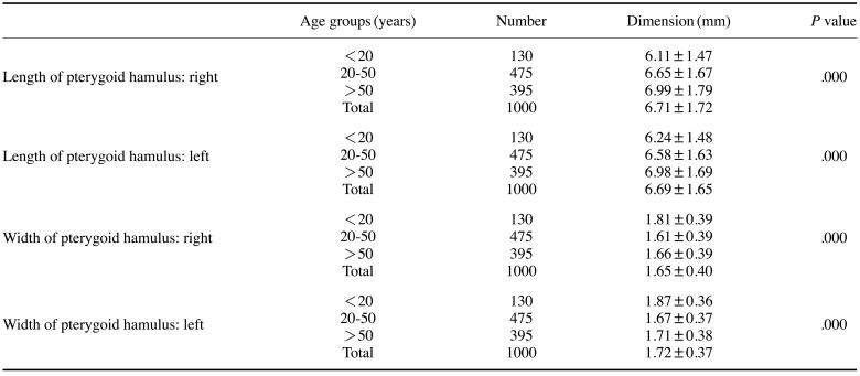
The length of the PH on the right side significantly increased from group 1 (6.11±1.47 mm), through group 2 (6.65±1.67 mm) to group 3 (6.99±1.79 mm) and on the left side from group 2 (6.58±1.63) to group 3 (6.98±1.70). The width of the PH significantly decreased from group 1 (1.81±0.39 mm) to group 2 (1.61±0.39 mm) on the right side, and similarly from 1.87±0.36 mm to 1.67±0.37 mm on the left side. PH length (7.18±1.81 mm on the right side and 7.10±1.72 mm on the left side) and width (1.68±0.38 mm on the right side and 1.74±0.36 mm on the left side) were significantly greater in males than in females.
Conclusion
The length of the PH increased with age, whereas width first decreased and then increased. Length and width measurements were significantly higher in males than in females. These findings will aid in the diagnosis of untraceable pain in the oropharyngeal region related to altered PH morphology.
Keywords: Cone-Beam Computed Tomography, Sphenoid Bone, Age Groups, Sex
Introduction
The pterygoid hamulus (PH) is an extension at the inferior end of the medial pterygoid plate of the sphenoid bone. This hook-like process is of great importance for the functioning of several muscles, and contributes to the separation of the oral cavity from the nasal cavity during sucking and swallowing in the growth and development stages and through adulthood. Therefore, the position and length of the PH are important for these functions.1,2
Even though the PH, because of its peculiar morphology, is a noteworthy feature of the skull base, it still remains a relatively unexplored region on the anatomical map and very few studies have been conducted on its morphology. However, the PH is of interest to all disciplines that deal with this region as it is closely related to the maxilla and oropharynx.3
Proficiency regarding the morphology of this structure is helpful for the interpretation of imaging and provides valuable information for the differential diagnosis of pain in the oral cavity and pharynx with no associated etiological factors. Elongation of the PH is associated with a rare syndrome (pterygoid hamulus syndrome), which shows various and complex symptoms in the palatal and pharyngeal regions and also causes pain and discomfort, especially while swallowing.3 When positive findings are observed in the hamular area, this syndrome should be considered in the differential diagnosis.
Therefore, this study was performed to establish reference standards for PH dimensions according to age and sex using cone-beam computed tomography (CBCT) among patients treated at a single institution in India.
Materials and Methods
The study was approved by the institutional review board (approval no. EC-PG-01/OMR/2016). The CBCT scans of 1000 patients (493 males and 507 females), referred to the Department of Oral Radiology were assessed retrospectively from May 2017 to May 2019. The study data were divided into 3 age groups (group 1: <20 years, group 2: 20–50 years, and group 3: >50 years) to facilitate the detection of differences in the dimensions of the PH according to age group.
CBCT images covering the posterior maxillary region, irrespective of the age and sex of patients, were included in the study. CBCT images where borders of the PH were not traceable clearly, images with streak artifacts that obscured the region of interest, and images from patients with a history of maxillofacial trauma involving PH fracture, traumatic removal of maxillary third molars, pathology in the posterior maxillary region, and bone disorders such as osteoporosis or skeletal asymmetries were excluded from the study. Out of 1426 scans obtained from the Department of Radiology, 426 CBCT scans were excluded by applying the above criteria.
All the CBCT scans retrieved from department's archives were obtained using KODAK imaging software version 9000C 3D (Carestream Health Inc, Rochester, NY, USA). All images were taken following a standard protocol for patient positioning and exposure parameter settings, operated at 80 kVp, 5 mA, the required field of view (FOV), a voxel size of 200 µm×200 µm×200 µm, and an image acquisition time of 10.8 s. The CBCT images were obtained in the Digital Imaging and Communications in Medicine (DICOM). Images were viewed on a HP Compaq LE 1911 with a 19-inch VGA LCD display (Hewlett Packard Company, Palo Alto, CA, USA) at a resolution of 1280×1024 pixels using the Kodak dental imaging software (ver. 6.12.10.0; Carestream Health Inc., Rochester, NY, USA).
The images were analyzed by 3 oral and maxillofacial radiologists with at least 5 years of experience in evaluating and reporting CBCT scans. Care was taken to ensure that all the investigators were reasonably matched with regards to their qualification and experience. Before commencement of the study, discussions were held among the investigators regarding how the measurements would be taken and to make sure that the investigators were thoroughly aware of the normal anatomy of the PH region. All images were viewed in coronal sections in a dark and quiet room, with no source of external light. The investigators were allowed to adjust the brightness and contrast of the scans.
The length and width of the PH were measured in coronal sections (Fig. 1A). The length was measured as the distance from the junction of the medial pterygoid plate with the PH to the tip of the PH (Fig. 1B). The greatest length between the reference points was measured. The width was taken as the distance between the most prominent points on the mesial and lateral aspects of the PH, as was done by Oz et al. (Fig. 1C).4
Fig. 1. A. The pterygoid hamulus is seen on a coronal cone-beam computed tomographic section (white arrow). B. The length of the pterygoid hamulus is measured from the base of the medial pterygoid to the tip of pterygoid hamulus. C. The width of the pterygoid hamulus is measured at the most prominent part of the pterygoid hamulus on either side.
Three measurements were made by each investigator and the average value was considered. Each of the investigators evaluated every CBCT scan independently on 3 different occasions.
The data obtained were compiled in a spreadsheet in MS Office Excel (v. 2010; Microsoft Corp., Redmond, WA, USA). The data were subjected to statistical analysis using IBM SPSS version 20.0 (IBM SPSS Statistics for Windows, IBM Corp., Armonk, NY, USA) software. Descriptive statistics, such as patients' age and sex, were recorded. One-way analysis of variance (ANOVA) was used to determine whether there were any statistically significant differences among the mean values of the 3 independent groups, but information about which specific groups showed statistically significant differences from each other could not be deduced using this test. Therefore, to determine which specific groups differed significantly from each other, the post hoc Tukey honest significant difference (HSD) test was done. Length and width were compared across different age groups using 1-way ANOVA. Post hoc test was done for multiple comparisons among different age groups using the Tukey HSD test. Length and width were compared according to sex using the t-test. Inter-examiner reliability was checked using 1-way ANOVA to determine whether any statistically significant differences existed among the observations of all 3 investigators. The intra-class correlation coefficient (ICC) was also calculated to assess intra-examiner reliability. For all the statistical tests, a P value<0.05 was considered to indicate statistical significance. With 5% α error and 20% β error, the power of the study was 80%.
Results
In the present study, 1,000 CBCT scans from 493 male and 507 female patients were assessed. CBCT scans were analyzed in coronal sections to measure the length and width of the PH on the right and left sides. The age of the patients ranged from 6 to 86 years, with a mean age of 43.5±18.3 (standard deviation) years.
Intra-investigator reliability, as measured using the ICC, was significantly higher for investigator 2 for all the parameters than for investigator 1 and 3. A statistically significant difference was seen in the ICC values for the right-side length of the PH for investigator 1 and the left-side length of the PH for investigator 3. Inter-investigator reliability, as assessed using 1-way ANOVA, showed no significant differences among the investigators in the mean measurements of PH width on the right side and PH length on both sides (P>0.05). The mean values of the measurements made by investigators 1, 2, and 3 were used as the final data for further analysis.
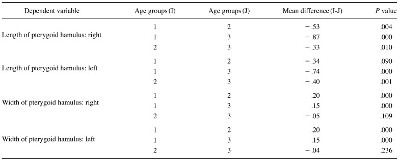
A highly significant relationship was observed between age and PH length using one-way ANOVA. As age advanced, the length of the PH increased (Table 1). This increase in length was statistically significant for the right side (6.11±1.47 mm in group 1, 6.65±1.67 mm in group 2, and 6.99±1.79 mm in group 3), and a steady increase in PH length with age was also found on the left side. This increase was significant from group 2 to group 3 and between group 1 and group 3 (Table 2).
Table 1. Comparison of length and width of the pterygoid hamulus across different age groups using 1-way analysis of variance.
Table 2. Post hoc tests for multiple comparisons among age groups using the Tukey honest significant difference test.
1: <20 years, 2: 20–50 years, 3: >50 years
The width of the PH first significantly decreased from group 1 to group 2 (from 1.81±0.39 mm to 1.61±0.39 mm on the right side and from 1.87±0.36 mm to 1.67±0.37 mm on the left side), and then a non-significant increase occurred from group 2 to group 3 (1.66±0.39 mm on the right side and 1.71±0.38 mm on the left side) (Tables 1 and 2).
The length and width of the PH were compared according to sex using the t-test. The length of the PH was statistically significantly longer in males than females on both sides. In males, the length was 7.18±1.81 mm on the right side and 7.10±1.72 mm on the left side, whereas in females, it was 6.26±1.48 mm on the right side and 6.30±1.47 mm on the left side. The width of the PH was greater in male patients than in female patients. It was 1.68±0.38 mm on the right side and 1.74±0.36 mm on the left side in males, and 1.63±0.41 mm on the right side and 1.69±0.37 mm on the left side in females. However, the width of the PH did not show a significant difference by sex (Table 3).
Table 3. Comparison of the length and width of the pterygoid hamulus on both sides according to sex using the t-test.
When comparing the length and width measurements of PH on the right and left sides, the width of PH was significantly higher on the left side in group 2. No significant difference was found for the length of the PH on both sides in any age groups. The width of the PH was greater for both males and females on the left side, but no such difference was found for the length of the PH on both sides.
Discussion
The position and morphology of the PH, especially its length, are of great importance for the functioning of muscles that are located in the region. Alterations in the morphology of the PH or associated structures can cause symptoms common to other diseases, such as pain when chewing or swallowing; edema and erythema in the posterior region of the palate; and ear pain, hearing loss, and autophony. These lesions can be misdiagnosed as temporomandibular disorders or glossopharyngeal neuralgia.5
It is of the utmost importance for dentists to have a thorough knowledge regarding the PH and its anatomical and functional relationships with neighboring structures for the accurate diagnosis, prevention, and management of diseases in the oropharyngeal region. However, few studies have been reported in the literature on PH morphology, and the extant studies have been conducted in different populations with varying results.
The present study was conducted to establish reference standards for the dimensions of the PH in accordance with age and sex in the Indian population, which may help in the diagnosis of untraceable pain in the oropharyngeal region related to altered PH morphology.
In our study, the length of the PH increased as age advanced. The increase in PH length was significant between age group 1 (<20 years) and group 3 (>50 years) on the right side and between age group 2 (20–50 years) and age group 3 (>50 years) on the left side. Contrasting results have been reported in the literature regarding age-related changes in the length of the PH. The study of Putz and Kroyer1 on a German population reported that the PH increased in length to the beginning of adulthood, and thereafter remained unchanged throughout life. However, Krmpotić-Nemanić et al.3 in Croatia and Orhan et al.2 in Turkey reported that older patients had shorter PHs than younger patients. Krmpotić-Nemanić et al.3 found that children (0–9 years) had the shortest PH length (3.6 mm), and that the PH length then increased in adults (21–59 years) to 6.9 mm and finally decreased significantly in the elderly group (60–100 years) to 5.0 mm. The researchers proposed that in adults, the factors responsible for lengthening of the PH could be the attachment of the buccopharyngeal raphe and its mechanical load-ing during chewing and swallowing. In infants, the PH is short, but then elongates to finally reach the upper part of the buccopharyngea raphe, which is fixed to the buccinatoria crista of the mandible. The loss of teeth and the resultant lack of biomechanical loading on the PH lead to shortening of the hamulus in old age.3 Similar to our findings, Komarnitki et al.6 found that the length of the PH increased with age.
The width of the PH in the present study significantly decreased from group 1 to group 2 and then increased non-significantly from group 2 to group 3 on both sides. Only 2 studies2,7 in the literature have evaluated the width of the PH across different age groups. Romoozi et al.7 divided the study population into 3 age groups (15–29, 30–44, and 45–59 years) and found that the average width of the PH on both sides decreased with increasing age. Because there was a substantial difference in the age distribution from our study, a comparative evaluation could not be done. The width measurements across different age groups in the present study are in contrast to the findings of Orhan et al.2 They evaluated the width of the PH in 2 age groups (22–55 years and older than 55 years) and found a statistically non-significant difference between these age groups, with PH width decreasing slightly from 1.85±0.88 mm in the younger age group to 1.73±1.07 in the older age group. This difference in the width of the PH across different age groups could be attributed to the difference in the population under investigation.
In the present study, the length of the PH was found to be statistically significantly longer in males than in females. The PH was also wider in males than in females, but the findings for width were statistically non-significant. Similar to our results, Romoozi et al.7 also found that the average length of the PH on both sides was significantly higher in males (6.6 mm) than in females (6.4 mm). Orhan et al.2 did not find any significant difference in PH dimensions according to sex.
According to the study of Nerkar et al.8 on an Indian population, the mean length of the PH was significantly higher in males (7.56±0.5 mm and 7.72±0.5 mm on the right and left sides, respectively) than in females (6.62±0.3 mm and 6.78±0.3 mm on the right and left sides, respectively). Their study did not find any significant differences in width between males and females. Their findings for the length and width of PH are similar to our findings. To the best of our knowledge, theirs is the only other study that has investigated this issue in an Indian population.
No significant difference was found according to age in the length measurements between the right and left sides. However, the width was significantly greater on the left side in group 2. Likewise, the length of the PH showed no significant difference between the right and left sides according to sex. The width of the PH was greater on the left side than on the right side in both sexes. There is no consensus regarding the cause of the difference in width between the left and the right PH, although it can be hypothesized that the disturbances in the distribution of forces exerted by stomatognathic muscles may contribute to changes in the dimensions of the PH.
The findings regarding length in the present study are similar to those of earlier studies, and no study in the literature has reported significant differences in the width measurements between the left and right sides according to age or sex.
From the clinical point of view, it is worth noting that the PH, because of its tendon attachments and the “pulley” it provides for the tensor veli palatini, is certainly a potential source of irritation. A morphological abnormality of the PH may provoke pain through mechanical irritation of surrounding tissues, impaired contraction of the tensor veli palatini muscle, or fibrosis or inflammation of the tensor veli palatini bursa due to excessive pressure on the palatine aponeurosis. In these processes, the greater palatine, lesser palatine, facial, or glossopharyngeal nerves can be stimulated, which may cause pain in various head and neck regions.9,10,11
The main diagnostic dilemma regarding patients with pterygoid hamulus syndrome is that it is not associated with specific clinical symptoms.12 The rarity of pterygoid hamulus syndrome, in combination with the absence of specific clinical symptoms, poses a significant diagnostic challenge.
Due to the potential complications caused by morphological alterations of the PH, maxillofacial radiologists must assess radiographs (particularly CBCT) with a focus on the morphology of the PH, especially its length. The findings of this study therefore indicate that anatomical variations in the dimensions of the PH according to age and sex should be considered for an accurate diagnosis of untraceable pain in the oropharyngeal region. The signs and symptoms that result from these changes may be difficult to diagnose if the anatomy is not taken into consideration.
The principal limitation of this study is that it was conducted among patients treated at a single institution. To establish more definitive reference standards for PH dimensions, research should be conducted at multiple centers with a large study population.
This retrospective study did not allow follow-up to ascertain or diagnose whether patients with an elongated PH presented with any symptoms or signs of pterygoid hamulus syndrome. Prospective research could be conducted to evaluate the possible association of pterygoid hamulus syndrome with variations in the dimensions of the PH.
In future studies, PH dimensions should be assessed in a broader population, since establishing reference standards for the dimensions of the PH according to age and sex parameters could provide a valuable tool for determination of age in the forensic sciences. No studies in the literature have utilized the dimensions of the PH for forensic age determination, which could be a promising topic for exploration in future studies.
Footnotes
Conflicts of Interest: None
References
- 1.Putz R, Kroyer A. Functional morphology of the pterygoid hamulus. Ann Anat. 1999;181:85–88. doi: 10.1016/S0940-9602(99)80099-5. [DOI] [PubMed] [Google Scholar]
- 2.Orhan K, Sakul BU, Oz U, Bilecenoglu B. Evaluation of the pterygoid hamulus morphology using cone beam computed tomography. Oral Surg Oral Med Oral Pathol Oral Radiol Endod. 2011;112:e48–e55. doi: 10.1016/j.tripleo.2011.02.038. [DOI] [PubMed] [Google Scholar]
- 3.Krmpotić-Nemanić J, Vinter I, Marusić A. Relations of the pterygoid hamulus and hard palate in children and adults: anatomical implications for the function of the soft palate. Ann Anat. 2006;188:69–74. doi: 10.1016/j.aanat.2005.05.005. [DOI] [PubMed] [Google Scholar]
- 4.Oz U, Orhan K, Aksoy S, Ciftci F, Özdoğanoğlu T, Rasmussen F. Association between pterygoid hamulus length and apnea hypopnea index in patients with obstructive sleep apnea: a combined three-dimensional cone beam computed tomography and polysomnographic study. Oral Surg Oral Med Oral Pathol Oral Radiol. 2016;121:330–339. doi: 10.1016/j.oooo.2015.10.032. [DOI] [PubMed] [Google Scholar]
- 5.Dupont JS, Jr, Brown CE. Comorbidity of pterygoid hamular area pain and TMD. Cranio. 2007;25:172–176. doi: 10.1179/crn.2007.027. [DOI] [PubMed] [Google Scholar]
- 6.Komarnitki I, Mankowska-Pliszka H, Ungier E, Dziedzic D, Grzegorczyk M, Tomczyk A, et al. Functional morphometry of the pterygoid hamulus. A comparative study of modern and medieval populations. Anthropol Rev. 2019;82:389–395. [Google Scholar]
- 7.Romoozi E, Razavi SH, Barouti P, Rahimi M. Investigating the morphologic indices of the hamulus pterygoid process using the CBCT technique. J Res Med Dent Sci. 2018;6:240–244. [Google Scholar]
- 8.Nerkar A, Gadgil R, Bhoosreddy A, Shah K, Varma S. Cone-beam computed tomography study of morphometric evaluation of pterygoid hamulus. Int J Recent Adv Multidiscip Res. 2017;4:2678–2682. [Google Scholar]
- 9.Kronman JH, Padamsee M, Norris LH. Bursitis of the tensor veli palatini muscle with an osteophyte on the pterygoid hamulus. Oral Surg Oral Med Oral Pathol. 1991;71:420–422. doi: 10.1016/0030-4220(91)90420-h. [DOI] [PubMed] [Google Scholar]
- 10.Sasaki T, Imai Y, Fujibayashi T. A case of elongated pterygoid hamulus syndrome. Oral Dis. 2001;7:131–133. [PubMed] [Google Scholar]
- 11.Sattur AP, Burde KN, Goyal M, Naikmasur VG. Unusual cause of palatal pain. Oral Radiol. 2010;27:60–63. [Google Scholar]
- 12.Komarnitki I, Skadorwa T, Chloupek A. Radiomorphometric assessment of the pterygoid hamulus as a factor promoting the pterygoid hamulus bursitis. Folia Morphol (Warsz) 2020;79:134–140. doi: 10.5603/FM.a2019.0049. [DOI] [PubMed] [Google Scholar]


