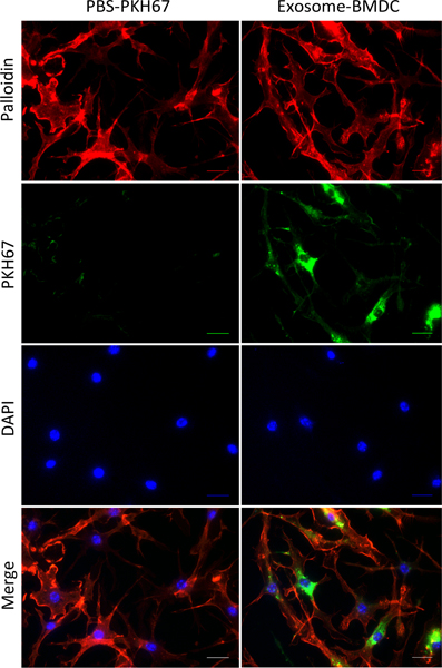
Figure 2.
Incorporation of exosomes in BMDCs. Exosomes isolated from culture supernatants were dissolved in PBS, incubated with PKH67 (green fluorescent dye) for 5 minutes and then washed with 1% BSA in PBS (Exosome+ BMDC). Both exosomes and the PBS control (PBS+ PKH67) were treated with BMDCs incubated on chamber slides. After 24 hours of incubation, the PKH67-labeled exosomes were taken up by BMDCs (red: F-actin; green: PKH67; blue: DAPI; scale bar: 50 μm).

