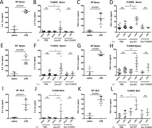
Figure 5.
TLR4KO mice gained responsiveness to LPS after the i.v. injection of exosomes from WT BMDCs. Measurement of TNFα and IL-6 in the serum (A-D), spleen (E-H) and mesenteric lymph nodes (MLN) (I-L) of WT and TLR4KO mice after the injection of exosomes and LPS (the results are expressed as the mean ± SEM of measurements from N=6–12 per group, and each dot represents a measurement from one animal; *p<0.05, **p<0.01, ***p<0.001).

