Abstract
The COVID‐19 pandemic has generated great interest in reviving an old intervention technology, particularly for air disinfection—ultraviolet germicidal irradiation (UVGI). Since UVGI was developed and refined more than 80–90 years ago, the ultraviolet source of choice has been almost exclusively the low‐pressure mercury vapor discharge lamp. Today, with new lamp technologies, there has been significant interest in the application of ultraviolet light‐emitting diodes and excimer lamps that emit in the UV‐C (180–280 nm) spectral band. This paper reviews these competing technologies with the aim of giving a sound basis for decisions on how to choose and install UV systems for disinfection of air and surfaces given the COVID‐19 pandemic.
For the three main types of UVGI technologies discussed in the report, the most interesting is the Excimer lamps source, particularly that formed between krypton and chloring, KrCl*. The reason for the interest in this source, still in development, is that at the strong emission wavelength at about 222 nm, the penetration depth of the skin is so short that the radiation does not reach the live part of the skin. Thus, it has the potential of being safely used to inactive viruses, such as that causing Covid‐19, in the presence of humans.
![]()
Introduction
Germicidal ultraviolet (GUV) emission or radiation encompasses a continuous range of UV wavelengths from 200 to 280 nm, which upper limit is identical to UV‐C region as defined by the CIE. The lower limit for UV‐C is defined as 100 nm, but due to difficulty of material transmission and the creation of ozone below 200 nm, the lower practical limit for UV‐C is usually set at 200 nm. In practice, not all wavelengths in the UV‐C region are used as predominant wavelengths are determined by the type of lamp or solid‐state source, with or without filters, as needed for safety. The major focus of this report is the GUV source technologies that can be used to disinfect both the room air and surfaces. Low‐pressure mercury (Hg) lamps have been used for this purpose for over 90 years and dominate the germicidal UV market. However, the rapidly evolving technology of excimer lamps, particularly KrCl*, and LEDs that emit in the UV‐C region are finding important applications.
From an electrical efficiency perspective, low‐pressure mercury lamps are most efficient in generating GUV and have a life of 6–8000 h (about one year of continuous use). Much of that efficiency is lost, however, when Hg fixtures used in the upper room are louvered to prevent occupant overexposure. Currently, LEDs are least efficient, and the efficiency goes down as the wavelength is reduced. Life of these lamps is not known well at this point in time but claims of 10 000 h are made by producers. KrCl* lamps are intermediate in efficiency and can potentially be used directly in occupied rooms. They generally have a 3000‐hour lamp life. KrBr* lamps, which emit strongly at 207 nm have also been investigated (1, 2).
More complex sources such as both continuous and pulsed xenon arc lamps are used for special applications, in particular studies where large amounts of UV are needed. Often pulsed xenon sources are used for scientific studies where the UV output from a pulsed xenon source is put through a monochrometer to study the effect, for example, virus inactivation, as a function of wavelength. Pulsed Xe lamps may be necessary if very rapid disinfection is required (3).
Medium‐pressure Hg lamps are used almost exclusively for water purification. Such sources have been used for that purpose for quite sometime and are the best sources for that purpose.
All the above technologies—continuously emitting or pulsed—are comparably effective for disinfection given that the effect of wavelength is taken into account through actinic weighting of the irradiance.
Sources for Air and Surface Disinfection
Low‐pressure Mercury Lamps
Historically, low‐pressure mercury lamps (LPM) have been used for disinfecting air, water and surfaces for nearly a century. Matthew Luckiesh, in a book published in 1946, describes LPM UV Germicidal systems developed by GE for disinfecting water, air and surfaces (4).
Currently, LPM lamps are still the most practical and most efficient method of generating germicidal radiant energy. These lamps are electrically the same as fluorescent lamps of corresponding sizes and wattages and require essentially the same auxiliary equipment (ballasts). They differ physically from fluorescent lamps in that they contain no phosphor and are constructed with a special kind of glass that transmits about 80 percent of the UV energy generated by the Hg discharge at 253.7 nm. Figure 1 shows two examples of low‐pressure Hg germicidal lamps.
Figure 1.
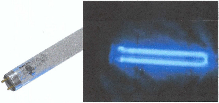
Two examples of germicidal low‐pressure Hg lamps.
The efficiency of LPM lamps depends on the amount of energy that is contained in the Hg 253.7‐nm emission for a given input wattage applied. For tubular LPM lamps, similar to fluorescent lamps, this efficiency will increase with the length of the discharge as end losses due to electrode interactions become a smaller portion of the total power input. Further, the efficiency has an intrinsic maximum at an optimum Hg vapor pressure on the order of 1 Pa for tubular lamps with a diameter of about 1 inch (T8). The optimum Hg pressure is reached when the liquid Hg temperature (cold spot) is about 40°C for such lamps filled with a rare gas, usually Argon, at a pressure of 100–400 Pa. As the Hg pressure increases the number of Hg* (metastable excited state) increases, so the number of photons emitted also increases. (Anyone trying to use a fluorescent lamp outside in winter is aware that there is not much light initially.) However, as the temperature increase past the optimum (~40°C) the number of Hg atoms increases exponentially leading to absorption of the photon by another Hg atom which, among other things, cause a loss of the Hg*, that is, no 253.7‐nm photon. If one continues heating the lamp until the liquid Hg is all in the vapor phase, you have a medium‐pressure Hg lamp (see below).
In a given application, the actual steady‐state efficiency of a LPM luminaire is likely below the optimum value. How much can be determined by taking emission measurements at regular intervals, say every 30–60 s, until steady‐state emission is obtained. The difference between the peak and steady‐state emission is the luminaire design loss. If such luminaires are operated in a very hot environment, say a room in the tropics without air conditioning, the effective Hg cold spot temperature will likely be well over 40°C and the efficiency of the unit significantly below its rating.
Amalgams of Hg and other materials are often used for LPM lamps in hot environments or where it is desired to drive the lamp at higher power. In this case, the optimum Hg pressure is reduced as a function of temperature, and the optimum temperature is now ~ 60°C rather than ~ 40°C.
The effective life for germicidal LPM lamp is usually significantly shorter than that of fluorescent lamps, as maintaining the transmission of the Hg 253.7 nm line through the glass over life is the main life‐limiting mechanism. Depending on the glass quality, the emission of the 253.7 nm line declines to about 70 % of its initial value in about 7000 h.
Low‐pressure mercury germicidal lamps using cold‐cathode electrodes are also available in various sizes, usually for shorter, smaller‐diameter lamps such as those typically are used in wands. Their operating characteristics are similar to those of hot‐cathode lamps, but their starting mechanisms are different.
Figure 2 shows a typical measurement of a GE G30T8, a germicidal low‐pressure Hg lamp (R. S. Bergman, unppublished data). The strength of the emission at the various wavelengths between 200 and 800 nm is plotted logarithmically. The special UV glass transmits about 80% of the generated 253.7‐nm radiation, which is about 45 % of the total electrical power delivered to the lamp, that is, nearly half the input power from the lamp is available for UV disinfection. Referring to Fig. 2, by summing the radiated power over the UV and visible region we find that approximately 85% is emitted at 253.7 nm (usually rounded to 254 nm). Of the remainder a, about 3% is emitted in UV lines and 7% in the visible lines; about 4% is the Bremsstrahlung continuum radiation peaking at about 300 nm, that is, photon emission due to collisional deceleration of electrons in an electric field. In the UV‐B region, the combined lines and Bremsstrahlung add to about 3% of the total emission. While emission in the UV‐B is small compared to the total, the UV‐B emission penetrates much deeper into the skin than does the 254‐nm emission; as such the UV‐B emission is likely an important part of the photobiological safety concern with LPM lamps.
Figure 2.
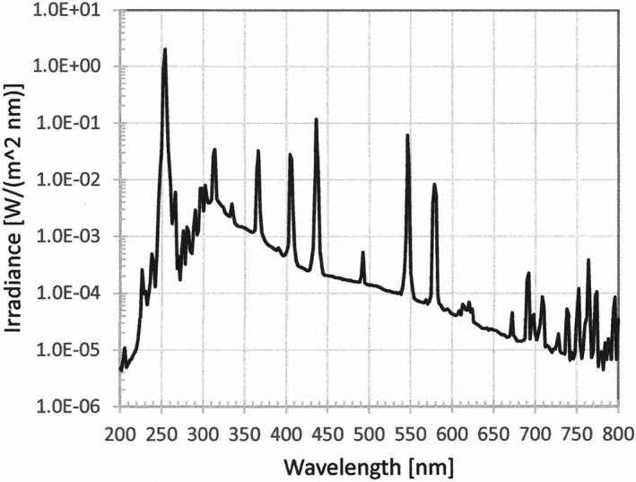
UV and visible irradiance spectrum for a germicidal low‐pressure Hg lamp (G30T8); the radiance is plotted logarithmically.
Excimer Lamps
Excimer lamps are quasimonochromatic sources that emit over a wide range of the UV region depending on the gas used. In such a gas discharge, which normally has no dimer molecules, a dimer molecule is formed between an excited state atom (normally the lowest and most stable state) and a neutral atom, which then dissociates by emitting a high energy photon, for example, XeXe* dissociates to 2 Xe atoms plus a 172‐nm photon (in the vacuum UV region). (5) Rare gas dimers have been used as the basis of excimer lasers for quite sometime. It has also been known for sometime that the combination of a rare gas and a halogen produces dimers that dissociate via emission of a photon.
Krypton‐chloride excimer (KrCl*) lamps have been shown to produce significant emission in the UV‐C region in a narrow wavelength band around 222 nm. The advantage of KrCl*is that the deactivation efficiency of bacteria and viruses at 222 nm appears to be roughly the same as emission at 270–280 nm while the effect of the emission on human skin appears to be much reduced compared even to the 253.7‐nm mercury emission (6, 7). Thus, these sources are being considered for disinfection of air and surfaces in occupied areas. However, in the above studies a small but significant emission at around 259 nm and higher, on the order of 20 to 100 times smaller than the main 222‐nm emission, became a concern. A concerted effort at Columbia University and other institutions to reduce the longer‐wavelength emission through use of special multi‐layer filters. The normalized spectral irradiance of a KrCl* lamp in the UV region is shown in Fig. 3 with and without a special filter designed to drastically reduce the irradiance above 230 nm (8).
Figure 3.
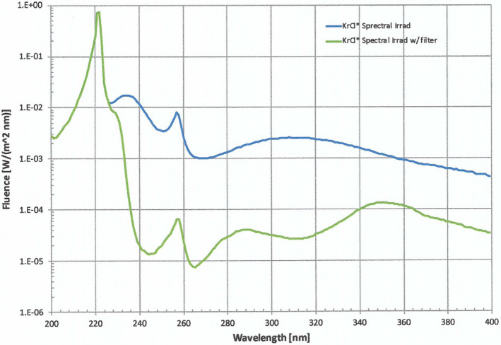
Spectral irradiance of a KrCl* lamp as a function of wavelength in the UV region with and without a filter to reduce the irradiance above 230 nm.
Note that the width of emission line at 50% of peak (HBW) of the 222‐nm emission is on the order of 5 nm. As will be shown later, this is less than the HBW of UV‐C LEDs.
A capacitive discharge is most often used to form the plasma or discharge of the excimer lamp. Such lamps can take multiple forms from a tubular structure to a flat panel and with power levels from 10 to a few hundred watts. At this time, excimer sources are being used in specific environments, but their presence in the disinfection marketplace is still very limited in comparison to that of germicidal LPM lamps, and there is little experience yet with any widespread use. However, the potential benefit of using these sources where humans are present will likely spur photobiological studies as well as luminaire commercial development.
UV‐C LEDs
There are several companies producing LEDs that emit in the longer‐wavelength portion of the UV‐C region, generally at 265–275 nm but some are being made and sold that have their output at 255 nm. Such LEDs are being tested for both upper air and surface disinfection. Where validated, UV‐C LEDs could be used for both targeted surface disinfection and upper room air disinfection (Fig. 4).
Figure 4.
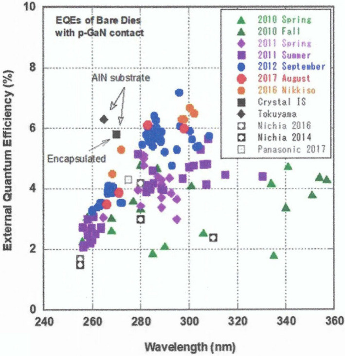
FEQE mapping of AlGaN‐based LED dies with p‐GaN contact layer. The data are mainly for LED dies on planar sapphire from our group (colored). EQEs of LED dies from elsewhere (uncolored) using AlN substrates and sapphire substrates with a p‐GaN contact layer are also shown. Solid uncolored symbols indicate the use of an AlN bulk substrate, where “_” indicates with encapsulation and “u” indicates no encapsulation. The EQEs in the case of an AlN substrate (“_” and “u”) were achieved with a roughened light extraction surface. Uncolored open marks represent LED dies with p‐GaN layer fabricated on planar sapphire reported by other groups. All of our data (colored) were obtained without boosting light extraction efficiency (LEE) using encapsulation or a sapphire lens (9).
The life of UV‐C LEDs is usually claimed to be at least 10 000 h, comparable or slightly better than LPM lamps. Since LEDs are directional emitters, this provides an advantage for the use UV‐C LEDs in upper room UVGI systems as it simplifies the system design, as the need for complex reflectors and possibly some of the baffling louvers may not be required as much simpler lenses or reflectors can be used with the LEDs to produce luminaires with the nearly horizontal emission needed for upper room UVGI. Thus, while the best efficiency of UV‐C LEDs in the 265–275 nm region is currently around 4 %, that is, an order of magnitude below the efficiency of the 253.7‐nm emission from a low‐pressure mercury lamps, the actual difference in delivered irradiance may not be that much lower than LPM in a UVGI system with louvers. Upper room air systems using LEDs are being tested currently for disinfection of tuberculosis in other parts of the world (10), and with the emergence of the SARS‐CoV‐2 virus, the use of LEDs in various disinfection configurations is now a reality.
A potential disadvantage of the UV‐C LEDs operating at a peak wavelength around 270 is that some of the LED emission on the high‐wavelength side spills over into the UV‐B region. UV‐C LEDs made with a peak wavelength around 270 nm have a HBW of about 15 nm and a 30 nm bandwidth at 10 % of the peak emission. Thus, about 20 percent of the LED output winds up in the UV‐B region. Further, the actual peak wavelength of LED is not that well controlled so a shift in peak wavelength of 5 nm in either direction is not unexpected. On top of that, the peak wavelength shifts to higher wavelengths as the junction temperature increases. If UV‐C LED devices are desired that do not have any emission in the UV‐B, then a peak design of about 260–265 nm would be needed.
LEDs offer the following advantages: flexible form factor, intensity tunability, wavelength choices and no start‐up or cool‐down time. Further, both visible‐ and UV‐emitting emitting LEDs can be put together in a single luminaire. Another advantage of LEDS is that they can be rapidly pulsed, if needed, since the radiation emitted from LEDs responds to the controlling electrical current waveform in microseconds.
A major disadvantage of LED’s is heat dissipation. Thus, thermal management of the luminaires containing many LEDs must be included in the design of the luminaire. For example, if it is desired to an Upper Room UVGI using LEDs that produced 5 watts of UV‐C output, one would need 250 watts, 250 1‐W LEDS, with assumed efficiency of 2% (half of light lost in keeping radiation in upper room). This would create a significant thermal management problem for the luminaire design to keep LED junctions at a reasonable temperature. Note that the radiant flux, life and reliability of LEDs decrease significantly with increasing temperature.
As noted above, almost all of the currently available germicidal UV‐C LEDs operate with a peak output in the 265 to 270‐nm region. Producing UV‐C LEDs at 255 nm or even lower would be desirable. UV‐C LEDs at a peak wavelength between 255 and 260 nm are being produced and put into germicidal luminaires. I do not have access to data on these devices, but I expect that the best efficiency would be around 0.2 %, but with rapid development in the fields such LEDs will likely become more efficient and may prove to become important devices that in the long run replace the LPM lamps.
Ozone
A potential disadvantage of UV emission below 240 nm is that ozone is produced. Thus, current LPM lamps and LEDs are not a concern as they produce ozone below the background ozone level, which is on the order of 0.01 ppm (parts per million). The type of glass/quartz used in for the lamp, even for LPM lamps, is important as emission from the Hg 185‐nm line would create large amounts of ozone. The glass or quartz material used is intended to absorb the 185‐nm emission keeping it from entering the atmosphere.
Not much information about ozone generation has been reported by most manufactures of KrCl* lamps, although it is usually assumed that it is below OSHA guidelines. OSHA guidelines are based on the ACGIH TLV’s (11). The maximum permissible exposure level (PEL) based on time‐weighted average exposure for 8 h at 0.1 ppm for light work and 0.05 ppm for heavy work. Measurements for ozone from a KrCl* luminaire systems have been made by USHIO (12) which show that the produced ozone depends strongly on the power of the lamp, as is expected, and also on the lamp and luminaire design. For lower power sources, the ozone levels generated are well below the OSHA levels. However, for lamp designs of a few hundred watts, depending also on the lamp/luminaire design, the amount of ozone generated will create levels above the OSHA limits unless the air in the room is well ventilated. Thus, for KrCl* lamps, particularly for high power systems, active air ventilation needs to be considered as part of the system design.
Other GUV Sources
Medium‐pressure Mercury Lamps
Medium‐pressure mercury lamps are almost exclusively used in water purification. Such lamps are usually made of quartz tubes of various lengths and diameters depending on the power required with tungsten electrodes. For water treatment, ozone generation is not a problem, but likely will help purification. The mercury content is controlled so that all the mercury evaporates during operation giving a mercury pressure on the order of 10 mm Hg (~1000 Pa). If the liquid mercury content is high enough so that not all of it is evaporated, the result is a high‐pressure mercury lamp rather than a medium‐pressure lamp. Typical output for medium‐pressure Hg lamps is on the order of 100 W cm−2 as compared to about 0.03 W cm−2 for a germicidal low‐pressure mercury lamp (also a fluorescent lamp). While the efficiency of the 253.7‐nm emission is significantly reduced and broadened due to reabsorption of the radiant energy, as can be seen in Fig. 5, the broadened self‐absorbed 253.7‐nm emission is nevertheless the major source of germicidal radiation.
Figure 5.
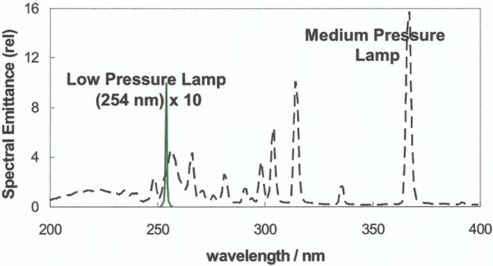
Comparison of low‐pressure mercury lamp (see Fig. 2) and a typical medium‐pressure mercury lamp.
Xenon Lamps
Xenon (Xe) arc lamps, which can be continuous operating short‐arc lamps or long‐arc lamps or pulsed lamps, generate substantial UV radiation. Such lamps are usually made with quartz envelopes. Xenon lamps can be used to create ozone if pure quartz glass is used but designs for most other purposes contain a doping agent in the quartz envelope to prevent emission below 200 nm.
Continuous operating short‐arc xenon lamps are filled to a very high pressure, usually on the order of 30 atmospheres, and produce a very intense short arc at the tip of the cathode. This intense radiation is fairly continuous through the visible region although line structures are easily observable. High power versions—up to 15 kW—of a short‐arc xenon lamp are used for movie projection, and low‐wattage versions were used as headlights.
Long‐arc versions which are similar to medium‐pressure mercury lamps and have multiple uses, for example, sterilization and UV curing. These lamps are filled at pressures near one atmosphere and operate in a similar manner to medium‐pressure mercury lamps.
Pulsed xenon lamps come in great varieties from the flash lamp on a camera to tower warnings for aircraft. The current pulse is usually short, on the order of microseconds, and the instantaneous power delivered during the pulse can be megawatts. The spectrum during the pulse thus becomes a quasi‐continuum in the UV and visible regions. Figure 6 shows the broadened spectrum of the pulsed lamp, particularly in the UV‐C region, as compared to the emission from a continuous operating xenon lamp. Thus, pulsed xenon lamps can simulate the emission spectrum of sunlight. Because the emission from a xenon lamp, particularly a pulsed xenon lamp, is very hazardous to both skin and eyes, direct viewing should be precluded. Further, in some cases, where the lamp contains xenon significantly above one atmosphere, the lamp needs to be encased, as rupture of the envelope is a possibility.
Figure 6.
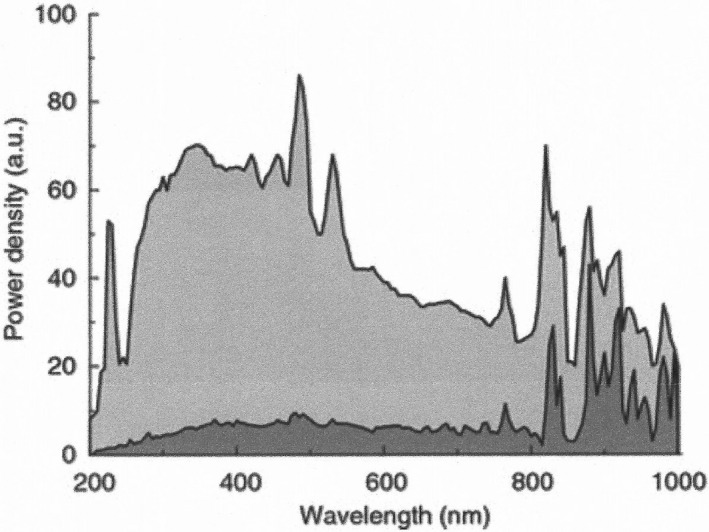
Difference in spectral characteristics between continuous (dark shading) and pulsed modes (light shading).
Biography
Rolf S. Bergman is currently an independent consultant (sole proprietor) in lighting technology and measurements. After graduating with a Ph.D. in Electrical Engineering in 1972 from the University of Minnesota, Dr. Bergman worked for over twenty‐eight years at GE Lighting, all at Nela Park, Cleveland, OH, both as an individual contributor and manager in lamp technology. While at GE Lighting, he was involved in new product and process development, measurement capability and industry standards. Dr. Bergman was named Chief Scientist, Lamp Technology in 1992, a position he held until retirement in 2001. Currently, among other consulting work, he serves as an assessor of lighting laboratories for accreditation to NVLAP, an accrediting body organized at the National Institute of Standards and Technology (NIST). Dr. Bergman served as President of the CIE/USA National Committee from 11/2003 to 11/2008. He chaired the CIE technical committee that produced a global standard for photobiological risk evaluation of lamps. He serves as a member of the IESNA Technical Procedures and Photobiology Committee, and he is a member of CORM, an industry group that advises NIST on measurement needs in US industry. While at GE, he was the author or co‐author of 19 US Patents and published about 20 Journal articles with an additional 20 to 30 internal GE reports.

This article is part of a Special Issue dedicated to the topics of Germicidal Photobiology and Infection Control.
References
- 1. Buonanno, M. , Randers‐Pehrson G., Bigelow A. W., Trivedi S., Lowy F. D., Spotniktz H. M., Hammer S. M. and Brenner D. J. (2013) 207 UV light – A promising tool for safe low‐cost reductio of surgical site infections. I: In‐vitro studies. PLoS One 8, e76968. [DOI] [PMC free article] [PubMed] [Google Scholar]
- 2. Buonanno, M. , Stanislauskas M., Ponnaiya B., Bigelow A. W., Randers‐Pehrson G., Shuryak I., Smilenov L., Owens D. M. and Brenner D. J. (2016) 207 UV light – A promising tool for safe low‐cost reductio of surgical site infections. II: In‐vivo safety studies. PLoS One 11(6), e0138418. [DOI] [PMC free article] [PubMed] [Google Scholar]
- 3. Wang, T. , MacGregor S. J., Anderson J. G. and Woolsey G. A. (2005) Pulsed ultra‐violet inactivation spectrum of Escherichia coli . Water Res. 39(13), 2921–2925. [DOI] [PubMed] [Google Scholar]
- 4. Luckiesh, M. (1946) Applications of Germicidal, Erythemal and Infrared Energy. D Van Nordstrand Company, New York. [Google Scholar]
- 5. Basov, N. G. et al (1970) Zh. Eksp. Fiz. I Tekh. Pis’ma. Red. 12, 473. [Google Scholar]
- 6. Welch, D. , Buonanno M., Grilj V., Shuryak I., Crickmore C., Biogelow A. W., Randers‐Pehrson G., Johson G. W. and Brenner D. J. (2018) Far‐UVC light: a new tool to control the spread of airborne‐mediated microbial diseases. Sci. Rep. 8, 2752. [DOI] [PMC free article] [PubMed] [Google Scholar]
- 7. Woods, J. A. , Evans A., Forbes P. D., Coates P. J., Gardner J., Valentine R. M., Ibbotson S. H., Ferguson J., Fricker C. and Moseley H. (2015) The effect of 222‐nm UVC phototesting on healthy volunteer skin: a pilot study. Photodermatal. Photoimmunal. Photomed. 31(3), 159–166. [DOI] [PubMed] [Google Scholar]
- 8. Figure 3 curtesy of Ewan Edie from a prepublication by Eadie, E. , Barnard I. M. R., Ibbotson S. H. and Wood K.. Extreme exposure to filtered far‐UVC: A case study. In press. [DOI] [PMC free article] [PubMed]
- 9. End TB Transmission Initiative (2020) Maintenance of Upper‐Room Germicidal Ultraviolet (GUV) Air Disinfection Systems for TB Emission Control. Copenhagen: United Nations Office for Project Services (UNOPS). http://www.stoptb.org/wg/ett/
- 10. Threshold Limit Values for Chemical Substances and Physical Agents & Biological Exposure Indices, ACGIH (2009) p 46.
- 11. Claus, H. unpublished white paper on ozone generation. In press.
- 12. Figure 4 taken from Nagasawa, Y. and Hirano A. (2018). A review of AlGaN‐based deep‐ultraviolet light‐emitting diodes on sapphire. Appl. Sci. 8, 1264. [Google Scholar]


