Superficial siderosis (SS) is a neurodegenerative condition caused by hemosiderin deposition on the surface of the brain, cranial nerves, and spinal cord. SS is an exceptionally rare condition characterized by a triad of hearing loss, ataxia, and myelopathy. The most common etiologies that lead to SS are trauma, previous surgical procedures, dural tears, and tumors of the central nervous system.1 In our letter published on-line November 4, 2010, in the American Journal of Neuroradiology, we demonstrated the efficacy of deferiprone (Ferriprox), a lipid-soluble iron chelator, for reducing hemosiderin deposition through MR imaging of a single patient on deferiprone for 1.5 years.2 On the basis of this case report, we conducted a pilot trial of 10 patients with SS by using 30 mg/kg/day of deferiprone and found that 4 showed MR imaging changes in as little as 3 months.3 As the only treatment to demonstrate any efficacy, deferiprone is emerging as the standard of care for the treatment of SS. With approval of deferiprone by the US Food and Drug Administration in October 2011, we launched an observational trial of deferiprone in superficial siderosis to characterize the potential clinical benefit of hemosiderin chelation (www.clinicaltrials.gov, identifier NCT01284127).
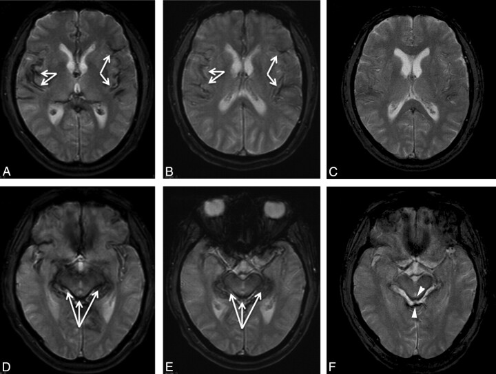
The single subject with SS in the initial case report remained on deferiprone during this time for a total of 3 years 2 months. Clinically, this patient's hearing loss and ataxia, which started 1 year before taking deferiprone, have resolved. He currently has no symptoms. A third MR imaging revealed marked reduction in hemosiderin deposition compared with the original scan and the first follow-up (Fig 1).
Fig 1.
Axial T2* brain MR imaging performed before deferiprone therapy (TR/TE, 800/26 ms; A and D) compared with similarly positioned axial T2* MR imaging after 1.5 years of deferiprone therapy (TR/TE, 560/26 ms; B and E) and 3.2 years of deferiprone therapy (TR/TE, 800/26 ms; C and F). Arrows in A and B show hemosiderin hypointensities along the pial surface of the temporal lobe that are now nearly absent in C. In the midbrain, arrows in D and E show hypointensities around the ventral surface of the brain stem that are now much reduced in F (arrowheads).
Deferiprone is unique among iron chelators in that it is lipid-soluble and can penetrate the blood-brain barrier to target hemosiderin in the central nervous system.4 Because it is currently FDA-approved for thalassemia, we are successfully using this drug off-label for the treatment of superficial siderosis at a dose of 1000 mg twice daily, but only 5 days per week to prevent total body iron depletion. We monitor blood counts for agranulocytosis weekly and monthly for liver toxicity and zinc deficiency. Twice yearly MRI confirms the effective chelation of hemosiderin by deferiprone. In our experience, with this patient and others, the clinical response can take many months to years to manifest, depending on the volume and extent of hemosiderin deposition at presentation. Although SS affects fewer than 200 individuals worldwide, we are hopeful that these patients may improve with deferiprone chelation therapy.
References
- 1.Levy M, Turtzo C, Llinas RH. Superficial siderosis: a case report and review of the literature. Nat Clin Pract Neurol 2007;3:54–58 [DOI] [PubMed] [Google Scholar]
- 2.Levy M, Llinas RH. Deferiprone reduces hemosiderin deposits in the brain of a patient with superficial siderosis. AJNR Am J Neuroradiol 2011;32:E1–2 [DOI] [PMC free article] [PubMed] [Google Scholar]
- 3.Levy M, Llinas RH. Pilot safety trial of deferiprone in 10 subjects with superficial siderosis. Stroke 2012;43:120–24 [DOI] [PubMed] [Google Scholar]
- 4.Fredenburg AM, Sethi RK, Allen DD, et al. The pharmacokinetics and blood-brain barrier permeation of the chelators 1,2 dimethly-, 1,2 diethyl-, and 1-[ethan-1′ol]-2-methyl-3-hydroxypyridin-4-one in the rat. Toxicology 1996;108:191–99 [DOI] [PubMed] [Google Scholar]


