Abstract
Background
The first case of corona virus disease (COVID‐19) was detected in South Australia on 1 February 2020. The Royal Adelaide Hospital (RAH) is the state's designated quarantine hospital.
Aim
To determine the characteristics, outcomes and predictors of outcomes for hospitalised patients with coronavirus disease (COVID‐19) within the RAH.
Methods
We performed a retrospective audit of 103 patients diagnosed with COVID‐19 who were discharged from the RAH between 14 February and 21 May 2020. We collected demographic, clinical and laboratory data through an audit of electronic medical records. The main outcome measures were: (i) the need for oxygen supplementation; (ii) need for intensive care unit (ICU) care; and (iii) death in hospital.
Results
The median age of patients was 60 years (range 19–85). A total of 55 (53%) patients was male. All patients were independent at baseline; 37 (36%) patients suffered from hypertension. Cardiovascular disease, respiratory disease and diabetes were present in fewer than 19 (18%) patients. Obesity was present in 24 (23%) patients; 39 (38%) patients required supplemental oxygen, 18 (17%) required ICU care and 4 (4%) patients died. Older patients were significantly more at risk of oxygen requirement (median 68 vs 57.5 years, P < 0.01), ICU admission (median 66.5 vs 60 years, P = 0.04) and death (median 74.5 vs 60 years, P = 0.02). We did not find a statistically significant association between gender, body mass index and poor outcomes. Lactate dehydrogenase (LDH) was the only parameter at admission associated with oxygen requirement, ICU care and death. Peak LDH, aspartate aminotransferase, alanine aminotransferase, C‐reactive protein and neutrophil lymphocyte ratio were significantly associated with oxygen requirement, ICU admission and death (P < 0.05 for all of the above laboratory markers).
Conclusions
Although our sample size was small, we found that certain comorbidities and laboratory values were associated with poor outcomes. This occurred in a setting where care was not influenced by limited hospital and intensive care beds.
Keywords: pandemic, infectious disease, public health, coronavirus infection, Australia
Introduction
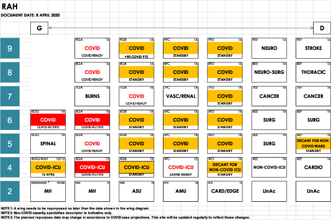
The first case of COVID‐19 caused by the severe acute respiratory syndrome coronavirus‐2 (SARS‐CoV‐2) was detected in South Australia on 1 February 2020. As of 21 May 2020, there have been over 7.1 million cases of COVID‐19 diagnosed worldwide and 7081 confirmed cases in Australia. 1 South Australia has a population of approximately 1.7 million people, 2 with 439 laboratory‐diagnosed cases of SARS‐CoV‐2 3 as of 21 May 2020. The majority of patients with COVID‐19 requiring inpatient care have been managed at the Royal Adelaide Hospital (RAH). The 800‐bed RAH is the state's designated quarantine hospital. The RAH was designed for management of emerging pathogens. All inpatient beds are single rooms with their own bathroom and toilet. In the general ward area, there are 16 cohorted inpatient negative pressure rooms with an anteroom and 80 beds for which ‘pandemic mode’ can be activated. Activation of the pandemic mode in these rooms allows for 100% outside air and a relative negative pressure environment (Appendix I, hospital map).
Several international studies have focussed on the outcomes of patients requiring intensive care unit (ICU) admission. Once admitted to the ICU, outcomes are generally poor. At least 50% of patients admitted to the ICU die and the outcomes are worse for those requiring mechanical ventilation. 4 , 5 , 6 , 7 Our primary objective is to describe the characteristics and outcomes of hospitalised patients with COVID‐19 in the RAH. The secondary objectives are to identify demographic, clinical and laboratory factors associated with risk of developing an oxygen requirement, subsequent ICU admission and death. Emerging therapies for at‐risk groups may reduce ICU admission, mechanical ventilation and poor outcomes.
Methods
Study population and case acquisition
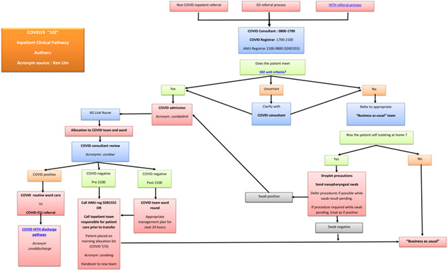
In the initial phase of the pandemic, all confirmed cases of COVID‐19 were admitted to hospital for quarantine purposes, independent of clinical care needs. All patients were initially admitted and managed by the infectious diseases unit. This model of care was not sustainable as case numbers increased. To manage surge capacity a new model of care (unit 102) was developed to create multi‐disciplinary medical teams for the dedicated care of COVID‐19 cases. General medicine, infectious diseases and thoracic medicine units were the first inpatient units to care for COVID‐19 patients under the 102 model from 20 March 2020. The RAH COVID‐19 management plan for surge capacity set out designated areas in the emergency department (ED), ICU and general wards areas for inpatient care. Persons under investigation who met predefined clinical and epidemiological criteria and confirmed COVID‐19 cases were admitted under unit 102. All patients were transferred from the ED to a designated COVID‐19 ward. If SARS‐CoV‐2 testing was negative, the patient was moved out of these dedicated wards (Appendix II, 102 flow sheet). Patients who tested positive and needed hospital admission were observed closely for evidence of organ dysfunction, particularly respiratory failure. Early escalation to ICU occurred if more than 4 L of oxygen through nasal cannula was required to maintain adequate oxygenation above 93% on room air.
Our study population included all patients who met both of the following criteria: (i) discharged from the infectious diseases unit or unit 102 between the dates of 14 February 2020 and 21 May 2020; and (ii) positive reverse transcriptase polymerase chain reaction (PCR) for SARS‐CoV‐2. Patients were excluded if they were never an inpatient being either directly triaged to our Hospital in the Home (HITH) programme or discharged to HITH after an initial assessment in the ED.
Data collection
The RAH uses an electronic medical record (Sunrise Clinical Manager; Allscripts). Patient demographics, co‐morbidities, dates of critical time points, epidemiological factors relating to COVID‐19 acquisition, radiological investigations, laboratory values and outcomes were collected on an excel database manually. This research protocol was approved by the RAH ethics committee. There was no external funding for this research. All authors had full access to data used in the study.
Clinical definitions
During the data collection period, several protocols were in place but many aspects of patient care were left to the discretion of the treating physician. Cardiovascular and respiratory comorbidities were broadly defined from the patients' recorded past medical history. We defined chronic kidney disease (CKD) stages according to Kidney Disease Improving Global Outcomes (KDIGO) criteria. 8 The majority of patients did not have an arterial blood gas performed; hence, a surrogate marker for respiratory failure was the use of supplemental oxygen. We did not differentiate between Types 1 and 2 respiratory failure. A minority of patients underwent computed tomography (CT) of the chest. Changes were classified as ‘typical’ if peripheral ground glass opacities were present. Known clinical and laboratory markers of severity from prior studies 4 , 5 , 6 , 7 , 8 were recorded from all patients when performed during their inpatient admission. The SARS‐CoV‐2 test was incorporated into the pre‐existing respiratory viral panel. All patients tested for SARS‐CoV‐2 were concurrently tested for adenovirus, influenza A and B, parainfluenza, human metapneumovirus, mycoplasma, rhinovirus, respiratory syncytial virus and bordetella pertussis by PCR. Co‐infection was defined as the concurrent detection of a virus or bacteria by nucleic acid amplification test or the isolation of an organism considered significant by the treating clinician on a sample that was collected as part of routine clinical care.
Statistical analysis
We used descriptive statistics for values that were not being compared. Values are reported as medians with interquartile range (IQR) or medians with range. Where comparisons between outcomes were examined, we used Fisher's exact test or Mann−Whitney U‐test where appropriate. Significance value was set at 0.05.
Results
Of the 103 inpatients with laboratory‐confirmed SARS‐CoV‐2, 28 were admitted under infectious disease and 75 were admitted under unit 102. The majority of patients were admitted post‐laboratory diagnosis of SARS‐CoV 2 in the community. Twelve were diagnosed during hospital admission under unit 102. In the audit period following activation of unit 102 on 20 March, there were 637 admissions under unit 102. Of the 637 admissions, 226 patients were discharged from unit 102, 79 of whom had confirmed COVID‐19. Four of these were readmissions following their index admission. Three hundred and twenty‐two patients were transferred to other general medical units after testing negative for SARS‐CoV‐2. The most common diagnoses in this group were community‐acquired pneumonia (114 patients) and exacerbation of chronic obstructive airways disease (32 patients). The remaining 89 patients were transferred to other medical subspecialty or surgical units.
The RAH had a well developed plan for bed flow and surge capacity as outlined in Appendix I. This plan maximised the features of the unique single‐bedded institution and used negative pressure rooms where available. At the peak of admissions, four wards were used as designated COVID‐19 wards. At the peak of COVID‐19 related admissions, hospital activity at the RAH dropped from 7811 episodes of care in March to 6129 episodes of care in April. 9
Demographic data are shown in Table 1. Median age was 60 years (IQR 51–69) and 55 (53%) were male. Hypertension was the most common comorbidity, with most prescribed an angiotensin‐converting enzyme (ACE) inhibitor or angiotensin receptor blocker (ARB). Major cardiovascular and respiratory illnesses were infrequent and diabetes was present in 18 (17%) patients. Six (6%) patients were immunosuppressed, of which two had chronic lymphocytic leukaemia, and four were on immunosuppressive medications for other established indications. Weight and height were available for 71 patients. Twenty‐four (33%) of these patients were obese (body mass index > 30). Two (2%) patients had CKD Stage 3 or greater. All patients were independent at baseline and the vast majority acquired COVID‐19 from overseas or cruise ship travel.
Table 1.
Clinical and epidemiological characteristics of admitted patients (n = 103)
| Characteristic | n (%) |
|---|---|
| Age, median (IQR) (years) | 60 (51–69) |
| Place of residence | |
| Home | 103 (100) |
| Supported living | 0 (0) |
| Residential care facility | 0 (0) |
| Acquisition of COVID‐19 | |
| Cruise ship | 36 (35) |
| Overseas travel | 50 (49) |
| Interstate travel | 1 (1) |
| Confirmed community contact | 13 (13) |
| Unknown contact | 3 (3) |
| Sex | |
| Male | 55 (53) |
| Female | 48 (47) |
| Comorbidities | |
| Cardiovascular | 16 (16) |
| Respiratory | 19 (18) |
| Hypertension | 37 (36) |
| ACE inhibitor/ARB use | 33 (32) |
| Diabetes | 18† (17) |
| CKD (stage 3B) | 2 (1.9) |
| Immunosuppressed | 6 (5.8) |
| BMI >30 (where known, n = 71) (kg/m²) | 24 (14) |
| DNR order | 7 (6.7) |
2 = insulin dependent.
ACE, angiotensin‐converting enzyme; ARB, angiotensin receptor blocker; BMI, body mass index; CKD, chronic kidney disease; DNR, do not resuscitate.
Results for relevant biochemistry and blood picture parameters are recorded in Table 2. CT of the chest was performed on 25 (24%) patients and demonstrated bilateral peripheral ground glass opacities in all cases. Two patients had CT evidence of lobar consolidation and two patients suffered pulmonary emboli; one had a large pulmonary embolus in the right main pulmonary artery, and the other had a left lower lobe segmental embolus.
Table 2.
Results at admission and peak values
| Laboratory values for all patients, median (IQR) | Admission | Peak or Nadir values | Reference range |
|---|---|---|---|
| LDH (U/L) | 269.5 (218.8–323.5) | 323 (255.8–439) | 120–250 |
| AST (U/L) | 34.5 (27–47.3) | 53 (33.8–89.5) | 0–45 |
| ALT (U/L) | 42.9 (26–50) | 75.9 (32–99) | 0–55 |
| CRP (mg/L) | 10.1 (2.8–42.4) | 25.6 (3.8–93.8) | 0–8 |
| Platelet count (×109/L) | 209 (175–244) | 192 (149–228)† | 150–450 |
| Lymphocyte count (×109/L) | 1.3 (0.95–1.7) | 1.1 (0.77–1.6)† | 1.1–3.5 |
| Neutrophil: lymphocyte (NLR) | 2.3 (1.6–4.1) | 3.5 (2.3–5.4)† | |
| Eosinophil count (×109/L) | 0.02 (0.01–0.05) | 0.01 (0.01–0.04)† | 0.02–0.5 |
| Computed tomography of the chest (n = 25), n (%) | |||
| Bilateral peripheral ground glass opacities | 24 (96) | ||
| Lobar consolidation | 3 (12) | ||
| Pulmonary embolism | 2 (8) | ||
Nadir values.
ALT, alanine aminotransferase; AST, aspartate aminotransferase; CRP, C‐reactive protein; IQR, interquartile range; LDH, lactate dehydrogenase.
Thirty‐nine (38%) patients required oxygen therapy. In these patients, median oxygen saturation was 93% while inspiring room air immediately prior to commencing oxygen therapy. Eighteen (17%) patients required ICU admission, of whom eight were mechanically ventilated and four (50%) died. Nasal high flow oxygen was administered to 13 ICU patients, seven of whom progressed to mechanical ventilation. One patient admitted to ICU required cardiac monitoring only. All patients who did not need mechanical ventilation were able to be discharged from ICU back to the ward. Five patients required renal replacement therapy and nine required cardiovascular support with inotropic medication. Four patients required rehabilitation prior to return home, the remaining 95 patients were discharged directly home.
Patients presented to hospital at a median of 7 days after symptom onset. Patients who were managed on the ward were discharged at a median of 12 days from the onset of illness with a median hospital length of stay (LOS) of 3 days. Those requiring ICU care were admitted to ICU at a median of 3 days after hospital admission. They were discharged or died at a median of 19 days after onset of illness. Their median hospital LOS was 13.1 days.
At admission, only lactate dehydrogenase (LDH) was significantly associated with oxygen requirement, ICU admission and death. Admission lymphocyte count, platelet count, eosinophil count and neutrophil lymphocyte ratio (NLR) were associated with oxygen requirement and the need for ICU admission. Peak C‐reactive protein (CRP), aspartate aminotransferase, alanine aminotransferase, LDH and the NLR at lymphocyte nadir were significantly associated with the development of oxygen requirement, ICU admission and death (Tables 3,4). Of note, lactate was only requested in 32 patients and troponin T in 21 patients. We therefore did not attempt to analyse clinical outcomes using these tests as risk factors. Twenty‐three patients only had one blood test performed during their admission.
Table 3.
Age, BMI and laboratory values
| Laboratory value, median (IQR) | Outcome | P‐value | |
|---|---|---|---|
| Yes | No | ||
| Oxygen supplementation | |||
| n | 39 | 64 | |
| Age (years) | 68 (55–72) | 57.5 (50.3–63.8) | <0.01 |
| BMI (kg/m²) | 29.1 (24.8–33.7) | 26.7 (22.9–31.2) | 0.24 |
| Peak LDH (U/L) | 455 (373–539) | 266 (235–318) | <0.01 |
| Peak AST (U/L) | 85 (55–127) | 37 (28–58) | <0.01 |
| Peak ALT (U/L) | 85 (53–129) | 41 (26.3–68) | <0.01 |
| Peak CRP (mg/L) | 120 (62.7–180) | 6.3 (1.9–24.7) | <0.01 |
| Nadir platelet count (×109/L) | 160 (125–195) | 203 (177–239) | <0.01 |
| Nadir lymphocyte count (×109/L) | 0.78 (0.5–1.2) | 1.23 (0.9–1.7) | <0.01 |
| Neutrophil: lymphocyte (at lymphocyte nadir) | 6.3 (4.1–13.4) | 2.2 (1.7–3.2) | <0.01 |
| Nadir eosinophil count (×109/L) | 0 (0.0–0.01) | 0.03 (0.01–0.06) | <0.01 |
| ICU admission | |||
| n | 18 | 85 | |
| Age (years) | 66.5 (57.5–74.3) | 60 (51–67.5) | 0.04 |
| BMI (kg/m²) | 27.6 (24.1–30.9) | 27.2 (23.9–31.6) | 0.78 |
| Peak LDH (U/L) | 479 (416.3–673.8) | 295 (245–381) | <0.01 |
| Peak AST (U/L) | 99 (77.5–166) | 42.5 (31–70.8) | <0.01 |
| Peak ALT (U/L) | 95.5 (62.8–183.3) | 53 (28–83.5) | <0.01 |
| Peak CRP (mg/L) | 143.3 (83.8–199.2) | 11.8 (3.3–60.3) | <0.01 |
| Nadir platelet count (×109/L) | 167.5 (107–195) | 194 (159–236) | 0.05 |
| Nadir lymphocyte count (×109/L) | 0.61(0.43–1.1) | 1.2 (0.9–1.6) | <0.01 |
| Neutrophil: lymphocyte (at lymphocyte nadir) | 9.4 (5.4–21.2) | 2.6 (1.8–4.0) | <0.01 |
| Nadir eosinophil count (×109/L) | 0 (0–0.003) | 0.02 (0.01–0.05) | <0.01 |
| Death | |||
| n | 4 | 99 | |
| Age (years) | 74.5 (65–75.8) | 60 (51–68) | 0.02 |
| BMI (kg/m²) | 25.7 (NA)† | 27.4 (24.2–31.4) | 0.57 |
| Peak LDH (U/L) | 682 (497–1130) | 316.5 (254.5–426) | <0.01 |
| Peak AST (U/L) | 301.5 (120.8–595.5) | 51.5 (33–80.5) | <0.01 |
| Peak ALT (U/L) | 132 (72.5–403.8) | 55 (30–93) | 0.03 |
| Peak CRP (mg/L) | 121 (96.9–154.6) | 23 (3.8–84) | 0.03 |
| Nadir platelet count (×109/L) | 130 (96–186) | 192 (150–228) | 0.05 |
| Nadir lymphocyte count (×109/L) | 0.5 (0.3–1.8) | 1.1 (0.78–1.6) | 0.15 |
| Neutrophil: lymphocyte (at lymphocyte nadir) | 22 (10.6–30.4) | 2.96 (10.6–30.4) | <0.01 |
| Nadir eosinophil count (×109/L) | 0 (0–0) | 0.01 (0–0.04) | <0.01 |
P‐values calculated with Mann−Whitney U‐test.
Insufficient data to calculate IQR.
ALT, alanine aminotransferase; AST, aspartate aminotransferase; BMI, body mass index; CRP, C‐reactive protein; IQR, interquartile range; LDH, lactate dehydrogenase.
Table 4.
Admission values
| Laboratory value, median (IQR) | Outcome | P‐value | |
|---|---|---|---|
| Yes | No | ||
| Oxygen supplementation | |||
| n | 39 | 64 | |
| Admission LDH (U/L) | 317 (274–409) | 250 (209–283) | <0.01 |
| Admission AST (U/L) | 40 (33–55) | 31 (25–42) | <0.01 |
| Admission ALT (U/L) | 35 (27–64) | 29.5 (24–46) | 0.01 |
| Admission CRP (mg/L) | 42.4 (14.8–87.4) | 4.3 (1.4–13) | <0.01 |
| Admission platelet count (×109/L) | 186 (137–243) | 219 (187–247.8) | <0.01 |
| Admission lymphocyte count (×109/L) | 1.04 (0.7–1.6) | 1.4 (1.1–1.8) | <0.01 |
| Neutrophil: lymphocyte (at admission) | 4.05 (2.3–7.2) | 2.0 (1.4–3.2) | <0.01 |
| Admission eosinophil count (×109/L) | 0.01 (0–0.03) | 0.03 (0.01–0.09) | <0.01 |
| ICU admission | |||
| n | 18 | 85 | |
| Admission LDH (U/L) | 349.5 (281.5–425.3) | 259.5 (218–309.8) | <0.01 |
| Admission AST (U/L) | 39.5 (31.8–56) | 33 (26.3–44) | 0.19 |
| Admission ALT (U/L) | 34 (27.8–57.3) | 32 (24–48) | 0.36 |
| Admission CRP (mg/L) | 44.5 (23.2–136.6) | 7.6 (1.8–29.5) | <0.01 |
| Admission platelet count (×109/L) | 183 (146.8–211) | 218 (178–247.5) | 0.02 |
| Admission lymphocyte count (×109/L) | 0.81 (0.7–1.6) | 1.4 (1.1–1.7) | <0.01 |
| Neutrophil: lymphocyte (at admission) | 5.3 (2.5–8.0) | 2.2 (1.4–3.5) | <0.01 |
| Admission eosinophil count (×109/L) | 0 (0–0.02) | 0.03 (0.01–0.06) | <0.01 |
| Death | |||
| n | 4 | 99 | |
| Admission LDH (U/L) | 353 (297–484) | 266.5 (218–318) | 0.05 |
| Admission AST (U/L) | 40 (33.5–55.5) | 34 (26.8–47.3) | 0.36 |
| Admission ALT (U/L) | 30.5 (26.5–48.8) | 32 (25–50) | 0.10 |
| Admission CRP (mg/L) | 29.6 (14.9–109.6) | 9.2 (2.3–42.4) | 0.17 |
| Admission platelet count (×109/L) | 173 (131.3–205.8) | 213 (175–245) | 0.14 |
| Admission lymphocyte count (×109/L) | 0.7 (0.7–3.5) | 1.3 (1.0–1.7) | 0.25 |
| Neutrophil: lymphocyte (at admission) | 5 (1.5–11) | 2.3 (1.6–4.1) | 0.30 |
| Admission eosinophil count (×109/L) | 0 (0–0.06) | 0.03 (0.01–0.05) | 0.28 |
P‐values calculated with Mann−Whitney U‐test.
ALT, alanine aminotransferase; AST, aspartate aminotransferase; CRP, C‐reactive protein; IQR, interquartile range; LDH, lactate dehydrogenase.
Several comorbidities were associated with poor outcomes. Cardiovascular disease, diabetes and the use of ACE inhibitors or ARB were significantly more prevalent in those with the need for oxygen supplementation, ICU admission and among those who died. Although the number of deaths in our cohort was low, there were significant differences in the mortality rate of those with COVID‐19 infection and cardiovascular disease, hypertension, ACE inhibitor or ARB use and diabetes. All immunosuppressed patients survived to discharge (Table 5).
Table 5.
Relative risk of clinical risk factors and outcomes
| Risk factor | Clinical outcome | Relative risk | P‐value | |
|---|---|---|---|---|
| Yes | No | |||
| Oxygen supplementation, n (%) | ||||
| n | 39 | 64 | ||
| Male gender | 24 (62) | 31 (48) | 1.4 | 0.23 |
| Hypertension | 20 (51) | 17 (27) | 1.9 | 0.02 |
| ACE/ARB use | 19 (49) | 14 (22) | 2.0 | 0.01 |
| Cardiovascular disease | 11 (28) | 5 (8) | 2.1 | 0.01 |
| Respiratory disease | 9 (23) | 10 (16) | 1.3 | 0.43 |
| Diabetes | 9 (23) | 5 (8) | 1.9 | 0.04 |
| Immunosuppression | 4 (10) | 2 (3) | 1.9 | 0.20 |
| ICU admission, n (%) | ||||
| n | 18 | 85 | ||
| Male gender | 14 (78) | 41 (48) | 3.1 | 0.04 |
| Hypertension | 11 (61) | 26 (31) | 2.8 | 0.03 |
| ACE/ARB use | 10 (56) | 23 (27) | 2.7 | 0.03 |
| Cardiovascular disease | 6 (33) | 10 (12) | 2.7 | 0.03 |
| Respiratory disease | 5 (28) | 14 (16) | 1.7 | 0.32 |
| Diabetes | 6 (33) | 8 (9) | 3.2 | 0.02 |
| Immunosuppression | 1 (6) | 5 (6) | 0.95 | 1 |
| Death, n (%) | ||||
| n | 4 | 99 | ||
| Male gender | 3 (75) | 52 (53) | 2.6 | 0.62 |
| Hypertension | 4 (100) | 32 (32) | NA | 0.01 |
| ACE/ARB use | 4 (100) | 29 (29) | NA | 0.01 |
| Cardiovascular disease | 3 (75) | 13 (13) | 16.3 | 0.01 |
| Respiratory disease | 1 (25) | 18 (18) | 1.5 | 0.56 |
| Diabetes | 3 (75) | 9 (9) | 19 | <0.01 |
| Immunosuppression | 0 | 6 (6) | 0 | 1 |
P‐values calculated with Fisher's exact test.
ACE, angiotensin‐converting enzyme; ARB, angiotensin receptor blocker.
Antimicrobials were prescribed to 38 patients. The most common indication was empiric treatment for community‐acquired pneumonia. Six patients suffered from viral co‐infections. Six separate patients had various bacterial pathogens isolated from blood, sputum or urine cultures. Glucocorticoids were prescribed in two patients for no longer than 3 days of their admission. Both of these patients required ICU care and one died.
Discussion
Our single‐centre study is unique as the majority of South Australia's COVID‐19‐related admissions were to the RAH. Our designated team of emergency staff, physicians and intensivists operated within a closely regulated paradigm of care. This allowed our patients to be cared for by a team with growing expertise managing COVID‐19 illness.
These patients, although predominantly middle aged and elderly, likely represent a healthier cohort than other international cohorts as all patients were independent and most had been able to travel overseas without assistance. Nearly all cases had been acquired from travel or through known contacts. Widespread testing in South Australia with an overall low prevalence would indicate widespread community transmission is negligible. As of 21 May 2020, over 87 000 tests have been performed in South Australia, which accounts for 5% of the population. Less than 1% of these tests have returned a positive result. 3 Overall hospital activity dropped by approximately 20% 10 during the peak period of admissions, which allowed adequate staffing of unit 102, ED and ICU. All of these factors have likely played a role in our comparatively low mortality rate 1 and good functional outcomes.
The clinical course of our patients is similar to those reported in previous cohorts. 4 , 5 , 9 Patients required hospitalisation at approximately 1 week from onset of symptoms. This time frame is similar among patients requiring either ward care or ICU care; however, those requiring ICU care had presented to hospital earlier in the course of their disease. Our patients had a longer ICU length of stay compared to other centres. 7
Comorbidities associated with ICU admissions or deaths were similar to other international cohorts. 5 , 6 , 7 , 9 The most important comorbidities that we observed associated with clinical deterioration were diabetes and cardiovascular disease. Both hypertension and ARB/ACE inhibitor use were more common in those who died compared with those who survived to discharge. Early studies in China linked the use of ACE inhibitors or ARB with poor outcomes. 4 However, other larger studies have shown either no association of the use of these medications with severity and mortality, 11 or a possible benefit. 12 Our protocols, in accordance with published guidelines, 13 did not include specific advice regarding cessation or continuation of these medications. In our cohort, it would be difficult to make any conclusions due to the low numbers of patients and the fact that all but four hypertensive patients were already using these medications at the point of admission. We note that all four patients who died were already using these medications at the time of admission. Age was associated with oxygen requirement and death, a finding that is consistent with other centres. 4 , 5 , 6 , 7 However, we did not observe a statistically significant increased risk of adverse outcomes with obese patients. Male patients were more likely to require ICU admission but not oxygen supplementation or death. We could not draw any conclusions about the presence of CKD as only two patients had CKD more advanced than Stage 3. It is interesting to note that none of the deaths and only one of the ICU admissions suffered from pre‐existing immunosuppression.
Lymphopenia has been a consistent finding, which correlates with disease severity. 4 Lagunas‐Rangel performed a meta‐analysis of six studies, which allowed the calculation of NLR. This confirmed the findings of multiple prior smaller studies that the NLR was associated with severe disease and mortality. 14 Yang et al. suggested that a threshold NLR of 3.3 was predictive of severe disease in a single‐centre cohort of patients in Wuhan. 15 In our cohort, progressively worse outcomes appeared to be linked with rising NLR. A raised CRP has been linked with poor outcomes including the development of severe pulmonary disease. 16 In our cohort, patients with mildest disease not requiring oxygen therapy had a median peak CRP of six compared with a median peak CRP of 120 in those requiring oxygen. Although not specific to COVID‐19, the above abnormalities may be useful for identifying patients who are at risk for deterioration. We note that in our cohort, the peak abnormalities of results occurred when patients had already deteriorated. Admission values were less useful predictors of outcomes.
The case fatality rate among our ICU patients was lower than other centres. 5 , 6 , 7 This may well have been due to the health attributes of our patient population. We also had a relatively low threshold for acceptance into ICU (those needing more than 4 L supplementation). Once patients required mechanical ventilation, mortality was very high (50%).
Our study has several limitations. The nature of our cohort makes it difficult to draw any conclusions that are relevant for areas where widespread community transmission has occurred. Multiple physician groups cared for our patients; the choice of investigation and management were driven by physician preference and evolving scientific data from around the world, rather than as part of a standardised care pathway. As a result, many tests that have been associated with poor outcomes were not performed, such as d‐dimer, troponin T and procalcitonin. We recognise that routine use of these investigations currently has uncertain clinical utility. Radiological investigation was mostly limited to plain radiographs with few CT performed. Sporadic off‐label use of antiviral medications such as hydroxychloroquine occurred outside of a clinical trial setting. Trends of laboratory results may have assisted with developing risk prediction models; however, many of our patients only had one set of results available. We did not report post‐discharge outcomes.
Conclusion
The present study and its population context suggest that in order to predict poor outcomes, factors at all levels need to be addressed. If new cases are limited to healthy returning travellers, the risk profile of this population may be lower than that of the general population of a similar age. At a community level, adequate case finding should occur such that patients with clinical and laboratory risk factors can be monitored for signs of deterioration. If identified, these patients may be isolated to prevent inadvertent spread into the vulnerable elderly population. Once cases are identified and needing admission, those with risk factors for ongoing deterioration should be identified. Guidelines for best practice of COVID‐19 as well as comorbidities in the context of COVID‐19 illness need to be developed; in particular, the management of hypertension, diabetes and cardiovascular disease. We can only speculate that the model of care in our state which concentrates expertise of care to one centre, may have contributed to good outcomes. Perhaps it is the substantial proportion of ‘well’ patients with risk factors for deterioration who should be the target for new interventions before they develop severe disease that results in hospitalisation and subsequent poor outcomes.
Hospital map

Unit 102 clinical pathway

Funding: None.
Conflict of interest: None.
References
- 1. Johns Hopkins University & Medicine . Coronavirus Resource Centre. 2020 [cited 2020 May 21]. Available from URL: https://coronavirus.jhu.edu/map.html
- 2. Australian Bureau of Statistics . 2016 Census Quickstats. 2016 [cited 2020 Apr 22]. Available from URL: https://quickstats.censusdata.abs.gov.au/census_services/getproduct/census/2016/quickstat/4?opendocument
- 3. Government of South Australia . Covid‐19 Dashboard and Daily update. 2020 [cited 2020 Apr 22]. Available from URL: https://www.covid-19.sa.gov.au/home/dashboard
- 4. Zhou F, Yu T, Du R, Fan G, Liu Y, Liu Z et al. Clinical course and risk factors for mortality of adult inpatients with COVID‐19 in Wuhan, China: a retrospective cohort study. Lancet 2020; 395: 1054–62. [DOI] [PMC free article] [PubMed] [Google Scholar]
- 5. Bhatraju P, Ghassemieh B, Nichols M, Kim R, Jerome KR, Nalla AK et al. Covid‐19 in critically ill patients in the Seattle region – case series. N Engl J Med 2020; 382: 2012–22. [DOI] [PMC free article] [PubMed] [Google Scholar]
- 6. Richardson S, Hirsch J, Narasimhan M, Crawford JM, McGinn T, Davidson KW et al. Presenting characteristics, comorbidities, and outcomes among 5700 patients hospitalized with COVID‐19 in the New York City area. JAMA 2020; 323: 2052–9. [DOI] [PMC free article] [PubMed] [Google Scholar]
- 7. Intensive Care National Audit and Research Centre (ICNARC) . ICNARC report on COVID‐19 in critical care, April 24 2020. Napier House, 24 High Holborn, London WC1V 6AZ, London, 2020.
- 8. KDIGO 2012 clinical practice guideline for the evaluation and management of chronic kidney disease. Kidney Int. Suppl. 2013; 3: 5–14. [DOI] [PubMed] [Google Scholar]
- 9. Wang D, Hu B, Hu C. Clinical characteristics of 138 hospitalized patients with 2019 novel coronavirus‐infected pneumonia in Wuhan, China. JAMA 2020; 323: 1061–9. [DOI] [PMC free article] [PubMed] [Google Scholar]
- 10. Health Roundtable . 2020 [cited 2020 Sep 11]. Available from URL: https://www.healthroundtable.org/
- 11. Li M, Wang M, Chen B, Zhang H, Deng A. Association of renin‐angiotensin system inhibitors with severity of risk of death in patients with hypertension hospitalized for coronavirus disease 2019 (COVID‐19) infection in Wuhan, China. JAMA Cardiol 2020; 5: 825–30. [DOI] [PMC free article] [PubMed] [Google Scholar]
- 12. Bean D, Kraljevic Z, Searle T, Bendayan R, Pickles A, Folarin A et al. Treatment with ACE‐inhibitors is associated with less severe SARS‐Covid‐19 infection in a multi‐site UK acute hospital trust. Eur J Heart Fail 2020; 22: 967–974. [DOI] [PMC free article] [PubMed] [Google Scholar]
- 13. De Simone G. Position statement of the ESC Council on hypertension on ACE‐inhibitors and angiotensin receptor blockers. [cited 2020 May 21]. Available from URL: https://www.escardio.org/Councils/Council‐on‐Hypertension‐(CHT)/News/position‐statement‐of‐the‐esc‐council‐on‐hypertension‐on‐ace‐inhibitors‐and‐ang
- 14. Lagunas‐Rangel F. Neutrophil‐to‐lymphocyte ratio and lymphocyte to C reactive protein ratio in patients with severe coronavirus disease 2019 (COVID‐19): a meta‐analysis. Journal of Medical Virology 2020; 92: 1733–4. [DOI] [PMC free article] [PubMed] [Google Scholar]
- 15. Yang A, Liu J, Tao W, Li H. The diagnostic and predictive role of NLR, d‐NLR and PLR in COVID‐19 patients. Int Immunopharmacol 2020; 84: 106504. [DOI] [PMC free article] [PubMed] [Google Scholar]
- 16. Wang L. C‐reactive protein levels in the early stage of COVID‐19. Med Mal Infect 2020; 50: 332–4. [DOI] [PMC free article] [PubMed] [Google Scholar]


