What are Prototheca?
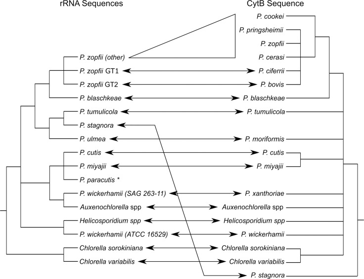
Members of the genus Prototheca are nonphotosynthetic algae, closely related to the well-known green algal genus Chlorella. This “genus” encompasses the additional genera Auxenochlorella and Helicosporidium (Fig 1). Together, this collection of genera is referred to as the AHP lineage, though relationships within the lineage remain unclear [1–5]. Analyses based on the mitochondrial cytochrome b sequence suggest this lineage may include Chlorella species (Fig 1), which might accordingly be called the CHAP lineage [1–5].
Fig 1. Probable relationships between species/genotypes within the Prototheca/Helicosporidium/Auxenochlorella/Chlorella lineage.
Left—a consensus cladogram built from analyses using predominantly ribosomal RNA (small subunit, internal transcribed spacer region, and D1/D2 region of the large subunit) sequence data [2–5]. Species names are defined by a mixture of assimilation profiles, growth conditions, and sequence data. Right–a cladogram built using a partial mitochondrial cytochrome b sequence [1]. Species names are defined by clustering and cytochrome b sequence similarity. Arrows between the trees indicate equivalent species, where renaming has occurred. All nodes shown are supported by bootstrap values greater than 70 in at least one analysis. Branch lengths are arbitrary. *Discovered recently and therefore not included in the cytochrome b analysis.
Why are we talking about an alga in PLOS Pathogens?
Lacking chlorophyll, Prototheca species are obligate heterotrophs, and six are opportunistic pathogens of vertebrates. Of particular interest are the former species Prototheca zopfii (specifically the lineage known as genotype 2, recently raised to species status as Prototheca bovis [1]) and the current species Prototheca wickerhamii, which are the main causative agents for cattle and human infections respectively [6,7]. P. bovis and P. wickerhamii also maintain the largest host ranges, including: cats, dogs, buffaloes, horses (P. bovis only), and goats (P. wickerhamii only).
Other pathogenic algae exist, though Prototheca are the most significant in terms of the number of infections and their predilection towards humans and domesticated animals. Members of the nonphotosynthetic genus Helicosporidium are pathogens of invertebrates, particularly insects [8]. Photosynthetic algae (Chlorella and Desmodesmus) have also been reported to infect mammals [9,10].
How do Prototheca infections occur?
Infections in cattle typically present as mastitis (inflammation of the mammary tissue). Infections are usually subclinical—detectable only through raised somatic cell counts and the presence of Prototheca in milk—but clinical mastitis has both acute and chronic presentations, both of which reduce milk yield [6,11]. Acute mastitis is associated with raised temperature, pain, and swelling, while chronic mastitis is associated with permanent damage to alveoli and mammary parenchyma. Infections are typically restricted to the mammary tissue, but rare cases of systemic infection have been reported [12].
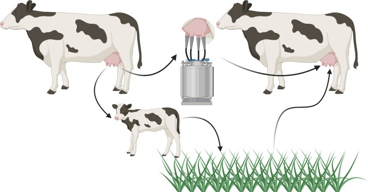
Prototheca appears responsible for a nonnegligible proportion of bovine mastitis cases (1.2% to 13.3% of Polish samples, 11.2% of Italian herds affected [6,13,14]), though the transmission cycle is currently speculative (Fig 2). Entry is probably through the teat orifice, from contaminated milking equipment or environmental sources [11]. Prototheca cells in milk may return to the environment in the faeces of calves [15]. There is currently no economically viable treatment, and spontaneous recovery is rare to nonexistent [11]. Consequently, affected cows are culled, representing a large economic and animal welfare burden.
Fig 2. Possible infection cycle for Prototheca bovis.
An udder infection results in the presence of Prototheca cells in milk. These cells may be ingested by calves and excreted into the environment. Contaminated milk may also result in Prototheca cells being present on milking machinery, representing a more direct method of transmission. Reentry into an udder is likely through the teat orifice contacting contaminated surfaces. Created with BioRender.com.
Presentation and prognosis of human infections are highly dependent on the site of infection, which is in turn strongly influenced by the host’s immune status. The majority of infections are restricted to localised skin lesions, but joint infection (particularly the elbow) and disseminated infection (affecting a range of organs, each with unique presentations) are more prevalent in immunocompetent and immunocompromised individuals, respectively [16]. Almost all human infections become chronic, with death usually associated with disseminated infections [17]. Human infections probably occur through environmental contamination of a wound, with little evidence for direct human-to-human transmission [16]. Reports of infection from contaminated dairy products exist but are infrequent. Given the differences between human and cattle Prototheca pathogens, contaminated dairy is unlikely to pose a public health risk.
Human and cattle infections have been reported from all permanently settled continents except, currently, in cattle in Africa; likely reflecting diagnostic challenges rather than a true absence of the pathogen [6,16]. Historically, infections by Prototheca species have been diagnosed through a combination of histopathology, morphology, and culturing methods, with biochemical assimilation assays providing species-level resolution. However, protothecal infections have been misdiagnosed (usually as fungal infections) or missed (in subclinical cases) obscuring the true prevalence and distribution of infections. More widespread use of molecular techniques, including PCR-restriction fragment length polymorphism (RFLP) assays and matrix-assisted laser desorption/ionization time-of-flight (MALDI-ToF) mass spectrometry, will hopefully improve this situation [3,18].
Unfortunately, few environmental studies have taken place for Prototheca. Those that have almost always investigate the surroundings of potential hosts. Our understanding of the natural ecology of Prototheca species is therefore lacking. P. wickerhamii is the most abundant species in human sewage [19], and species formerly identified as P. zopfii (either P. bovis or Prototheca ciferrii [1]) tend to be the most abundant around cattle [6], but the abundance of any species away from their preferred host remains unknown. In environments where Prototheca have been identified, they are present in a variety of contexts including: tree slime flux; rivers and ponds; mud; faeces/sewage (human, cattle, pigs, dogs); food (human and cattle); and industrial waste [16,19]. Prototheca have been found to colonise animals nonpathogenically and transiently [19,20].
How are algae capable of causing disease?
The mechanisms by which algae are able to infect hosts and cause disease are currently unknown. Adaptations that facilitate Prototheca pathology in particular are also unknown. Genomic and proteomic approaches to identify virulence factors have been stymied by a lack of supporting information. Typically, only single genomes per species are being published, further complicating identification of relevant virulence determinants from individual variation.
Protothecal infections typically develop over months, indicating Prototheca’s ability to survive or evade host immunity is a key component of their pathology. To this end, Prototheca cells are able to survive digestion by macrophages and appear to replicate within the phagolysosome [21]. Furthermore, P. bovis has been shown to kill phagocytic cells even after phagocytosis was blocked [22]. Killing was restricted to phagocytic host cells, and the former P. zopfii genotype 1 (raised to P. ciferrii [1]) could not kill these cells, suggesting that specific host-directed toxin(s) may be important for the virulence of P. bovis.
One possibility is that, rather than bespoke virulence factors, Prototheca may exploit environmental adaptations for “accidental virulence,” as has been proposed for other eukaryotic pathogens [23]. The presence of closely related endosymbiotic algal species may suggest one such exaptation. The genera Chlorella and Auxenochlorella both contain species that are known endosymbionts of organisms such as the ciliate protist Paramecium bursaria and the cnidarian Hydra viridis [24–26].
Current models require Chlorella to survive digestion by their prospective host to establish endosymbioses [24]. Additionally, Chlorella endosymbionts exist in a wide range of host protists and invertebrates [24,27]. This may indicate that processes that enable endosymbionts to survive digestion in one organism may be generalisable to other hosts. If mechanisms to survive digestion facilitate either endosymbiosis or parasitism, we may expect some pathogens to be closely related to endosymbionts—as occurs within the AHP/CHAP algal lineage. Desmodesmus, an unrelated pathogenic alga, also has close endosymbiotic relatives [9,28].
Another feature potentially predisposing Prototheca to pathology is the ability to form biofilms in isolation [29,30], while biofilm formation in Chlorella is limited without a microbial community [31]. Biofilms have been proposed to play important roles in immune evasion and drug resistance of many pathogens, and biofilm formation appears to correlate with pathogenicity in Prototheca species [29,30,32]. Biofilms appear to increase the resistance of species formerly identified as P. zopfii (likely P. bovis) against various sanitizers, potentially enhancing transmission by preventing removal from contaminated surfaces [33]. Peripheral blood mononuclear cells produce IL-6, an early pro-inflammatory cytokine, in response to planktonic P. wickerhamii but not P. wickerhamii biofilms, thus potentially enhancing immune evasion [29].
What do we still not know?
Quite a lot. The fundamental differences between pathogenic and environmental species, if any, remain unknown. We do not know if P. bovis and P. wickerhamii, which are relatively distantly related species within the lineage (Fig 1), use similar mechanisms for pathogenesis.
Prototheca host preferences are poorly understood. For example, P. wickerhamii and P. bovis dominate human and cattle infections, respectively, but seem equally prevalent in buffaloes [34]. The reason for the uniquely aggressive progression of Prototheca infection in dogs, which is usually fatal, is also unknown [35].
From an evolutionary perspective, it is unclear whether Prototheca benefit from pathology. Dispersal in milk and faeces is a potential advantage for P. bovis from infecting cattle (Fig 2), but there is no obvious mechanism of egress for P. wickerhamii from humans.
What drives success or failure of an immune response against Prototheca, in any host, is unknown. Neutropenic cancer patients and transplant recipients are at particular risk, potentially highlighting the importance of neutrophils. By contrast, those with severely depleted CD4+ cell counts (as a result of HIV infection) are not as severely affected as one might expect, suggesting that T-cell responses may be less important [36].
Finally, our understanding of how Prototheca infections respond to treatment is insufficient. As the only pathogenic algae of note, treatment usually involves surgical removal and/or antifungal drugs with mixed efficacy. Unfortunately, in vitro susceptibility tests are poor predictors for success of existing antiprotothecal treatment [37]. There have been notable cases of treatment failure when isolates seemed susceptible or success when isolates seemed resistant, as well as unpredictable changes in susceptibility during the course of treatment [38,39]. Recent in vitro work has revealed promising, novel algicidal treatments, but their in vivo efficacy remains to be seen [37,40,41].
Conclusions
Prototheca and their relatives represent a fascinating but poorly understood class of pathogens. A deeper understanding of their genomes and cell biology holds great potential, both in terms of improving the treatment of animal and human infections and in shedding light on principles that underlie pathogenesis in general.
Funding Statement
This work was supported by the Biotechnology and Biological Sciences Research Council (BBSRC) and University of Birmingham funded Midlands Integrative Biosciences Training Partnership (MIBTP) to CDS. The funders had no role in study design, data collection and analysis, decision to publish, or preparation of the manuscript.
References
- 1.Jagielski T, Bakuła Z, Gawor J, Maciszewski K, Kusber WH, Dyląg M, et al. The genus Prototheca (Trebouxiophyceae, Chlorophyta) revisited: Implications from molecular taxonomic studies. Algal Res. 2019;43(March):101639. [Google Scholar]
- 2.Suzuki S, Endoh R, Manabe RI, Ohkuma M, Hirakawa Y. Multiple losses of photosynthesis and convergent reductive genome evolution in the colourless green algae Prototheca. Sci Rep. 2018;8(1):1–11. 10.1038/s41598-017-17765-5 [DOI] [PMC free article] [PubMed] [Google Scholar]
- 3.Jagielski T, Gawor J, Bakuła Z, Decewicz P, Maciszewski K, Karnkowska A. Cytb as a new genetic marker for differentiation of prototheca species. J Clin Microbiol. 2018;56(10):e00584–18. 10.1128/JCM.00584-18 [DOI] [PMC free article] [PubMed] [Google Scholar]
- 4.Masuda M, Hirose N, Ishikawa T, Ikawa Y, Nishimura K. Prototheca miyajii sp. nov., isolated from a patient with systemic protothecosis. Int J Syst Evol Microbiol. 2016;66(3):1510–20. 10.1099/ijsem.0.000911 [DOI] [PubMed] [Google Scholar]
- 5.Kunthiphun S, Endoh R, Takashima M, Ohkuma M, Tanasupawat S, Savarajara A. Prototheca paracutis sp. nov., a novel oleaginous achlorophyllous microalga isolated from a mangrove forest. Mycoscience. 2019;60(3):165–9. [Google Scholar]
- 6.Jagielski T, Krukowski H, Bochniarz M, Piech T, Roeske K, Bakuła Z, et al. Prevalence of Prototheca spp. on dairy farms in Poland–a cross-country study. J Microbial Biotechnol. 2019;12(3):556–66. [DOI] [PMC free article] [PubMed] [Google Scholar]
- 7.Kano R. Emergence of Fungal-Like Organisms: Prototheca. Mycopathologia. Netherlands: Springer; 2019. p. 1–8. 10.1007/s11046-019-00365-4 [DOI] [PubMed] [Google Scholar]
- 8.Tartar A. The non-photosynthetic algae Helicosporidium spp.: Emergence of a novel group of insect pathogens. Insects. 2013;4(3):375–91. 10.3390/insects4030375 [DOI] [PMC free article] [PubMed] [Google Scholar]
- 9.Fujimoto M, Inaba Y, Takahashi T, Nakanishi G, Muraosa Y, Yahiro M, et al. Image Gallery: Granulomatous dermatitis due to infection with the chlorophyllic green alga Desmodesmus. Br J Dermatol. 2018;179(4):e167. 10.1111/bjd.17007 [DOI] [PubMed] [Google Scholar]
- 10.Ramírez-Romero R, Rodríguez-Tovar LE, Nevárez-Garza AM, López A. Chlorella infection in a sheep in mexico and minireview of published reports from humans and domestic animals. Mycopathologia. 2010;169(6):461–6. 10.1007/s11046-010-9287-4 [DOI] [PubMed] [Google Scholar]
- 11.Milanov D, Petrović T, Polaček V, Suvajdžić L, Bojkovski J. Mastitis associated with Prototheca zopfii—An emerging health and economic problem on dairy farms. J Vet Res. 2016;60(4):373–8. [Google Scholar]
- 12.Thompson G, Silva E, Marques S, Müller A, Carvalheira J. Algaemia in a dairy cow by Prototheca blaschkeae. Med Mycol. 2009;47(5):527–31. 10.1080/13693780802566341 [DOI] [PubMed] [Google Scholar]
- 13.Zecconi A, Dell’Orco F, Rizzi N, Vairani D, Cipolla M, Pozzi P, et al. Cross-sectional study on the prevalence of contagious pathogens in bulk tank milk and their effects on somatic cell counts and milk yield. Ital J Anim Sci. 2020;19(1):66–74. [Google Scholar]
- 14.Krukowski H, Lassa H, Zastempowska E, Smulski S, Bis-Wencel H. Etiological agents of bovine mastitis in Poland. Med Weter. 2020;76(04):221–5. [Google Scholar]
- 15.Kurumisawa T, Kano R, Nakamura Y, Hibana M, Ito T, Kamata H, et al. Is bovine protothecal mastitis related to persistent infection in intestine? J Vet Med Sci. Japanese Society of Veterinary Science. 2018;80. p. 950–2. 10.1292/jvms.17-0710 [DOI] [PMC free article] [PubMed] [Google Scholar]
- 16.Lass-Flörl C, Mayr A. Human protothecosis. Clin Microbiol Rev. 2007;20(2):230–42. 10.1128/CMR.00032-06 [DOI] [PMC free article] [PubMed] [Google Scholar]
- 17.Todd JR, Matsumoto T, Ueno R, Murugaiyan J, Britten A, King JW, et al. Medical phycology 2017. Med Mycol. 2018;56(suppl_1):S188–204. 10.1093/mmy/myx162 [DOI] [PubMed] [Google Scholar]
- 18.Murugaiyan J, Ahrholdt J, Kowbel V, Roesler U. Establishment of a matrix-assisted laser desorption ionization time-of-flight mass spectrometry database for rapid identification of infectious achlorophyllous green micro-algae of the genus Prototheca. Clin Microbiol Infect. 2012;18(5):461–7. 10.1111/j.1469-0691.2011.03593.x [DOI] [PubMed] [Google Scholar]
- 19.Pore RS, Barnett EA, Barnes WC, Walker JD. Prototheca ecology. Mycopathologia. 1983;81(1):49–62. 10.1007/BF00443909 [DOI] [PubMed] [Google Scholar]
- 20.Pore RS, Shahan TA. Prototheca zopfii: Natural, transient, occurrence in pigs and rats. Mycopathologia. 1988;101(2):85–8. 10.1007/BF00452891 [DOI] [PubMed] [Google Scholar]
- 21.Marques S, Silva E, Carvalheira J, Thompson G. Short communication: In vitro antimicrobial susceptibility of Prototheca wickerhamii and Prototheca zopfii isolated from bovine mastitis. J Dairy Sci. 2006;89(11):4202–4. 10.3168/jds.S0022-0302(06)72465-1 [DOI] [PubMed] [Google Scholar]
- 22.Shahid M, Cobo ER, Chen L, Cavalcante PA, Barkema HW, Gao J, et al. Prototheca zopfii genotype II induces mitochondrial apoptosis in models of bovine mastitis. Sci Rep. 2020;10(1):1–10. 10.1038/s41598-020-57645-z [DOI] [PMC free article] [PubMed] [Google Scholar]
- 23.Casadevall A, Pirofski LA. Accidental virulence, cryptic pathogenesis, martians, lost hosts, and the pathogenicity of environmental microbes. Eukaryot Cell. 2007;6(12):2169–74. 10.1128/EC.00308-07 [DOI] [PMC free article] [PubMed] [Google Scholar]
- 24.Kodama Y, Fujishima M. Symbiotic Chlorella sp. of the ciliate Paramecium bursaria do not prevent acidification and lysosomal fusion of host digestive vacuoles during infection. Protoplasma. 2005;225(3–4):191–203. 10.1007/s00709-005-0087-5 [DOI] [PubMed] [Google Scholar]
- 25.Huss VAR, Holweg C, Seidel B, Rahat M, Kessler E. There is an ecological basis for host/symbiont specificity in Chlorella/Hydra symbioses. Endocytobiosis. Cell Res. 1994;46(1):35–46. [Google Scholar]
- 26.Darienko T, Pröschold T. Genetic variability and taxonomic revision of the genus Auxenochlorella (Shihira et Krauss) Kalina et Puncocharova (Trebouxiophyceae, Chlorophyta). De Clerck O, editor. J Phycol. 2015;51(2):394–400. 10.1111/jpy.12279 [DOI] [PubMed] [Google Scholar]
- 27.Gomaa F, Kosakyan A, Heger TJ, Corsaro D, Mitchell EAD, Lara E. One Alga to Rule them All: Unrelated Mixotrophic Testate Amoebae (Amoebozoa, Rhizaria and Stramenopiles) Share the Same Symbiont (Trebouxiophyceae). Protist. 2014;165(2):161–76. 10.1016/j.protis.2014.01.002 [DOI] [PubMed] [Google Scholar]
- 28.Gorelova OA, Baulina OI, Solovchenko AE, Chekanov KA, Chivkunova OB, Fedorenko TA, et al. Similarity and diversity of the Desmodesmus spp. microalgae isolated from associations with White Sea invertebrates. Protoplasma. 2015;252(2):489–503. 10.1007/s00709-014-0694-0 [DOI] [PubMed] [Google Scholar]
- 29.Kwiecinski J. Biofilm formation by pathogenic Prototheca algae. Lett Appl Microbiol. 2015;61(6):511–7. 10.1111/lam.12497 [DOI] [PubMed] [Google Scholar]
- 30.Morandi S, Cremonesi P, Capra E, Silvetti T, Decimo M, Bianchini V, et al. Molecular typing and differences in biofilm formation and antibiotic susceptibilities among Prototheca strains isolated in Italy and Brazil. J Dairy Sci. 2016;99(8):6436–45. 10.3168/jds.2016-10900 [DOI] [PubMed] [Google Scholar]
- 31.Irving TE, Allen DG. Species and material considerations in the formation and development of microalgal biofilms. Appl Microbiol Biotechnol. 2011;92(2):283–94. 10.1007/s00253-011-3341-0 [DOI] [PubMed] [Google Scholar]
- 32.Akers KS, Cardile AP, Wenke JC, Murray CK. Biofilm formation by clinical isolates and its relevance to clinical infections. Adv Exp Med Biol. 2015;830:1–28. 10.1007/978-3-319-11038-7_1 [DOI] [PubMed] [Google Scholar]
- 33.Gonçalves JL, Hwa In Lee S, de Paula Arruda E, Pedroso Galles D, Camargo Caetano V, Fernandes de Oliveira CA, et al. Biofilm-producing ability and efficiency of sanitizing agents against Prototheca zopfii isolates from bovine subclinical mastitis. J Dairy Sci. 2015;98(6):3613–21. 10.3168/jds.2014-9248 [DOI] [PubMed] [Google Scholar]
- 34.Capra E, Cremonesi P, Cortimiglia C, Bignoli G, Ricchi M, Moroni P, et al. Simultaneous identification by multiplex PCR of major Prototheca spp. isolated from bovine and buffalo intramammary infection and bulk tank. Lett Appl Microbiol. 2014;59(6):642–7. 10.1111/lam.12326 [DOI] [PubMed] [Google Scholar]
- 35.Stenner VJ, MacKay B, King T, Barrs VRD, Irwin P, Abraham L, et al. Protothecosis in 17 Australian dogs and a review of the canine literature. Med Mycol. 2007;45(3):249–66. 10.1080/13693780601187158 [DOI] [PubMed] [Google Scholar]
- 36.Carey WP, Kaykova Y, Bandres JC, Sidhu GS, Bräu N. Cutaneous Protothecosis in a Patient with AIDS and a Severe Functional Neutrophil Defect: Successful Therapy with Amphotericin B. Clin Infect Dis. 1997;25(5):1265–6. 10.1086/516974 [DOI] [PubMed] [Google Scholar]
- 37.Jagielski T, Niedźwiecka K, Roeske K, Dyląg M. 3-Bromopyruvate as an Alternative Option for the Treatment of Protothecosis. Front Pharmacol. 2018;9(APR):375. 10.3389/fphar.2018.00375 [DOI] [PMC free article] [PubMed] [Google Scholar]
- 38.Leimann BCQ, Monteiro PCF, Lazéra M, Ulloa Candanoza E, Wanke B. Protothecosis. Med Mycol. 2004;42(2):95–106. 10.1080/13695780310001653653 [DOI] [PubMed] [Google Scholar]
- 39.Yun CH, Jeong JH, Ryu HR, Kim JH, Baek JO, Lee JR, et al. Cutaneous protothecosis responds rapidly to voriconazole. Int J Dermatol. 2016;55(12):1373–7. 10.1111/ijd.13160 [DOI] [PubMed] [Google Scholar]
- 40.Jagielski T, Bakuła Z, Pleń M, Kamiński M, Nowakowska J, Bielecki J, et al. The activity of silver nanoparticles against microalgae of the Prototheca genus. Nanomedicine. 2018;13(9):1025–36. 10.2217/nnm-2017-0370 [DOI] [PubMed] [Google Scholar]
- 41.dos Anjos C, Sellera FP, Gargano RG, Lincopan N, Pogliani FC, Ribeiro MG, et al. Algicidal effect of blue light on pathogenic Prototheca species. Photodiagnosis Photodyn Ther. 2019;26(February):210–3. 10.1016/j.pdpdt.2019.04.009 [DOI] [PubMed] [Google Scholar]


