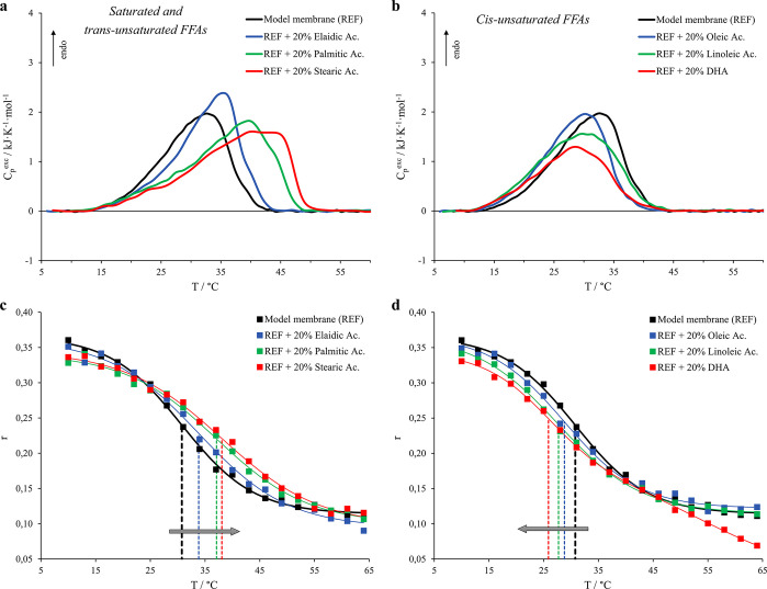
Figure 2.
Micro-DSC profiles for model membrane (5.7 DMPC:3.8 DPPS:0.5 DOPC, black curve) and vesicles with the addition of 20% FFAs and corresponding fluorescence anisotropy (r) of DPH trapped within the named systems. Thermograms are reported for membranes including saturated and trans-unsaturated FFAs in panel a, whereas the cis-unsaturated FFAs are shown in panel b. As for the fluorescence anisotropy (r) trends, the FFAs-free system is shown as black squares, whereas the FFAs considered are (c) elaidic acid (blue squares), palmitic acid (green squares), and stearic acid (red squares) and (d) oleic acid (blue squares), linoleic acid (green squares), and DHA (red squares). The dashed lines indicate the flex point of the sigmoidal fits with the respective colors. Probe:lipid molar ratio was 1:500.

