Abstract
The acronym PHACES stands for posterior fossa malformations, hemangiomas, arterial anomalies (cardiovascular or cerebrovascular), coarctation of the aorta/cardiac defects, eye abnormalities, and sternal defects. The characteristic dermatological clinical manifestation of PHACES syndrome is a segmental and extensive hemangioma, usually on the face. A combined therapy with 1,064 nm Nd-YAG/595-nm pulsed dye laser was performed in a young 15-year-old patient with PHACES syndrome, who presented a hemangioma on the left side of the face, located in the periorbital region. A first session with Nd-YAG laser (2,5 mm spot size, fluence 100 J/cm2, pulse duration 7 ms) for the treatment of teleangectasias and subsequently, three treatment sessions with pulsed dye laser (12 mm spot size, fluence 7 J/cm2, pulse duration 0,5 ms, repetition rate 0,6 Hz), once every 2 months, were performed. No postoperative complications were recorded, except for transient purpura after the pulsed dye laser sessions. The vascular lesion had a decrease in size bigger than 75%, and these results was maintained 6 months after the last treatment. Combined therapy Nd- YAG/pulsed dye laser is an effective and noninvasive procedure for hemangiomas in patients with PHACES syndrome.
Key words: Dye laser, hemangioma, PHACES syndrome
Introduction
PHACES syndrome is a neurocutaneous condition characterized by posterior fossa malformations, hemangioma, arterial anomalies, coarctation of the aorta/cardiac defects, eye abnormalities, and sternal malformations. PHACES syndrome is a nonhereditary condition and its etiology and pathogenesis are not fully known. Fetal and embryonic developmental defects have been suggested.1
PHACES syndrome predominantly affects the female sex (M:F = 1:9) and it is observed in 2% to 3% of infantile haemangiomas (IHs) cases. IHs associated with PHACES syndrome are typically extensive (>5cm in diameter) and segmental, and they can present as teleagectasias, solitary lesions, papules or confluent plaques.2 Hemangiomas in PHACES syndrome are most commonly located on the face, but cases involving other regions, such as occipital area, trunk, upper thoracic and proximal upper limb regions have also been described.
The treatment of the syndrome requires a multidisciplinary approach involving the cardiologist, neurologist and dermatologist. Systemic propranolol and steroids, surgery and laser therapy can be used to treat hemangiomas.3 595nm pulsed dye laser (PDL) and 1064 nm Nd:YAG-laser represent two laser systems very effective in the treatment of vascular anomalies such as HI. The combined use of these two lasers have been proposed in the treatment of HI, with good clinical results.4
Case Report
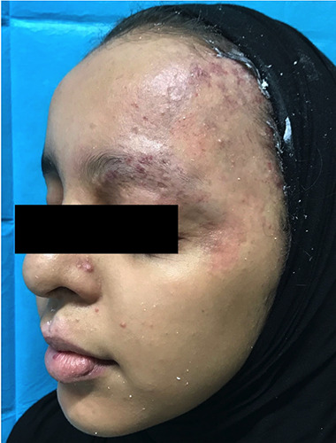
In this study we present the case of a 15 years-old girl affected by PHACES syndrome. At the age of one y.o., the patient underwent corrective surgery for aortic coarctation. Physical examination showed IH covering the left periocular region. Patient reported that IH appeared two months after birth, it grew for the first year of life and only partially spontaneously regressed up to current form. Cranial computed tomography documented the presence of angiomatous formation localized mainly on the lateral side of the eye which extended to the tear gland and to the ipsilateral eyelid region. Magnetic resonance angiography did not detect malformative alterations of the examined districts. Extracutaneous manifestations included headache, myopia and glaucoma.
Informed consent regarding the possible risks such as scars, discoloration, or hyperpigmentation of the procedures and the use of photographs for scientific reasons was obtained. This study was approved by ethical committee Calabria Centro with reference number 373/2019. Facial hemangioma was treated with 1,064 nm Nd:YAG laser Synchro Replay (Deka Medical Lasers, Italy )followed by 595-nm PDL Synchro VasQ (Deka Medical Lasers, Italy) the first used on telangiectasias and the second on the entire surface of the hemangioma respectively. Device characteristics are reported in Table 1.
The parameters used were 2,5 mm spot size, fluence 100 J/cm2, pulse duration 7 ms for Nd:YAG laser, and 12 mm spot size, fluence 7 J/cm2, pulse duration 0,5 ms, repetition rate 0,6 Hz for PDL.
A first session of Nd:YAG laser treat ment was performed without prior application of a local anesthetic with follow up and retouching after 40 days. The first treatment session with PDL, preceded by the application of topical anesthetic (Lidocaine/ Tetracaine cream), was performed 40 days after the treatment session with Nd:YAG laser. A total of three PDL treatment sessions, once every 2 months, and a single session of Nd:YAG laser were performed; the size and appearance of the IH gradually and slowly improved after each treatment session. Antibiotic cream was applied topically to the treated area after each session. During the laser treatment, no severe side effects were observed; transient purpura lasting less than 15 days occurred after PDL treatment sessions. Photographs were taken before and at the end of all treatments ; the results were assessed by evaluating the reduction in IH size and classified as 0-25% (I), 26-50% (II), 51-75% (III) and 76-100% (IV). Postoperatively, the patient was seen at 4 weeks, and 6 months after last treatment.
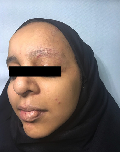
Four weeks after the last treatment, the hemangioma showed a strong improvement (IV) in size, thickness, and appearance, and the result remained stable after 6 months (Figures 1 and 2).
Discussion and Conclusions
Several types of lasers can be used for management of HIs, including argon laser, PDL and Nd:YAG laser,5 acting on intravascular oxyhemoglobin and resulting in vascular injury.
PDL produce pulses of visible light at a wavelength of 585 or 595 nm, which is primarily absorbed by oxyhemoglobin, destroying blood vessels selectively and keeping the overlying skin intact.
PDL represents the gold standard therapy for vascular lesions such as superficial hemangiomas, port-wine stains, and telangiectasias but it has also proven effective for treating vascular dependent lesions, or nonvascular lesions. In fact, PDL acts selectively on the abnormal vessels of the lesions to be treated and causes selective thrombosis with the consequent destruction of the supply of nutrients to the lesions.6
While PDL reaches 0.75-1 mm in depth, Nd:YAG laser, whose 1064 nm wavelength light is absorbed by oxyhemoglobin, melanin and water, is able to penetrate 5-6 mm deep into the tissues, representing the most efficient vascular laser system in terms of skin penetration.7,8 Therefore, combined therapy using Nd:YAG-laser and PDL is very effective in the treatment of vascular lesions. and it is important to start the treatment with the Nd:YAG because PDL therapy causes post-treatment edema, which could influence the effectiveness of the treatment with Nd:YAG, since its light is also absorbed by the water. Transient purpura is the main side effect that occurs immediately after PDL treatment and correlates with the effectiveness of the treatment; this visible side effect disappears within 1-2 weeks and then the reduction in the number and size of vessels will start.9
Table 1.
Device characteristics.
| Technical specifications | Synchro VasQ DEKA | Synchro Replay DEKA |
|---|---|---|
| Wavelength | Dye Laser 595 nm | Nd:YAG Laser 1064 nm |
| Spot size (mm) | 12-Mar | 2.5-24 |
| Max fluence J/cm2 | 33 | 1500 |
| Pulse duration (ms) | 0.3-40 | 0.2-300 |
| Number of pulses per shot | 1 | up to 3 |
| Emission control | Foot and finger switch | Foot and finger switch |
| Repetition rate | up to 1 Hz | up to 10 Hz |
Figure 1.

Facial hemangioma before treatment.
Figure 2.

Facial hemangioma 6 months after therapy.
Although different recent works report the effectiveness of combined PDL and Nd:YAG-laser in the treatment of hemangiomas, 4 this is to our knowledge the first time that this combination technique is proposed in the treatment of this rare cutaneous condition. Our results demonstrate that Nd:YAG laser in conjunction with PDL is a well-tolerated and effective therapy in patients with facial hemangiomas in the context of PHACES syndrome, and represents a promising alternative to other medical and surgical treatments. In fact, the different wavelengths of light emitted by these two laser systems manage to reach vascular structures at different levels of depth within the tissues, and their synergistic action allows to obtain excellent aesthetic results, with minimal incidence of side effects.
Funding Statement
Funding: None.
References
- 1.Haggstrom AN, Lammer EJ, Schneider RA, et al. Patterns of infantile hemangiomas: new clues to hemangioma pathogenesis and embryonic facial development. Pediatrics 2006;117:698-703 [DOI] [PubMed] [Google Scholar]
- 2.Chiller KG, Passaro D, Frieden IJ. Hemangiomas of infancy: clinical characteristics, morphologic subtypes, and their relationship to race, ethnicity, and sex. Arch Dermatol 2002;138:1567-76 [DOI] [PubMed] [Google Scholar]
- 3.Winter PR, Itinteang T, Leadbitter P, Tan ST. PHACE syndrome--clinical features, aetiology and management. Acta Paediatr 2016;105:145-53. [DOI] [PubMed] [Google Scholar]
- 4.Hartmann F, Lockmann A, Grönemeyer LL, et al. Nd:YAG and pulsed dye laser therapy in infantile haemangiomas: a retrospective analysis of 271 treated haemangiomas in 149 children. J Eur Acad Dermatol Venereol 2017;31:1372-9. [DOI] [PubMed] [Google Scholar]
- 5.Tanzi EL, Lupton JR, Alster TS. Lasers in dermatology: four decades of progress. J Am Acad Dermatol 2003;49:1–31. [DOI] [PubMed] [Google Scholar]
- 6.Nisticò S, Campolmi P, Moretti S, et al. Nonconventional Use of Flash-Lamp Pulsed-Dye Laser in Dermatology. Biomed Res Int 2016;2016:7981640. [DOI] [PMC free article] [PubMed] [Google Scholar]
- 7.Cannarozzo G, Negosanti F, Sannino M, et al. Q-switched Nd:YAG laser for cosmetic tattoo removal. Dermatol Ther 2019;32:e13042. [DOI] [PubMed] [Google Scholar]
- 8.Del Duca E, Zingoni T, Bennardo L, et al. Long-Term Follow-Up for QSwitched Nd:YAG Treatment of Nevus of Ota: Are High Number of Treatments Really Required? A Case Report. Photobiomodul Photomed Laser Surg 2020. Aug 5. [DOI] [PubMed] [Google Scholar]


