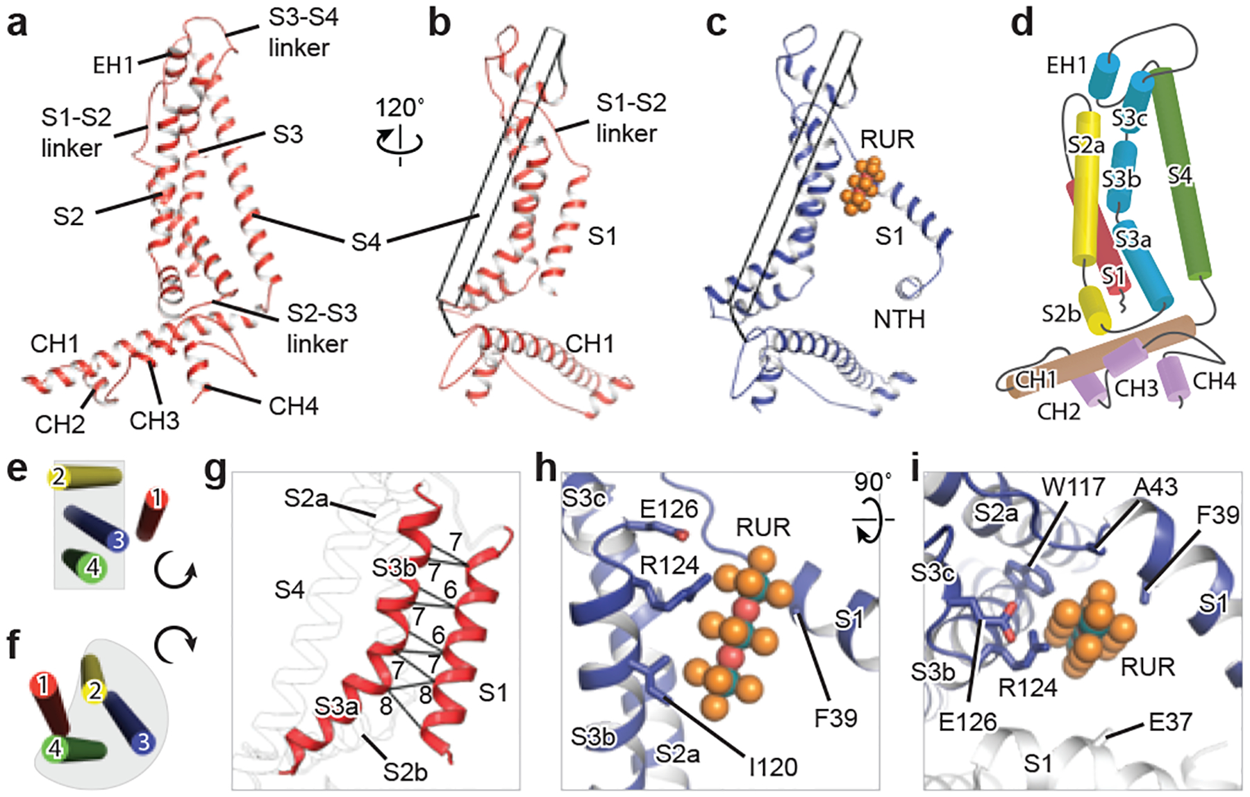
Fig. 2 |. A single subunit of CALHM2, and RUR-binding site.

a–c, Cartoon representation of EDTA–CALHM2hemi (a, b) and RUR–CALHM2 (c). The S4 helix in b and c is shown as a transparent tube for clarity. d, Domain organization of EDTA–CALHM2hemi. e, f, TMD organization of EDTA–CALHM2hemi (e) and connexin 43 (f) viewed from the extracellular side. g, The loose contacts between the S1 and S3 helices in EDTA–CALHM2hemi. Distances (in Å) between Cα of adjacent residues in helices S1 and S3 are labelled. h, i, RUR-binding site.
