Abstract
Purpose
This study aimed to evaluate and analyze the feasibility and the oncological and functional outcomes of limb salvage surgery in extremity soft tissue sarcomas (ESTS) and bone tumors invading vessels.
Methods
Materials and This single-center retrospective analysis included patients with ESTS encasing or invading major blood vessels that were treated by limb salvage surgery with vascular resection and reconstruction between January 1995 and December 2019. Patients with contiguous involvement of major blood vessels and nerves and patients requiring amputation were excluded from the study.
Results
A total of 24 vessels (14 arteries and 10 veins) in 14 patients were reconstructed. Ten (71.4%) patients underwent both arterial and venous reconstruction, and four (28.6%) underwent only arterial reconstruction. Reconstruction was performed with a reversed saphenous vein (RSV) graft in 12 patients and with a synthetic graft (SG) in the other 12 patients. At a median follow-up of 27 months, RSV grafts were patent in 10 of 12 (83.3%) vessels and SGs were patent in 6 of 12 (50.0%) vessels (log-rank test, P=0.083). Out of 14 arteries and 10 veins, 11 arteries and 5 veins were patent, respectively. No patient developed local recurrence, and 2 (14.3%) patients developed distant metastases. Limb salvage rate was 13/14 (92.9%). The mean Musculoskeletal Tumor Society score was 83.3%. The 5- and 10-year overall survival rates were 80% and 50%, respectively.
Conclusion
Limb salvage surgery in ESTS with vascular resection and reconstruction is feasible and provides favorable oncological and functional outcomes.
Keywords: Sarcoma, Bone neoplasms, Vascular surgical procedures, Limb salvage, Reconstructive surgery
INTRODUCTION
Soft tissue and bone sarcomas are rare malignant neoplasms that account for less than 1% of all cancers diagnosed annually in the United States [1]. According to the Madras Metropolitan Tumor Registry, bone sarcomas and soft tissue sarcomas account for 1.1% and 1.2% of all cancers in women and 0.9% and 1.1% of all cancers in men, respectively [2]. Sarcomas involve adjacent blood vessels in 5% of cases [3]. Amputation was previously considered to be the treatment of choice for sarcomas located in the pelvis and extremities that encased or invaded large-caliber vascular structures [3]. Changes in the management of these complex cases were suggested and implemented by Fortner et al. [4], who described the earliest series of en bloc resection for sarcomas invading vascular structures. This procedure was reported to be oncologically safe with satisfactory local control [5,6].
Patients having extremity soft tissue sarcomas (ESTS) or bone sarcomas with vascular involvement require multimodal management to achieve limb salvage. Limb salvage surgery offers significantly better functional outcomes and quality of life compared to amputation, even in patients with distant metastasis [1]. In the present study, we review our experience with en bloc resection of the tumor and blood vessels followed by immediate reconstruction to achieve limb salvage.
MATERIALS AND METHODS
This study retrospectively analyzed ESTS cohorts from January 1995 to December 2019. Patients with ESTS encasing vessels (arteries, veins, or both) requiring vessel resection and reconstruction to achieve satisfactory margins were included in the study. The exclusion criteria comprised patients with ESTS involving both nerves and vessels and those requiring amputation due to intraoperative vessel injury. The medical records of eligible patient cohorts were reviewed, and all relevant clinical details including demographic profiles, histological variants, treatments, and disease outcomes were collected and analyzed.
All patients were clinically evaluated by a specialized musculoskeletal oncologist and underwent preoperative magnetic resonance imaging for local staging of the tumor and for identifying tumor invasion of major vessels (arteries, veins, or both). Consultation with a vascular surgeon was done for all patients potentially requiring vessel resection and reconstruction. Core needle biopsy was performed for the tissue diagnosis, and an appropriate metastatic workup was performed based on the histology. All patients underwent en bloc resection of the tumor and its involved vessels. Modular prosthesis reconstruction was performed for bone tumors.
Factors influencing the choice of vascular graft for reconstruction were a) length and caliber of the resected vessel; b) availability of a suitable venous graft in the contralateral limb; and c) age. We used a reversed saphenous vein graft harvested from the contralateral limb for small- and medium-sized vessels (tibial and popliteal) and synthetic grafts (SGs) for large vessels (femoral). Vascular grafts were anastomosed in an end-to-end manner. The distal pedal flow was monitored with a continuouswave Doppler after anastomosis.
Antithrombotic medication was started postoperatively on the same day. All patients were started on parenteral low molecular weight heparin (Fragmin [Dalteparin Sodium]; Pfizer Ltd., Mumbai, India), which was continued until discharge. Patients were also started on antiplatelets (Ecosprin [Aspirin]; USV Private Ltd., Mumbai, India). As a protocol, oral antithrombotics (Acitrom [Acenocoumarol]; Abbott Ltd., Mumbai, India) were administered for 3 months, irrespective of the type of graft used.
Follow-ups were performed every month for 1 year, every 3 months for the next 2 years, every 6 months for the subsequent 2 years, and annually thereafter. Routine examination was performed with a handheld Doppler, and duplex ultrasound (DUS) was performed if findings were suggestive of a thrombus. Routine DUS was performed at 1- and 6-month intervals after surgery and after 6 months only in the event of absent peripheral pulses or unsatisfactory findings on handheld Doppler. Functional outcomes were calculated using the Musculoskeletal Tumor Society (MSTS) score at the 6-month follow-up.
Statistical analysis was performed with SPSS software ver. 24.0 (IBM Corp., Armonk, NY, USA). The Kaplan–Meier method was used to estimate overall survival (OS) using the log-rank test. Results were considered significant if the P-value was <0.05 with a confidence interval of 95%. This study was approved by the Institute Ethics Committee, bearing the number “IEC/2020/July08.”
RESULTS
Fourteen patients with ESTS underwent en bloc tumor resection with major vascular resection and reconstruction. The mean age was 33 years (range, 13-63). Patient characteristics are listed in Table 1. The primary tumor was in the lower limb in 12 (85.7%) patients and in the upper limb in 2 (14.3%) patients. The most common etiology was osteosarcoma (n=5, 35.7%), followed by synovial sarcoma (n=4, 28.6%) and fibromatosis (n=2, 14.3%).
Table 1.
Patient characteristics, adjuvant treatment, and follow-up (n=14)
| Patient characteristic | Number (%) |
|---|---|
| Sex, male | 6 (42.9) |
| Site, upper limb/lower limb | 2/12 |
| Location, thigh/leg/arm | 11/1/2 |
| Histology | |
| Osteosarcoma | 5 (35.7) |
| Synovial sarcoma | 4 (28.6) |
| Fibromatosis | 2 (14.3) |
| Liposarcoma | 1 (7.1) |
| Malignant peripheral nerve sheath tumor | 1 (7.1) |
| Giant cell tumor | 1 (7.1) |
| Grade, high/intermediate/low/undetermined | 8/1/1/4 |
| Limb salvage/amputation | 13/1 |
| Adjuvant and perioperative therapy | |
| Adjuvant chemotherapy+radiotherapy | 4 (28.6) |
| Perioperative chemotherapy | 6 (42.9) |
| Adjuvant radiotherapy | 1 (7.1) |
| No additional therapy | 3 (21.4) |
| Local recurrence | 0 |
| Distant metastasis | 2 (14.3) |
| Alive/dead | 10/4 |
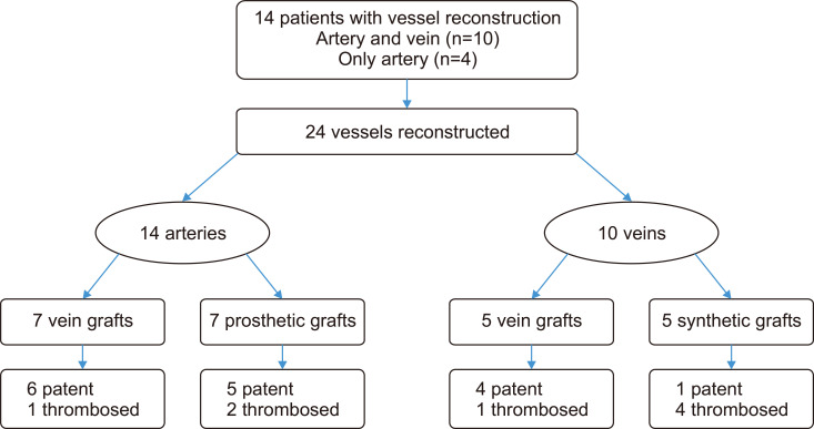
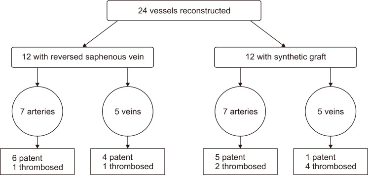
Ten patients underwent both arterial and venous reconstruction, and four patients underwent arterial reconstruction only (total of 24 vessels: 14 arteries and 10 veins). The most common vessels resected were the femoral artery and vein (7 each). Autologous (great saphenous vein) grafts and prosthetic (polytetrafluoroethylene) grafts were used to reconstruct 12 vessels each. Details of vessels resected and reconstructed, conduit type, and patency rates are described in Table 2 and in Fig. 1, 2. The resected margins were clear in all patients on histopathological examination. The types of vascular involvement included 18 (75.0%) cases of type 1 (vessel encasement without vessel involvement), 1 (4.2%) case of type 2 (contiguous vessel wall invasion), and 5 (20.8%) cases of type 3 (intact adventitia with discontinuous tumor emboli in the vessel lumen). Multimodal therapy was performed in 11 of the 14 (78.6%) patients. Six patients received perioperative chemotherapy alone (five osteosarcomas and one primary synovial sarcoma of bone). Four patients (three synovial and one malignant peripheral nerve sheath tumour) received adjuvant chemoradiotherapy. One patient (fibromatosis) received adjuvant radiation therapy. Three patients did not receive any adjuvant therapy (fibromatosis, well-differentiated liposarcoma, and giant cell tumor of the bone). The mean time to start adjuvant therapy was 42 days (Table 1).
Table 2.
Characteristics of vessels and the graft used
| Name of vessel | Total number (P/T) | Reconstructed with RSV (P/T) | Reconstructed with SG (P/T) | Total | P-value |
|---|---|---|---|---|---|
| Artery (n=14) | |||||
| Femoral | 7 (7/0) | 2 (2/0) | 5 (5/0) | ||
| Popliteal | 4 (2/2) | 3 (2/1-amputation) | 1 (0/1) | Total 14 arteries, 11 patent (6 RSV, 5 SG), 3 thrombosed (1 RSV, 2 SG) | Artery vs. vein graft patency: 0.143 |
| Brachial | 2 (1/1) | 1 (1/0) | 1 (0/1) | ||
| Posterior tibial | 1 (1/0) | 1 (1/0) | 0 (0/0) | ||
| Vein (n=10) | |||||
| Femoral | 7 (3/4) | 3 (2/1) | 4 (1/3) | ||
| Popliteal | 2 (1/1) | 1 (1/0) | 1 (0/1) | Total 10 veins, 5 patent (4 RSV, 1 SG),5 thrombosed (1 RSV, 4 SG) | RSV vs. SG patency: 0.083 |
| Posterior tibial | 1 (1/0) | 1 (1/0) | 0 (0/0) | ||
| Total | 24 (16/8) | 7 arteries (6/1) 5 veins (4/1) |
7 arteries (5/2) 5 veins (1/4) |
||
P/T, patent/thrombosed; RSV, reversed saphenous vein; SG, synthetic graft.
Fig. 1.
Flow chart according to the reconstructed vessels and their patency.
Fig. 2.
Flow chart according to the conduit of the reconstructed vessels and their patency.
Graft thrombosis was classified as early (within 1 month of surgery) or delayed (1 to 6 months of surgery). Early graft thrombosis developed in 5 (35.7%) patients: both arterial and venous thrombosis in 2 (14.3%) patients, only venous thrombosis in 2 (14.3%) patients, and only arterial thrombosis in 1 (7.1%) patient. For both arterial and venous thrombosis, thrombectomy and fasciotomy for compartment syndrome was performed in one patient, and thrombectomy alone was performed in the other, who subsequently developed dry gangrene of the toe. Two patients with venous thrombosis were managed with antithrombotic agents: one patient achieved subsequent recanalization; however, the other continued antithrombotic therapy due to persistent venous thrombosis and eventually required debridement and secondary suturing for marginal flap necrosis. One patient with only arterial thrombosis developed extensive blisters in the lower limb. Reoperation revealed full-thickness muscle necrosis, and above-knee amputation was performed. At the 1-month follow-up, one vein remained thrombosed.
Delayed graft thrombosis at 6 months developed in eight (57.1%) patients. Five (35.7%) patients had venous thrombosis with good collateral flow. These five patients, along with one patient with early thrombosis, were managed with antithrombotics. Two patients had arterial thrombosis with good collateral flow, and hence, were managed conservatively. The mean interval to arterial and venous thrombi formation was 2.6 months and 2 months, respectively. Three patients developed lymphedema, which was managed with compression stockings.
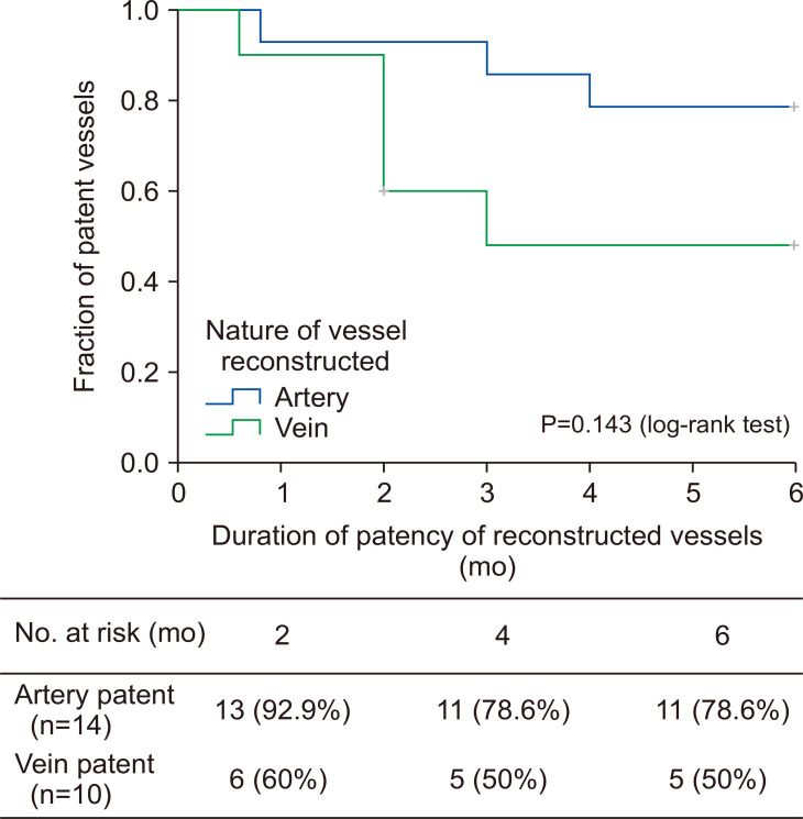
Limb salvage was achieved in 13 (92.9%) patients. The long-term patency rate for arteries was 78.6% and that for veins was 50% (P=0.143; Fig. 3). No patient had local recurrence; however, two patients developed distant metastases at 16 and 78 months. At a median follow-up of 2 years, four (28.6%) patients had died, two each due to malignancy and antecedent causes. The 5-year disease-free survival rate of patients with both arterial and venous reconstruction was 92%, and that of only arterial reconstruction was 87% (P=0.458). The 5-year OS rates were not significantly different (94% vs. 88%, P=0.538). The 5- and 10-year OS rates for the entire cohort were 80% and 50%, respectively. The mean MSTS score was 83.3% for the entire cohort.
Fig. 3.
The long-term patency rate for arterial reconstruction was 78.6%, and that for venous reconstruction was 50%, which was statistically insignificant (P=0.143).
DISCUSSION
Until 20 years ago, amputation was the only reasonable treatment for sarcomas of the lower limbs [7]. Fortner et al. [4] reported the first successful series of vascular resection and reconstruction as a part of tumor excision to achieve limb salvage. Abbas et al. [8] and Enneking et al. [9] reported no significant differences in oncological outcomes between amputation and limb salvage groups. Locally advanced tumors involving major vascular structures were considered an indication for limb salvage [5]. Advancements in surgical skill and imaging modalities have made en bloc resection of the tumor with affected vascular structures possible [5,10], Vascular reconstructions after en bloc resection of the tumor and vessels were successfully performed despite difficult surgery, longer operating time, and limited patency (in some cases) [1]. Vascular surgeons were often involved as “reconstructive surgeons” during these procedures because of their unique skills in dealing with vessels [1].
Arterial reconstruction is clearly indicated following resection as the limb is unlikely to survive due to ischemia; however, the literature recommending venous reconstruction is less robust [3]. Tsukushi et al. [11] reported no significant differences in terms of lymphedema, complications, or postoperative function between groups receiving arterial and venous reconstruction and groups receiving arterial reconstruction only. Schwarzbach et al. [5] reconstructed both arteries and veins in all cases, of which 42% of patients developed venous thrombosis, and they presented either asymptomatically or with well-tolerated edema. Umezawa et al. [12] reported that the mean duration of limb edema after surgery was greater in patients who underwent only arterial reconstruction (117 days) than in patients who underwent arteriovenous reconstruction (58.8 days).
At our center we prefer to reconstruct non-occlusive veins. Even if the vein graft eventually develops a thrombus, the flow through the graft provides time for sufficient collaterals to develop. Therefore, phlebolymphedema in patients with a venous graft might be much less than in patients without venous reconstruction. We do not reconstruct veins that are completely obstructed as the collateral flow is already established, and reconstruction might not add further benefit.
Patency rates of arterial and venous reconstruction vary across studies, but patency is generally better in arterial reconstruction. Park et al. [1] reported an arterial patency of 78.6% and a venous patency of 61.5%, without a significant difference in occlusion rate (P=0.37). Nishinari et al. [13] reported patency rates of arterial and venous reconstruction as 93.1% and 79.3%, respectively (P=0.63). The long-term patency rates for arterial and venous reconstructions in our study were 78.5% and 50%, respectively (P=0.143).
Vascular substitutes can either be autologous vein grafts or prosthetic grafts. An autologous vein graft is the preferred conduit because its patency is superior to that of a SG [3,4]. Leggon et al. [14] reported patency rates of autologous vein grafts for arterial and venous reconstruction to be 88% and 75%, respectively, and those for SGs to be 83% and 56%, respectively. Park et al. [1] reported patency rates of 85.7% and 16.7% for autologous vein grafts and SGs, respectively (P<0.001). Our study reported patency rates of 83.3% for autologous vein grafts and 50% for SGs (P=0.083).
Another issue is oncologic safety. Surgery for advanced cancer with vascular invasion is complex and requires expertise and a trained multidisciplinary team. There is a risk of tumor spill or compromised margins. Patients with vessel reconstruction must be monitored carefully during the postoperative period to identify complications, such as vascular thrombosis and compartment syndrome. Despite these challenges, limb salvage surgery is still considered to be the standard treatment for ESTS [6,12] and is feasible in the vast majority of cases. Many studies have reported limb salvage rates between 84.6% and 100% [5,11,13,15,16]. Our study had a limb salvage rate of 92.8%.
ESTS are aggressive tumors with frequent local recurrences due to compromised margins, which are reported to be as high as 65% [17,18]. Thus, en bloc resection is the mainstay of treatment for sarcomas involving blood vessels, to achieve negative margins and reduce local recurrence. Many studies have reported local recurrence rates of 14% to 47% [1,5,19]. Fortunately, there were no local recurrences in the present study.
Vascular resection and reconstruction leads to limb preservation, which ensures better functional outcomes and quality of life. The mean MSTS score in this study was 83.3%, which is comparable with scores in previous reports ranging from 70% to 81% [11,14,20]. Despite the locally advanced nature and possible aggressive biology of these sarcomas, satisfactory oncological outcomes and survival can be achieved in these patients. The 5-year OS in the current study was 80%, comparable with previous studies that reported an OS of 60% to 88% (Table 3) [12,13,21].
Table 3.
Comparison with other studies
| Parameter | Current study | Nishinari et al. [13] | Schwarzbach et al. [5] | Tsukushi et al. [11] |
|---|---|---|---|---|
| Total number of patients | 14 | 17 | 21 | 25 |
| Most common site of tumor | Lower limb | Lower limb | Lower limb | Lower limb |
| Most common histology | Osteosarcoma | Synovial sarcoma | Liposarcoma | Liposarcoma |
| Most common grade | High | High | High | High |
| Most common vessel involved | Femoral | Femoral | Femoral | Femoral |
| Conduit used, RSV/SG | 12/12 | 23/9 | 10/22 | 27/10 |
| Patent/thrombosed | 16/8 | 26/6 | 22/10 | 25/12 |
| Limb salvage (%) | 92.80 | 100 | 100 | 100 |
| 5-year overall survival rate (%) | 80 | 60 | 52 | 92 |
RSV, reversed saphenous vein; SG, synthetic graft.
This study has several limitations. The retrospective nature of the study and the small sample size pose a certain selection bias. The lack of objective surveillance protocols, such as ankle-brachial index or duplex ultrasonography, might have delayed the detection of vessel occlusion. In addition, the lack of regular imaging during follow-up might have underestimated the local recurrence rate.
CONCLUSION
Limb salvage surgery for ESTS and bone tumors invading vessels is feasible and provides good local control rates and favorable oncological and functional outcomes.
Footnotes
CONFLICTS OF INTEREST
The authors have nothing to disclose.
AUTHOR CONTRIBUTIONS
Concept and design: AR, VR, ACS. Analysis and interpretation: AR, VR, KM. Data collection: ACS. Writing the article: ACS. Critical revision of article: ACS, AR, VR. Final approval of article: AR, VR, KM. Statistical analysis: ACS. Obtained funding: none. Overall responsibility: all authors.
REFERENCES
- 1.Park D, Cho S, Han A, Choi C, Ahn S, Min SI, et al. Outcomes after arterial or venous reconstructions in limb salvage surgery for extremity soft tissue sarcoma. J Korean Med Sci. 2018;33:e265. doi: 10.3346/jkms.2018.33.e265. [DOI] [PMC free article] [PubMed] [Google Scholar]
- 2.Swaminathan R, Shanta V, Balasubramanian S, Sampath P. Madras Metropolitan Tumour Registry, technical report 2012-2014 [Internet] National Cancer Registry Program, Cancer Institute (WIA); Chennai: C 2016. [cited 2020 Sep 28]. Available from: http://www.iacr.com.fr/index.php?option=com_comprofiler&task=userprofile&user=952&Itemid=498 . [Google Scholar]
- 3.Lawrence W., Jr Concepts in limb-sparing treatment of adult soft tissue sarcomas. Semin Surg Oncol. 1988;4:73–77. doi: 10.1002/ssu.2980040114. [DOI] [PubMed] [Google Scholar]
- 4.Fortner JG, Kim DK, Shiu MH. Limb-preserving vascular surgery for malignant tumors of the lower extremity. Arch Surg. 1977;112:391–394. doi: 10.1001/archsurg.1977.01370040043007. [DOI] [PubMed] [Google Scholar]
- 5.Schwarzbach MH, Hormann Y, Hinz U, Bernd L, Willeke F, Mechtersheimer G, et al. Results of limb-sparing surgery with vascular replacement for soft tissue sarcoma in the lower extremity. J Vasc Surg. 2005;42:88–97. doi: 10.1016/j.jvs.2005.03.017. [DOI] [PubMed] [Google Scholar]
- 6.Nagarajan R, Neglia JP, Clohisy DR, Robison LL. Limb salvage and amputation in survivors of pediatric lower-extremity bone tumors: what are the long-term implications? J Clin Oncol. 2002;20:4493–4501. doi: 10.1200/JCO.2002.09.006. [DOI] [PubMed] [Google Scholar]
- 7.Bonardelli S, Nodari F, Maffeis R, Ippolito V, Saccalani M, Lussardi L, et al. Limb salvage in lower-extremity sarcomas and technical details about vascular reconstruction. J Orthop Sci. 2000;5:555–560. doi: 10.1007/s007760070005. [DOI] [PubMed] [Google Scholar]
- 8.Abbas JS, Holyoke ED, Moore R, Karakousis CP. The surgical treatment and outcome of soft-tissue sarcoma. Arch Surg. 1981;116:765–769. doi: 10.1001/archsurg.1981.01380180025006. [DOI] [PubMed] [Google Scholar]
- 9.Enneking WF, Spanier SS, Goodman MA. A system for the surgical staging of musculoskeletal sarcoma. Clin Orthop Relat Res. 1980;(153):106–120. doi: 10.1097/00003086-198011000-00013. [DOI] [PubMed] [Google Scholar]
- 10.Hohenberger P, Allenberg JR, Schlag PM, Reichardt P. Results of surgery and multimodal therapy for patients with soft tissue sarcoma invading to vascular structures. Cancer. 1999;85:396–408. doi: 10.1002/(SICI)1097-0142(19990115)85:2<396::AID-CNCR18>3.0.CO;2-O. [DOI] [PubMed] [Google Scholar]
- 11.Tsukushi S, Nishida Y, Sugiura H, Nakashima H, Ishiguro N. Results of limb-salvage surgery with vascular reconstruction for soft tissue sarcoma in the lower extremity: comparison between only arterial and arterovenous reconstruction. J Surg Oncol. 2008;97:216–220. doi: 10.1002/jso.20945. [DOI] [PubMed] [Google Scholar]
- 12.Umezawa H, Sakuraba M, Miyamoto S, Nagamatsu S, Kayano S, Taji M. Analysis of immediate vascular reconstruction for lower-limb salvage in patients with lower-limb bone and soft-tissue sarcoma. J Plast Reconstr Aesthet Surg. 2013;66:608–616. doi: 10.1016/j.bjps.2013.01.001. [DOI] [PubMed] [Google Scholar]
- 13.Nishinari K, Krutman M, Aguiar Junior S, Pignataro BS, Yazbek G, Zottele Bomfim GA, et al. Surgical outcomes of vascular reconstruction in soft tissue sarcomas of the lower extremities. J Vasc Surg. 2015;62:143–149. doi: 10.1016/j.jvs.2015.02.004. [DOI] [PubMed] [Google Scholar]
- 14.Leggon RE, Huber TS, Scarborough MT. Limb salvage surgery with vascular reconstruction. Clin Orthop Relat Res. 2001;387:207–216. doi: 10.1097/00003086-200106000-00028. [DOI] [PubMed] [Google Scholar]
- 15.Karakousis CP, Emrich LJ, Rao U, Krishnamsetty RM. Feasibility of limb salvage and survival in soft tissue sarcomas. Cancer. 1986;57:484–491. doi: 10.1002/1097-0142(19860201)57:3<484::AID-CNCR2820570314>3.0.CO;2-K. [DOI] [PubMed] [Google Scholar]
- 16.Imparato AM, Roses DF, Francis KC, Lewis MM. Major vascular reconstruction for limb salvage in patients with soft tissue and skeletal sarcomas of the extremities. Surg Gynecol Obstet. 1978;147:891–896. [PubMed] [Google Scholar]
- 17.Williard WC, Hajdu SI, Casper ES, Brennan MF. Comparison of amputation with limb-sparing operations for adult soft tissue sarcoma of the extremity. Ann Surg. 1992;215:269–275. doi: 10.1097/00000658-199203000-00012. [DOI] [PMC free article] [PubMed] [Google Scholar]
- 18.Karakousis CP, Proimakis C, Walsh DL. Primary soft tissue sarcoma of the extremities in adults. Br J Surg. 1995;82:1208–1212. doi: 10.1002/bjs.1800820919. [DOI] [PubMed] [Google Scholar]
- 19.Ramamurthy R, Soundrarajan JC, Mettupalayam V, Shanmugham S, Arumugam B, Periasamy S. Limb conservation in extremity soft tissue sarcomas with vascular involvement. Indian J Orthop. 2009;43:403–407. doi: 10.4103/0019-5413.54969. [DOI] [PMC free article] [PubMed] [Google Scholar]
- 20.Muramatsu K, Ihara K, Miyoshi T, Yoshida K, Taguchi T. Clinical outcome of limb-salvage surgery after wide resection of sarcoma and femoral vessel reconstruction. Ann Vasc Surg. 2011;25:1070–1077. doi: 10.1016/j.avsg.2011.05.009. [DOI] [PubMed] [Google Scholar]
- 21.Baxter BT, Mahoney C, Johnson PJ, Selmer KM, Pipinos II, Rose J, et al. Concomitant arterial and venous reconstruction with resection of lower extremity sarcomas. Ann Vasc Surg. 2007;21:272–279. doi: 10.1016/j.avsg.2007.03.005. [DOI] [PubMed] [Google Scholar]


