Abstract
Objective
Spinal meningioma is mostly benign, but they can exhibit neurological deficit. The relationship between neurological impairment and its radiographic findings, including intratumor magnetic resonance imaging (MRI) gadolinium enhancement and calcification in computed tomography (CT) scan, has not been studied. The purpose of this study was to investigate the association of preoperative image findings with neurological status in spinal meningioma.
Methods
Patients histologically diagnosed with spinal meningioma (n = 24), with an average age of 65.4 years, were included. The patients were classified into 2 groups, the homogeneous and heterogeneous groups, based on the contrast-enhanced T1-weighted MRI findings. Further, baseline demographics (age, sex, presence of preoperative paralysis [manual muscle testing 3 or worse neurological deficit in upper and/or lower limbs], tumor level, tumor length, and tumor occupation ratio), histological findings (Ki-67 index and histological subtypes), and CT findings (presence of intratumor calcification and Hounsfield unit [HU] value) were examined.
Results
Preoperative paralysis was observed in 33.3% (8 of 24) of the patients. These patients exhibited frequent heterogeneous contrast-enhanced MRI findings than those without preoperative paralysis (57.1% vs. 14.3%, p = 0.040). Further, preoperative paralysis did not associate with tumor level, tumor length, tumor-occupied ratio, Ki-67 index, and histological subtypes. The heterogeneous group showed 100% intratumor calcification and higher maximum HU than the homogeneous group (1,109.8 vs. 379.2, p = 0.001).
Conclusion
The heterogeneous contrast-induced MRI findings in the spinal meningioma were significantly associated with preoperative neurological impairment. Moreover, the intratumor contrast-deficient region in the heterogeneously enhanced tumors reflected marked calcification. The tumor hardness due to calcification may be related to preoperative neurological deficit.
Keywords: Spinal meningioma, Computed tomography, Magnetic resonance imaging, Calcification, Motor deficit
INTRODUCTION
Meningioma, one of the most common intra-canal spinal tumors, accounts for 24%–46% of spinal neoplasms [1-3]. The spinal meningioma typically occurs in the intradural extramedullary space of the thoracic level in middle-aged women. It is usually benign, slow-growing tumor, and more than 95% of the spinal meningiomas are classified as World Health Organization (WHO) grade I [3-6]. Due to its slow-growing nature, it can be incidentally diagnosed asymptomatic, but is often found to exhibit neurological deficit in the clinical settings. The incidence of motor weakness has been reported in 64%–93% cases [2,6,7]. Radiographically, magnetic resonance imaging (MRI) is the gold standard for diagnosis of spinal meningiomas. The typical signal intensity of spinal meningioma is iso- to slightly high in T2-weighted images (T2WI). Moreover, the T1-weighted image (T1WI), after intravenous injection of gadolinium-diethylenetriamine pentaacetate (Gd+), typically shows a homogeneous positive enhancement effect [8]. Histologically, the meningioma forms a characteristic “psammoma body,” in which the intratumor calcification is observed with computed tomography (CT) scan. However, few reports have described an association of the image findings with preoperative neurological status. We hypothesized that these characteristic image findings may correlate with tumor proliferation and could be a potential predictor of neurological impairment. Thus, the purpose of this study was to assess the relationship between the preoperative contrast-enhanced MRI/CT findings and neurological status of spinal meningioma.
MATERIALS AND METHODS
1. Patient Population
This study was conducted following the approval by Institutional Review Board of Kyoto University (IRB No. 1888). We retrospectively reviewed patients who were surgically treated and histologically diagnosed with spinal meningioma between September 2006 and November 2019. We included patients with intradural extramedullary meningioma, whereas those with dumbbell-type, intramedullary, or extradural types were excluded. With these inclusion and exclusion criteria, 24 patients were enrolled in the analysis. We collected following baseline demographic data: age, sex, body mass index, duration of symptom, and preoperative neurological findings. Further, we investigated the patient’s follow-up duration, recurrence rate at the final follow-up, and tumor resection procedure (Simpson grade [9]) based on their medical records.
2. Radiographic and Histological Evaluation
Based on the preoperative MRI, tumor level was classified into cervical, thoracic, and lumbar region. The tumor length was measured as the maximum diameter in the sagittal view of T2WI MRI image. The tumor occupation ratio was calculated as the ratio of tumor diameter to intradural space diameter in the axial view of T2WI MRI image at the level in which the tumor appeared largest. The tumor localization was determined as dorsal, ventral, and lateral type based on the tumor attachment to dura. The presence of intratumor calcification and the maximum of Hounsfield unit (HU) value inside the tumor were analyzed based on the CT findings. These radiographic analyses were reviewed by 2 independent spine surgeons (KO and TS). Additionally, the WHO grade, histological subtypes, and tumor proliferation index (Ki-67 index) were evaluated using the surgically resected specimens.
3. Analysis of Relationship Between Radiographic Findings and Motor Status
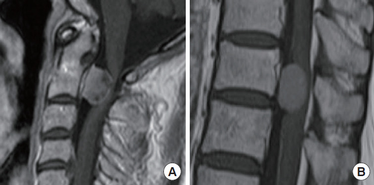
The patients who demonstrated grade 3 or worse preoperative neurological deficit with manual muscle testing were defined as having preoperative paralysis in the upper and/or lower limbs for cervical meningioma, while in the lower limbs for thoracic or lumbar meningioma. Additionally, the patients were distributed into 2 groups based on findings of the contrast-enhanced MRI as: the homogeneous group (those with homogeneous Gd+ enhancement effect) and heterogeneous group (those with heterogeneous Gd+ enhancement effect) (Fig. 1). The baseline characteristics were compared between patients in both the groups.
Fig. 1.

Contrast-enhanced T2-weighted image magnetic resonance imaging. (A) Heterogeneous enhancement group. (B) Homogeneous enhancement group.
4. Statistical Analysis
Data have been presented as mean ± standard deviation, unless otherwise specified. The chi-square test and Mann-Whitney U-test were used for analysis of the categorical and continuous variables, respectively. A p-value < 0.05 was considered statically significant. All statistical analyses were performed using the JMP pro 14 software (SAS Institute Inc., Cary, NC, USA).
RESULTS
The baseline demographics of patients (n = 24) have been summarized in Table 1. These comprised of 20 women and 4 men with an average age of 65.4 years at the time of surgery. Further, 33.3% (8 of 24) of the patients showed preoperative paralysis. All the patients experienced neurological improvement after surgery. Of the 21 patients who underwent contrast MRI, 71.4% demonstrated homogeneous enhancement effect, while 28.6% showed heterogeneous enhancement effect. Additionally, of 23 patients who underwent CT scan, intratumor calcification was observed in 65.2%. The mean HU value was 628.7 ± 423.4.
Table 1.
Patient demographic data (n=24)
| Variable | Value |
|---|---|
| Age (yr) | 65.4 ± 15.4 (16–87) |
| Sex | |
| Women | 20 (83.3) |
| Men | 4 (16.7) |
| Body mass index (kg/m2) | 21.4 ± 2.7 |
| Follow-up period (mo) | 59.9 ± 30.3 |
| Duration of symptoms (mo) | 17.5 ± 23.4 |
| Preoperative neurological status | |
| With paralysis (MMT ≤ 3) | 8 (33.3) |
| Without paralysis (MMT ≥ 4) | 16 (66.7) |
| Tumor level | |
| Cervical | 6 (25.0) |
| Thoracic | 17 (70.8) |
| Lumbar | 1 (4.2) |
| Tumor length (mm) | 18.2 ± 5.2 |
| Tumor occupation ratio (%) | 66.3 ± 14.5 |
| Tumor localization | |
| Dorsal | 8 (33.3) |
| Lateral | 10 (41.7) |
| Ventral | 5 (20.8) |
| Unable to classify due to massive tumor size | 1 (4.2) |
| Contrast MRI finding (n = 21) | |
| Heterogeneous | 6 (28.6) |
| Homogeneous | 15 (71.4) |
| Calcification in CT scan (n = 23) | |
| Positive | 15 (65.2) |
| Negative | 8 (34.8) |
| Maximum of Hounsfield unit value | 628.7 ± 423.4 |
| Histological subtype (n = 22) | |
| Meningothelial | 10 (45.5) |
| Psammomatous | 6 (27.3) |
| Transitional | 5 (22.7) |
| Clear cell | 1 (4.5) |
| Ki-67 index | 2.4 ± 1.9 |
| Surgical procedure | |
| Simpson grade 1 | 17 (70.8) |
| Simpson grade 2 | 7 (29.2) |
Values are presented as mean±standard deviation (range) or number (%).
MMT, manual muscle testing; MRI, magnetic resonance imaging; CT, computed tomography.
Next, we compared the characteristics of patients with preoperative paralysis versus those without paralysis (Table 2). The heterogeneous enhancement effect was observed in 57.1% of patients with preoperative paralysis versus 14.3% of patients without paralysis (p = 0.040). No significant association was observed between preoperative paralysis and tumor level, tumor length, tumor occupation ratio, Ki-67 index, and histological subtypes.
Table 2.
Patients with preoperative paralysis versus without paralysis
| Variable | With paralysis (n=8) | Without paralysis (n=16) | p-value |
|---|---|---|---|
| Age (yr) | 72.9 ± 11.3 | 61.7 ± 16.2 | 0.080 |
| Sex | 0.699 | ||
| Women | 7 (87.5) | 13 (81.3) | |
| Men | 1 (12.5) | 3 (18.8) | |
| Body mass index (kg/m2) | 21.1 ± 3.5 | 21.5 ± 2.2 | 0.327 |
| Duration of symptoms (mo) | 14.8 ± 6.9 | 18.8 ± 28.5 | 0.540 |
| Tumor level | 0.423 | ||
| Cervical | 1 (12.5) | 5 (31.3) | |
| Thoracic | 7 (87.5) | 10 (62.5) | |
| Lumbar | 0 (0) | 1 (6.3) | |
| Tumor length (mm) | 20.6 ± 5.3 | 17.2 ± 5.0 | 0.158 |
| Tumor occupation ratio (%) | 70.9 ± 11.7 | 64.1 ± 15.5 | 0.377 |
| Tumor localization | 0.870 | ||
| Dorsal | 3 (37.5) | 5 (31.3) | |
| Lateral | 3 (37.5) | 7 (43.8) | |
| Ventral | 2 (25.0) | 3 (18.8) | |
| Unable to classify due to massive tumor size | 0 (0) | 1 (6.3) | |
| T2WI spinal cord signal change | 5 (62.5) | 8 (50.0) | 0.562 |
| Contrast-enhanced MRI finding (n = 21) | 0.040* | ||
| Heterogeneous | 4 (57.1) | 2 (14.3) | |
| Homogeneous | 3 (42.9) | 12 (85.7) | |
| Calcification in CT (n = 23) | 0.101 | ||
| Positive | 7 (87.5) | 8 (53.3) | |
| Negative | 1 (12.5) | 7 (46.7) | |
| Histological subtype | 0.633 | ||
| Meningothelial | 3 (42.9) | 7 (46.7) | |
| Psammomatous | 3 (42.9) | 3 (20.0) | |
| Transitional | 1 (14.3) | 4 (26.7) | |
| Clear cell | 0 (0) | 1 (6.7) | |
| Ki-67 index | 2.7 ± 2.0 | 2.2 ± 2.0 | 0.329 |
T2WI, T2-weighted image; MRI, magnetic resonance imaging; CT, computed tomography.
The chi-square test and Mann-Whitney U-test were used for the categorical and continuous variables, respectively.
p < 0.05, statistically significant differences.
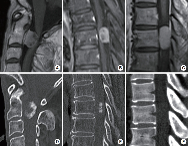
In the CT scan, the heterogeneous group showed significantly frequent intratumor calcification than in the homogeneous group (100% vs. 50.0%, p = 0.031) (Table 3). The maximum HU value also showed a similar trend (379.2 ± 195.4 vs. 1,109.8 ± 300.5, p = 0.001). Additionally, the intratumor contrast-deficient region in the typical contrast MRI of the heterogeneous enhancement group overlapped with the significantly calcified site observed in the CT scan (Fig. 2). There was specific calcification pattern according to the MRI enhancement. All of the calcification positives in the homogeneous group (7 of 14) showed diffuse-low-density calcification, while the heterogeneous group showed focal high- or heterogeneous-density calcification pattern. Table 4 shows the relationship between histological subtype and CT calcification. All of the psammomatous subtype demonstrated positive calcification, while no significant tendency was observed in other histological subtypes.
Table 3.
Relationship between contrast-enhanced MRI and CT findings
| Variable | Homogeneous group (n=14) | Heterogeneous group (n=6) | p-value |
|---|---|---|---|
| Calcification | 0.031* | ||
| Positive | 7 (50.0) | 6 (100) | |
| Negative | 7 (50.0) | 0 (0) | |
| Maximum of Hounsfield unit value | 379.2 ± 195.4 | 1,109.8 ± 300.5 | 0.001* |
| T2WI spinal cord signal change | 9 (60.0) | 4 (66.6) | 0.126 |
MRI, magnetic resonance imaging; CT, computed tomography; T2WI, T2-weighted image.
The chi-square test and Mann-Whitney U-test were used for the categorical and continuous variables, respectively.
p < 0.05, statistically significant differences.
Fig. 2.

Typical contrast-enhanced T2-weighted image magnetic resonance imaging and corresponding computed tomography (CT) images. (A, B) Heterogeneous enhancement group; the intratumor contrast-deficient region was consistent with the marked calcification site in the CT scan (D, E). (C) Homogeneous enhancement group. (F) No calcified region was detected in the CT scan.
Table 4.
Relationship between histological subtype and computed tomography calcification
| Calcification positive (n=15) | Calcification negative (n=8) | p-value | |
|---|---|---|---|
| Histological subtype | 0.290 | ||
| Meningothelial | 5 (33.3) | 5 (62.5) | |
| Psammomatous | 5 (33.3) | 0 (0) | |
| Transitional | 3 (13.3) | 2 (25.0) | |
| Clear cell | 0 (0) | 1 (12.5) | |
| Unknown | 2 (13.3) | 0 (0) |
The chi-square test was used.
DISCUSSION
Spinal meningioma, due to its slow growth, is diagnosed asymptomatic with a radiographic work-up, but it often manifests neurological deficit. The MRI is the gold standard for diagnosis, and the contrast-enhanced T1WI image with Gd+ shows the homogeneous enhancement effect [8]. Furthermore, the meningiomas may demonstrate characteristic intratumor calcification that can be observed in the CT scan. Studies have identified various factors explaining the postoperative functional outcomes or recurrence rate of spinal meningiomas, such as old age, tumor calcification, psammomatous histological subtype, high WHO grade, tumor localization anterior to spinal cord, large tumor size, incomplete tumor resection, and spinal cord signal changes in T2WI, which relate to poor postoperative outcome [5,10-13]. However, only few studies have predicted the preoperative neurological status. Here, we investigated the relationship between image findings and preoperative neurological impairment, where patients with heterogeneously enhanced tumor frequently showed preoperative neurological impairment.
Spinal meningioma typically demonstrates iso- or hyposignal intensity on T1WI, iso- or hypersignal intensity on T2WI, and enhancement effect with gadolinium in the MRI [8]. In our analysis, 28.5% patients demonstrated heterogeneous enhancement, in which tumors showed calcification in the CT scan. Also, the contrast-deficient sites were overlapped with markedly calcified lesion. Based on the intraoperative observations, the highly calcified regions were found to be extremely hard. We anticipate that the preoperative neurological impairment in the heterogeneously enhanced tumor may arise due to tumor hardness. Moreover, the incidentally diagnosed asymptomatic patient with intratumor calcification may develop neurological deficit in the follow-up period along with progression of the calcified region. From this perspective, early surgical intervention for asymptomatic patients with heterogeneously enhanced spinal meningioma might facilitate the functional outcome or improve postoperative prognosis. There was interesting calcification pattern according to the MRI enhancement. All of the calcification positives in the homogeneous group showed diffuse-low-density calcification, while the heterogeneous group showed focal high- or heterogeneous-density calcification pattern. This indicates that the homogeneous group with positive calcification could transition to heterogeneous if focal calcification is amplified.
Yamaguchi et al. [14] observed high tumor occupation ratio to be associated with preoperative motor weakness. In addition to this, we had anticipated that patients with preoperative neurological deficit might show high tumor proliferation rates (Ki-67). However, our analysis did not indicate any significant increase in tumor occupation ratio or Ki-67 levels in the patients with preoperative paralysis. This may be due to small sample size in the present analysis. We need to further investigate this in large case series. Nevertheless, according to our data, we believe that the association between the tumor and symptomatology should also be discussed in terms of the MRI enhancement/CT image findings.
Meningioma has been classified into 15 histological subtypes, and the psammomatous type represents the most common subtype of spinal meningioma [15]. It often forms the characteristic “psammoma body,” along with significant calcification observed in the CT scan. This intratumor calcification allows differentiating them from other spinal cord tumors, such as the spinal schwannoma. Liu et al. [8] reported that approximately 50% of the spinal meningioma show calcification in CT scan. In our analysis, calcification was observed in 65.2% of the patients. Moreover, tumors with high HU values showed poor enhancement effect in the contrast MRI.
After surgery or before discharge, all the patients with preoperative paralysis showed improvement. In addition, no postoperative neurological deterioration was observed in patients without paralysis. Previous reports have shown neurological improvement even in patients with severe paralysis after surgery [16,17]. On the other hand, Gilard et al. [10] have reported that the absence of preoperative neurological signs indicates postoperative poor neurological outcome. This discrepancy could be attributed to the technically demanding nature of the tumor resection. To avoid poor outcome, meticulous care should be taken in resecting the calcified meningioma in order not to damage the spinal cord. An ultrasonic bone curette was extremely helpful to debulk the hard, calcified tumor.
This study has some limitations. First, due to the retrospective nature and small sample size, previously reported possible prognostic factors for motor status, except the image findings, could not be detected. These may include the Ki-67 index, tumor occupation ratio, and tumor level (tumor in the thoracic region could be at risk of neurological deficit due to less tolerance for compression). Thus, a multivariate analysis with adequate statistical power needs to be conducted in the future. Furthermore, most of the histological evaluation was based on fragmented specimen and not on the en bloc resected tumor and, hence, may not reflect the true proliferation property of the tumor. Although, performing en bloc resection is often a difficult procedure. Second, this study had a relatively short follow-up duration. While the neurological improvement was confirmed in all the patients after surgery, long-term follow-up of the clinical outcome could not be obtained. Prospective study including long-term follow-up would allow to test the clinical relevance.
CONCLUSION
The heterogeneous contrast-induced MRI findings in the spinal meningioma were significantly associated with preoperative neurological impairment. Moreover, the intratumor contrast-deficient region in the heterogeneously enhanced tumors reflected marked calcification. The tumor hardness due to calcification may be related to preoperative neurological deficit.
Footnotes
The authors have nothing to disclose.
REFERENCES
- 1.Gelabert-González M, García-Allut A, Martínez-Rumbo R. Spinal meningiomas. Neurocirugia (Astur) 2006;17:125–31. [PubMed] [Google Scholar]
- 2.Engelhard HH, Villano JL, Porter KR, et al. Clinical presentation, histology, and treatment in 430 patients with primary tumors of the spinal cord, spinal meninges, or cauda equina. Journal of neurosurgery. Spine. 2010;13:67–77. doi: 10.3171/2010.3.SPINE09430. [DOI] [PubMed] [Google Scholar]
- 3.Westwick HJ, Shamji MF. Effects of sex on the incidence and prognosis of spinal meningiomas: a Surveillance, Epidemiology, and End Results study. Journal of neurosurgery. Spine. 2015;23:368–73. doi: 10.3171/2014.12.SPINE14974. [DOI] [PubMed] [Google Scholar]
- 4.Kshettry VR, Hsieh JK, Ostrom QT, et al. Descriptive epidemiology of spinal meningiomas in the United States. Spine. 2015;40:E886–9. doi: 10.1097/BRS.0000000000000974. [DOI] [PubMed] [Google Scholar]
- 5.Sandalcioglu IE, Hunold A, Müller O, et al. Spinal meningiomas: critical review of 131 surgically treated patients. Eur Spine J. 2008;17:1035–41. doi: 10.1007/s00586-008-0685-y. [DOI] [PMC free article] [PubMed] [Google Scholar]
- 6.Solero CL, Fornari M, Giombini S, et al. Spinal meningiomas: review of 174 operated cases. Neurosurgery. 1989;25:153–60. [PubMed] [Google Scholar]
- 7.Gezen F, Kahraman S, Canakci Z, et al. Review of 36 cases of spinal cord meningioma. Spine. 2000;25:727–31. doi: 10.1097/00007632-200003150-00013. [DOI] [PubMed] [Google Scholar]
- 8.Liu WC, Choi G, Lee SH, et al. Radiological findings of spinal schwannomas and meningiomas: focus on discrimination of two disease entities. Eur Radiol. 2009;19:2707–15. doi: 10.1007/s00330-009-1466-7. [DOI] [PubMed] [Google Scholar]
- 9.Simpson D. The recurrence of intracranial meningiomas after surgical treatment. J Neurol Neurosurg Psychiatry. 1957;20:22–39. doi: 10.1136/jnnp.20.1.22. [DOI] [PMC free article] [PubMed] [Google Scholar]
- 10.Gilard V, Goia A, Ferracci FX, et al. Spinal meningioma and factors predictive of post-operative deterioration. J Neurooncol. 2018;140:49–54. doi: 10.1007/s11060-018-2929-y. [DOI] [PubMed] [Google Scholar]
- 11.Maiti TK, Bir SC, Patra DP, et al. Spinal meningiomas: clinicoradiological factors predicting recurrence and functional outcome. Neurosurg Focus. 2016;41:E6. doi: 10.3171/2016.5.FOCUS16163. [DOI] [PubMed] [Google Scholar]
- 12.Schaller B. Spinal meningioma: relationship between histological subtypes and surgical outcome? J Neurooncol. 2005;75:157–61. doi: 10.1007/s11060-005-1469-4. [DOI] [PubMed] [Google Scholar]
- 13.Nakamura M, Tsuji O, Fujiyoshi K, et al. Long-term surgical outcomes of spinal meningiomas. Spine. 2012;37:E617–23. doi: 10.1097/BRS.0b013e31824167f1. [DOI] [PubMed] [Google Scholar]
- 14.Yamaguchi S, Menezes AH, Shimizu K, et al. Differences and characteristics of symptoms by tumor location, size, and degree of spinal cord compression: a retrospective study on 53 surgically treated, symptomatic spinal meningiomas. J Neurosurg Spine. 2020 Jan 31;:1–10. doi: 10.3171/2019.12.SPINE191237. [Epub]. [DOI] [PubMed] [Google Scholar]
- 15.Kleihues P, Louis DN, Scheithauer BW, et al. The WHO classification of tumors of the nervous system. J Neuropathol Exp Neurol. 2002;61:215–25. doi: 10.1093/jnen/61.3.215. discussion 226-9. [DOI] [PubMed] [Google Scholar]
- 16.Haegelen C, Morandi X, Riffaud L, et al. Results of spinal meningioma surgery in patients with severe preoperative neurological deficits. Eur Spine J. 2005;14:440–4. doi: 10.1007/s00586-004-0809-y. [DOI] [PMC free article] [PubMed] [Google Scholar]
- 17.Morandi X, Haegelen C, Riffaud L, et al. Results in the operative treatment of elderly patients with spinal meningiomas. Spine. 2004;29:2191–4. doi: 10.1097/01.brs.0000141173.79572.40. [DOI] [PubMed] [Google Scholar]


