Figure 2.
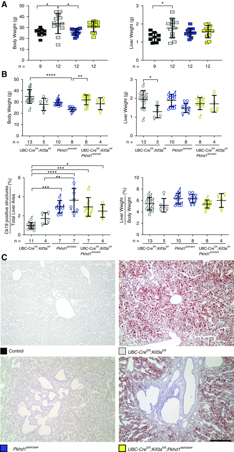
Loss of cilia results in increased body weight and hepatic steatosis. (A) Comparison of body and liver weights of mice with the indicated genotypes. Colored squares show corresponding genotypes in (A and C). Loss of cilia in Kif3a single knockouts (gray squares) resulting in a significant increase in body and liver weight compared with controls (black squares). (B) Graphical representation of the physiologic parameters of the different sexes used in this study. Male (♂) and female (♀) data for total body weight, liver weight, liver weight-body weight ratio, and cytokeratin-19 (CK19)–positive structures were analyzed for sex differences. Sex differences were only evident in the Kif3a (UbcCre; Kif3afl/fl) mutant animals when comparing liver weights. Multiple group comparisons were performed using one-way ANOVA followed by Tukey multiple comparison test and are presented as the mean±SD. *P=0.05; **P=0.01; ***P<0.001; ****P<0.001. (C) Hepatic steatosis was evident by the presence of fat droplets as identified by Oil Red O stain in the cilia-deficient mutants. Scale bar, 400 µM.

