Abstract
Purpose
To assess the effect of adrenocorticotropic hormone (ACTH) on plasma vascular endothelial growth factor (VEGF) levels in healthy children and adolescents and to inform future work on the effects of ACTH on VEGF in bone.
Methods
An Institutional Review Board-approved prospective study of 10 healthy subjects, ages 9–17, was conducted to assess the effect of ACTH on plasma VEGF levels. VEGF levels were collected at baseline and every 30 minutes for 3 hours. Cosyntropin (a synthetic ACTH analogue) was administered at a low-dose (1 μg) given at t=0 minutes and a high-dose (250 μg) given at t=60 minutes. A Friedman test was performed comparing baseline to peak VEGF levels after stimulation with low-dose and high-dose cosyntropin.
Results
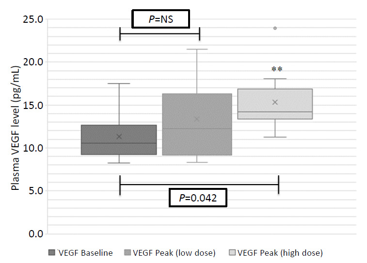
Peak plasma VEGF levels significantly increased after high-dose cosyntropin compared with baseline (P=0.042). Peak plasma VEGF levels did not significantly increase after low-dose cosyntropin compared to baseline.
Conclusions
To our knowledge, this is the first study to demonstrate that ACTH administration causes a significant increase in plasma VEGF levels in humans. This finding may have important implications in the protective effects of ACTH on bone. Decreased bone mineral density and adrenal suppression are common side effects of glucocorticoid use in pediatrics. VEGF increases vascularity and may play a role in reducing glucocorticoid-induced bone disease. Animal studies have shown that ACTH stimulates release of VEGF in osteoblasts, though this effect has yet to be evaluated in humans.
Keywords: Bone density, Glucocorticoids, Adrenocorticotropic hormone, Vascular endothelial growth factor, Adrenal insufficiency
Highlights
This is the first study showing adrenocorticotropic hormone causes a significant increase in plasma vascular endothelial growth factor in humans. This has important implications in the protective effects of ACTH on bone.
Introduction
Glucocorticoids are used in the treatment of many pediatric diseases, and up to 10% of children may require glucocorticoids in some formulation during childhood [1-3]. Long-term glucocorticoid therapy is associated with significant side effects, including weight gain, growth failure, secondary adrenal insufficiency due to adrenal suppression, and increased fracture risk with low bone mineral density (BMD). Osteoporosis is a serious consequence of chronic glucocorticoid therapy, leading to fractures in 30%–50% of patients, and 11%–40% of patients have some degree of osteonecrosis [3,4]. Glucocorticoids cause decreased bone formation and an increase in bone resorption while also reducing intestinal calcium absorption, increasing renal calcium excretion, and diminishing sex steroid and growth hormone production, which all contribute to decreased BMD and fragility fractures [2,3,5].
Glucocorticoid use also suppresses the hypothalamic-pituitary-adrenal axis, which inhibits release of adrenocorticotropic hormone (ACTH) from the pituitary gland and decreases adrenal cortisol production [2]. In addition to its main steroidogenic effect on the adrenal gland, like many of the pituitary hormones, ACTH has a direct influence on bone mass regulation, and steroid-induced suppression of ACTH may have adverse effects on bone independent of the direct glucocorticoid effect [2,6,7]. Animal and in vitro cell studies have suggested that ACTH plays a role in bone protection by promoting osteoblast differentiation and release of vascular endothelial growth factor (VEGF) [8-10]. VEGF plays an important role in angiogenesis and bone ossification and remodeling [5,11]. Thus, ACTH-induced stimulation of bone VEGF levels may help prevent glucocorticoid-associated osteonecrosis [8-10]. Currently, it is unknown whether ACTH stimulates VEGF release in humans. The aim of this proof-of-concept study was to demonstrate the acute effects of ACTH on systemic VEGF levels in healthy humans.
Materials and methods
1. Subjects
Ten healthy subjects, 9–17 years old, participated in this study. There were no sex, weight, or height criteria. Exclusion criteria included taking any medications other than over-the-counter medications (and nothing on the study day), any nontopical steroid use (including intravenous, oral, inhaled, and intranasal) or oral contraceptive pill use in the prior 6 months, or pregnancy at the time of the study (screened by a urine pregnancy test in all postmenarchal girls).
2. Testing protocol
Subjects were admitted to Clinical Research Services, which is part of the Nationwide Children’s Hospital Research Institute. Testing started about 8:30 AM, and fasting was not required. An intravenous catheter without tubing was placed in one arm for cosyntropin administration and blood sampling. At t=0 minutes, after baseline ACTH, cortisol, and VEGF levels were obtained, and 1 μg (low dose) cosyntropin (ACTH-[1-24], Cortrosyn, Amphastar Pharmaceuticals, CA, USA) was administered intravenously. Cortisol and VEGF levels were obtained at t=30 and 60 minutes. After the 60-minute blood draw, 250 μg (high dose) cosyntropin was administered intravenously. At t=90, 120, 150, and 180 minutes, cortisol and VEGF levels were again obtained.
The effect of 1-μg cosyntropin on VEGF was evaluated for 1 hour per the low-dose ACTH stimulation test protocol, followed by high-dose cosyntropin, to asses the VEGF responses to both physiological and supraphysiological doses of ACTH. The 1-μg dose is more physiologic and may reflect a more realistic amount of stimulation of VEGF and cortisol in response to normal daily stressors, while the 250-μg dose is supraphysiologic and can maximally stimulate the ACTH receptor response [12,13].
3. Laboratory analysis
Serum cortisol and ACTH were measured at our institutional clinical laboratory using a chemiluminescent microparticle immunoassay (Architect i2000SR, Abbott, Abbott Park, IL, USA) and a competitive chemiluminescent enzyme immunoassay (Immulite XPi, Siemens, Munich, Germany), respectively. The cortisol cutoff for the ACTH stimulation test is assay-specific, with new literature suggesting lower thresholds for newer assays than the classic threshold of 18 μg/dL (497 nmol/L) used by many institutions [14,15]. Internal validation performed by our institution's laboratory established lower thresholds of cortisol levels as compared with a reference laboratory with a new a peak cortisol cutoff of 15.5 μg/dL (428 nmol/L) (personal communication; Amy Pyle-Eilola, PhD). The precision of the cortisol assay is reflected by a coefficient of variation between 2.8%–3.3% for quality control materials with values between 4.0–32.2 μg/dL (110–888 nmol/L).
VEGF samples were drawn in ethylenediaminetetraacetic acid tubes and placed on ice for up to 90 minutes. They were then centrifuged at 2,500 g for 10 minutes (at 4℃). The supernatant was removed and centrifuged at 10,000 g for 10 minutes (at room temperature). The supernatant plasma was then removed again and stored at -80℃ until processed as a batch sample. Once all subject samples were completed, VEGF-A levels were measured by enzyme-linked immunosorbent assay via V-Plex Human VEGF Kit (Mesoscale Discovery, Rockville, MD, USA). The precision of the VEGF-A assay is reflected by a coefficient of variation between 2.3%–3.4% (intrarun) and 16.4%–17.8% (interrun) for quality control materials, with values between 18.8–294 pg/mL. Sensitivity of the assay is demonstrated with a lower limit of detection of 1.12 pg/mL with a range of 0.55–6.06 pg/mL [16].
4. Statistical analysis
Data analyses were performed using IBM SPSS Statistics ver. 22.0 (IBM Co., Armonk, NY, USA) and Systat 13 (Systat Software, Inc., San Jose, CA, USA). We performed a power calculation based on a study by Dassan et al. [17] that showed median baseline serum VEGF levels at 466 pg/mL with an interquartile range of 392–649 pg/mL and peak VEGF levels at 1,700 pg/mL with an interquartile range of 1,500–1,900 pg/mL. A 1-way within-subjects analysis of variance (a priori) with 9 participants across 3 time points would be sensitive to effect size=0.5 (medium effect size) with 80% power (alpha=0.05). For sample size calculation, G*Power 3.1.9.2 was employed.
Because of the small sample size, a related-samples Friedman analysis of variance by ranks was conducted to evaluate differences in medians among the 3 time points (baseline to peak plasma VEGF levels after stimulation with low-dose and high-dose cosyntropin). The low-dose peak VEGF level is greater at the 30- and 60-minute levels and the high-dose peak VEGF is the greatest at the 90- through 180-minute levels. Follow-up pairwise comparisons were conducted using Dunn-Bonferroni post hoc tests. Results are expressed as the mean ± standard deviation (SD) or standard error (SE) of the median. Pearson correlation coefficient was used to assess the relationship between cortisol and VEGF levels.
Results
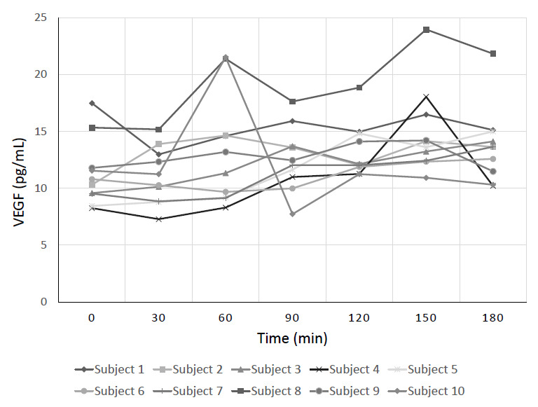
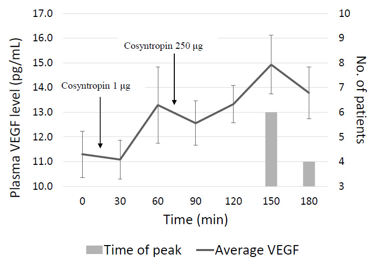
Eight female and 2 male subjects participated in the study (Table 1). The mean±SD age at the time of the study was 13.2±2.9 years (range 9.9–17.8 years). Fig. 1 shows the VEGF level at each time for every subject, and Fig. 2 shows the overall mean plasma VEGF levels at each time point. The highest peak VEGF level for each subject was at either the 150- or the 180-minute mark. The mean, median, and interquartile ranges for baseline and peak VEGF are shown in Fig. 3. Median peak VEGF levels at each time point were 10.55 pg/mL, 12.25 pg/mL, and 14.17 pg/mL, at baseline and after low- and high-dose cosyntropin, respectively. There was a significant difference in medians among the 3 time points (χ2 [2, N=10]=6.20, P=0.045). Follow-up pairwise comparisons were conducted and showed that the median high-dose peak plasma VEGF after high-dose cosyntropin was significantly greater than the median VEGF for baseline (P=0.042). The median peak plasma VEGF after low-dose cosyntropin did not differ significantly from the median VEGF for baseline or the high-dose peak (P=1.000 and P=0.353, respectively).
Table 1.
Subject demographics
| Subject No. | Sex | Age (yr) | Ethnicity | Weight (kg) | Height (cm) | BMI (kg/m2) | Pubertal status |
|---|---|---|---|---|---|---|---|
| 1 | Female | 17 | Caucasian | 71.9 | 169.9 | 24.91 | Postmenarche |
| 2* | Female | 12 | Asian-American | 53.0 | 169.7 | 18.40 | Premenarche |
| 3* | Female | 10 | Asian-American | 35.7 | 144.4 | 17.12 | Premenarche |
| 4 | Female | 17 | Caucasian | 78.9 | 165.2 | 28.91 | Postmenarche |
| 5 | Female | 15 | African-American | 60.0 | 155.7 | 27.22 | Postmenarche |
| 6† | Male | 15 | Caucasian | 70.4 | 181.2 | 21.44 | Pubertal |
| 7† | Male | 13 | Caucasian | 66.8 | 178.1 | 21.06 | Pubertal |
| 8 | Female | 9 | Caucasian | 42.4 | 153.1 | 18.09 | Premenarche |
| 9 | Female | 14 | Caucasian | 47.4 | 162.9 | 17.86 | Postmenarche |
| 10 | Female | 10 | Caucasian | 36.3 | 142.0 | 18.00 | Premenarche |
Pubertal status based on patient report.
BMI, body mass index.
Subject numbers 2 and 3 are sisters.
Subjects number 6 and 7 are brothers.
Fig. 1.

Paired vascular endothelial growth factor (VEGF) levels at each time point for every subject. Each subject’s plasma VEGF levels are shown at each time point denoted by a different symbol for each subject. Baseline VEGF was at t=0 minutes, the response to the low-dose (1 μg) cosyntropin was t=30 and 60 minutes, and the response to the high-dose (250 μg) cosyntropin was at t=90, 120, 150, and 180 minutes.
Fig. 2.

Mean and peak vascular endothelial growth factor (VEGF) at each time point. The solid line denotes the mean (±standard error) VEGF level of all of the subjects at each time point. Cosyntropin was given right after the t=0 and t=60-minute blood draws. The bars denote how many subjects had their highest peak VEGF level at that time point.
Fig. 3.

Baseline compared to peak vascular endothelial growth factor (VEGF) level after cosyntropin administration. Mean, median, and interquartile ranges for VEGF levels at baseline and peak after stimulation by low- and high-dose cosyntropin. NS, not significant. **Statistically significant increase from baseline (P=0.042). X denotes mean value.
Baseline ACTH levels ranged from 6.5–34.0 pg/mL (1.43–7.48 pmol/L), with a normal range of 6–48 pg/mL (1.32–10.6 pmol/L), except in subject 1 for whom the sample quantity was insufficient. The mean±SE cortisol level at baseline was 7.5±1.0 μg/dL (207.1±27.6 nmol/L), after low-dose cosyntropin was 16.5±0.8 μg/dL (455.6±22.1 nmol/L), and after high-dose cosyntropin was 24.2±0.9 μg/dL (667.9±24.8 nmol/L). Baseline VEGF levels did not significantly correlate with baseline cortisol levels (r=-0.113, P=0.756) nor did low-dose or high-dose peak VEGF correlate with low-dose and high-dose peak cortisol levels, respectively (low dose: r=0.169, P=0.641; high dose: r=0.158, P=0.663).
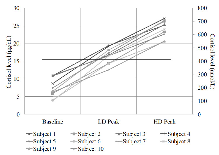
Baseline and peak cortisol levels after the low and high doses of cosyntropin for each patient are shown in Fig. 4. Only 7 of the 10 subjects had peak cortisol levels above 15.5 μg/dL (428 nmol/L) after low dose of cosyntropin. The 3 patients who did not pass the low-dose test had peak cortisol levels of 14.4, 14.4, and 12.6 μg/dL (397, 397, 347 nmol/L). The increment in cortisol levels following low-dose ACTH did not differ between the latter 3 subjects and those with peak responses greater than 15.5 μg/dL (8.4±1.2 μg/dL vs. 9.2±1.0 μg/dL, P=0.630). All 10 subjects had peak cortisol levels above 15.5 μg/dL (428 nmol/L) after high-dose cosyntropin. When comparing the mean peak VEGF levels of the subjects that passed the low-dose stimulation test to those that did not (due to a concern of false negative tests, secondary to technical errors, resulting in patients not receiving the full dose of 1-μg cosyntropin), there was no significant between-subject differences, with a P-value of 0.569. When the 3 subjects who did not pass the low-dose ACTH test were excluded, however, there became no significant difference in plasma VEGF levels of the other 7 subjects from baseline to peak (16.1±1.5 pg/mL) after high dose (P=0.102).
Fig. 4.

Baseline and peak cortisol levels for each subject. Baseline compared with peak cortisol for each patient after low-dose (LD) and high-dose (HD) cosyntropin administration. Solid horizontal line denotes a value of 15.5 μg/dL (428 nmol/L, passing cutoff for ACTH stimulation test). Subject numbers 2, 6, and 7 did not have a peak cortisol above 15.5 μg/dL (428 nmol/L) after the low-dose stimulation, but all subjects had a peak above 15.5 μg/dL (428 nmol/L) after the high-dose stimulation.
Discussion
To our knowledge, this is the first study to demonstrate the acute effect of ACTH on plasma VEGF levels in healthy children and adolescents, as shown by a significant increase in peak VEGF values from baseline after high-dose cosyntropin administration. Studies to date have shown ACTH-induced VEGF expression in bone cells in vitro or in animal studies [8-10], but this had not yet been demonstrated in humans. This finding may have important implications in maintaining bone health in patients on chronic glucocorticoid therapy who are at high risk for steroid-induced osteoporosis or osteonecrosis [2,3]. Zaidi et al. [8] reported that in a rabbit model, ACTH protects against glucocorticoid-induced osteonecrosis of bone by inducing VEGF production in osteoblasts, maintaining the fine vascular network that surrounds highly remodeling bone. Another animal study by Wang et al. [9], showing lower VEGF levels in femoral heads with osteonecrosis compared to controls, also supports the critical role of VEGF in steroid-induced osteonecrosis. Therefore, based on these positive effects of ACTH on bone in animal studies, it has been postulated by other authors that patients on chronic glucocorticoids may benefit from exogenous ACTH [8].
Glucocorticoid use is the most prevalent cause of secondary osteoporosis [2,3]. Glucocorticoids cause decreased function and apoptosis in osteoblasts, which leads to decreased bone formation while simultaneously decreasing apoptosis of osteoclasts causing increased bone resorption. Overall, this results in decreased BMD and increased fracture risk [2,3,5]. Another adverse effect of chronic glucocorticoid therapy is osteonecrosis, which is postulated to be caused by increased marrow fat, fat emboli, hypercoagulability, cell apoptosis, and vascular endothelial dysfunction [4]. Patients on chronic glucocorticoids are also at risk for suppressed ACTH and cortisol levels, secondary to negative feedback by the exogenous glucocorticoids on the hypothalamic-pituitary-adrenal axis [2].
ACTH has a direct effect on bone mass regulation that is mediated through the melanocortin-2 receptor (MC2R) expressed on osteoblasts and stimulates VEGF release and osteoblast proliferation [6-8]. VEGF consists of a family of glycoprotein cytokines that are responsible for angiogenesis, with VEGF-A (measured using the assay in this study) being the most abundant form [5]. VEGF maintains blood vessel density, thickness, and permeability, and is vital for endothelial cell survival [11]. The baseline VEGF levels in the subjects in our study are similar to those in other studies [18]. VEGF is found in the lungs, kidneys, heart, adrenals, bone, brain, and several other organs [8,11]. Within the adrenal, VEGF is expressed in both the zona glomerulosa and zona fasciculata. Its secretion is stimulated by ACTH, and it may protect against cortical adrenal atrophy in glucocorticoid use [19]. The chromaffin cells of the adrenal gland also produce small amounts of ACTH that can cause paracrine effects within the adrenal gland. VEGF is released within the adrenal in response to both the endocrine and paracrine effects of ACTH [19,20].
Within bone, VEGF plays an essential role in skeletal development, growth, remodeling, and repair through osteoblast differentiation and osteoclast recruitment. During bone formation and repair, osteogenesis and angiogenesis are tightly coupled processes. VEGF-induced angiogenesis facilitates bone formation by increasing delivery of oxygen, nutrients, and growth factors to the bone cells. These effects may be protective against osteonecrosis [5,8,9]. The potential positive effects of ACTH-induced VEGF release on bone are clinically evident in patients with ACTH-producing pituitary Cushing disease; these patients have less BMD loss than those with adrenal Cushing syndrome (with suppressed ACTH levels) [21,22]. In addition, ACTH concentrations in patients with pituitary Cushing disease correlate with lumbar BMD [22]. Moreover, due to feedback on the hypothalamic-pituitary-adrenal axis, patients with familial glucocorticoid deficiency with elevated ACTH levels have a higher BMD than age-matched controls [23]. While ACTH doses as low as 3 μg have been shown to maximally stimulate the adrenal gland [12], the supraphysiologic 250-μg cosyntropin is the standard dose that is used for maximum stimulation of the adrenal gland for the high-dose ACTH stimulation test [13]. The significant VEGF increase to this high level of ACTH may be clinically significant in cases like Cushing disease and familial glucocorticoid deficiency. These clinical observations provide evidence supporting the protective and anabolic effect of ACTH on bone and suggest a theoretical benefit of ACTH replacement to help preserve BMD in patients on chronic glucocorticoid therapy.
Another potential implication of the ACTH-induced VEGF release shown in our study relates to the use of the ACTH analogue, repository corticotropin injection (Acthar Gel) in the treatment of nephrotic syndrome. Acthar has been used in cases of nephrotic syndrome resistant to glucocorticoids alone; therefore, ACTH must function by other mechanisms than a supraphysiologic increase in cortisol levels leading to the usual immune suppression and anti-inflammatory effects of glucocorticoids [24,25]. ACTH has a direct effect on melanocortin receptors in the kidneys [24-26] and treatment with subcutaneous Acthar has been shown to decrease proteinuria and increase urinary VEGF levels in patients with diabetic nephropathy (through activation of melanocortin receptors in the glomerular cells) [27]. The antiproteinuric effect of VEFG is supported by the fact that bevacizumab, an anti-VEGF monoclonal antibody used in certain cancer treatments, causes proteinuria [28]. Therefore, VEGF may also play a role in the mechanism of ACTH treatment of nephrotic syndrome.
In this study, all 10 subjects were healthy adolescent volunteers and all 10 passed the high-dose ACTH stimulation test with a peak cortisol value of greater than 15.5 μg/dL (428 nmol/L), which confirms that none of them had primary adrenal insufficiency. However, the low-dose ACTH stimulation test produced false positive results in 3 healthy subjects who would have been misdiagnosed with secondary adrenal insufficiency had this test been performed for diagnostic purposes. Although the low-dose test is very sensitive in diagnosing cortisol deficiency secondary to steroid-induced adrenal suppression or ACTH deficiency, it has several technical limitations [29-32]. Cosyntropin is dispensed in 250-μg vials, which is used for the high-dose test but must be diluted to 1 μg for the low-dose. The dilution process may cause dilution error or loss of ACTH due to binding to intravenous tubing [29-32], and the ACTH administered may not be truly given to the patient; this can lead to false positive results, which have been reported in approximately 10%–20% of healthy adults [29,30]. However, although 3 subjects did not have “passing” low-dose peak cortisol levels, the incremental rise in cortisol in these subjects did not differ from the response in those who did surpass the 15.5 μg/dL (428 nmol/L) threshold. This change in cortisol is sufficient to rule out underlying adrenal insufficiency and to ensure that the proper dose of cosyntropin was both given and reached the patient. Further studies with a larger sample size could be used determine whether the increase in plasma VEGF after low-dose cosyntropin could be used to reflect a properly performed ACTH stimulation test (i.e., that the cosyntropin reached the patient).
Even though our study was limited by the small sample size, it is a proof-of-concept study demonstrating the effects of ACTH on systemic VEGF, which may inform future work on the effects ACTH has on VEGF in bone. Another limitation was the sequential ACTH stimulation tests, which may have obscured the peak VEGF after the low dose. VEGF could have continued rising after low-dose ACTH administration beyond 1 hour, but the high dose was given at 60 minutes. This also may have resulted in synergistic effects from both doses on peak VEGF levels. However, since cosyntropin has a half-life of about 15 minutes [13], there should not have been much carry-over effect after one hour when the high dose was given, especially since all subjects’ peak VEGF levels were at 150 or 180 minutes. Since VEGF can be stimulated in many different tissues, follow-up studies will be needed to assess the site-specific VEGF response. In addition, the supraphysiologic high-dose cosyntropin was shown to cause a statistically significant increase in VEGF levels, but future studies could also be done to determine the minimal dose of ACTH needed to stimulate VEGF release. Finally, cosyntropin was used in this study due to its ready availability and low cost, but it has a short half-life. Longer lasting ACTH preparations (Acthar, Synacthen) could be used in future studies to assess prolonged effects of ACTH on VEGF levels [33]. Further studies are also needed to elucidate the ACTH effect on VEGF in nephrotic syndrome.
In summary, this study was the first to demonstrate increased plasma VEGF levels in response to ACTH administration in humans. This finding may help elucidate the mechanism of bone-protective effects of ACTH. Future studies to assess whether ACTH can stimulate VEGF in patients taking chronic glucocorticoids and determine any relationship to BMD before and after ACTH administration in these patients will be an important next step to further evaluate the protective effects of ACTH on bone health. By showing early evidence that ACTH can stimulate VEGF release in humans, this study provides groundwork for future studies in this area and will be paramount for patients on chronic glucocorticoids.
Acknowledgments
The present study was supported by an intramural grant from Nationwide Children’s Hospital in Columbus, OH, USA (grant #2011511). We would like to thank Mary Cismowski, PhD, for assistance with the VEGF assays; Shelli Farley, RN, CCRC and Rachel Heffern, MSN, BSN, RN, CCRC, nurses at the Clinical Research Services; and Amy Pyle-Eilola, PhD, for assistance with the cortisol assay specifications.
Footnotes
Ethical statement
The study was approved by our institution's ethics committee, the Nationwide Children's Hospital Institutional Review Board (approval number: IRB17-00194). Written informed assent and consent was obtained from the subject and parents, respectively. The study protocol and consent were performed in accordance with the ethical standards of the Helsinki Declaration.
Conflicts of interest
No potential conflict of interest relevant to this article was reported.
References
- 1.Leonard MB. Glucocorticoid-induced osteoporosis in children: impact of the underlying disease. Pediatrics. 2007;119 Suppl 2:S166–74. doi: 10.1542/peds.2006-2023J. [DOI] [PubMed] [Google Scholar]
- 2.Aulakh R, Singh S. Strategies for minimizing corticosteroid toxicity: a review. Indian J Pediatr. 2008;75:1067–73. doi: 10.1007/s12098-008-0211-6. [DOI] [PubMed] [Google Scholar]
- 3.Mazziotti G, Angeli A, Bilezikian JP, Canalis E, Giustina A. Glucocorticoid-induced osteoporosis: an update. Trends Endocrinol Metab. 2006;17:144–9. doi: 10.1016/j.tem.2006.03.009. [DOI] [PubMed] [Google Scholar]
- 4.Xie XH, Wang XL, Yang HL, Zhao DW, Qin L. Steroid-associated osteonecrosis: Epidemiology, pathophysiology, animal model, prevention, and potential treatments (an overview) J Orthop Translat. 2015;3:58–70. doi: 10.1016/j.jot.2014.12.002. [DOI] [PMC free article] [PubMed] [Google Scholar]
- 5.Yang YQ, Tan YY, Wong R, Wenden A, Zhang LK, Rabie AB. The role of vascular endothelial growth factor in ossification. Int J Oral Sci. 2012;4:64–8. doi: 10.1038/ijos.2012.33. [DOI] [PMC free article] [PubMed] [Google Scholar]
- 6.Zaidi M, Sun L, Liu P, Davies TF, New M, Zallone A, et al. Pituitary-bone connection in skeletal regulation. Horm Mol Biol Clin Investig. 2016;28:85–94. doi: 10.1515/hmbci-2016-0015. [DOI] [PubMed] [Google Scholar]
- 7.Isales CM, Zaidi M, Blair HC. ACTH is a novel regulator of bone mass. Ann N Y Acad Sci. 2010;1192:110–6. doi: 10.1111/j.1749-6632.2009.05231.x. [DOI] [PubMed] [Google Scholar]
- 8.Zaidi M, Sun L, Robinson LJ, Tourkova IL, Liu L, Wang Y, et al. ACTH protects against glucocorticoid-induced osteonecrosis of bone. Proc Natl Acad S ci U S A. 2010;107:8782–7. doi: 10.1073/pnas.0912176107. [DOI] [PMC free article] [PubMed] [Google Scholar]
- 9.Wang G, Zhang CQ, Sun Y, Feng Y, Chen SB, Cheng XG, et al. Changes in femoral head blood supply and vascular endothelial growth factor in rabbits with steroid-induced osteonecrosis. J Int Med Res. 2010;38:1060–9. doi: 10.1177/147323001003800333. [DOI] [PubMed] [Google Scholar]
- 10.Tourkova IL, Liu L, Sutjarit N, Larrouture QC, Luo J, Robinson LJ, et al. Adrenocorticotropic hormone and 1,25-dihydroxyvitamin D3 enhance human osteogenesis in vitro by synergistically accelerating the expression of bone-specific genes. Lab Invest. 2017;97:1072–83. doi: 10.1038/labinvest.2017.62. [DOI] [PMC free article] [PubMed] [Google Scholar]
- 11.Behl T, Kotwani A. Exploring the various aspects of the pathological role of vascular endothelial growth factor (VEGF) in diabetic retinopathy. Pharmacol Res. 2015;99:137–48. doi: 10.1016/j.phrs.2015.05.013. [DOI] [PubMed] [Google Scholar]
- 12.Thaler LM, Blevins LS. The low dose (1-microg) adrenocorticotropin stimulation test in the evaluation of patients with suspected central adrenal insufficiency. J Clin Endocrinol Metab. 1998;83:2726–9. doi: 10.1210/jcem.83.8.5039. [DOI] [PubMed] [Google Scholar]
- 13.Hamilton DD, Cotton BA. Cosyntropin as a diagnostic agent in the screening of patients for adrenocortical insufficiency. Clin Pharmacol. 2010;2:77–82. doi: 10.2147/CPAA.S6475. [DOI] [PMC free article] [PubMed] [Google Scholar]
- 14.El-Farhan N, Pickett A, Ducroq D, Bailey C, Mitchem K, Morgan N, et al. Method-specific serum cortisol responses to the adrenocorticotrophin test: comparison of gas chromatography-mass spectrometry and five automated immunoassays. Clin Endocrinol (Oxf) 2013;78:673–80. doi: 10.1111/cen.12039. [DOI] [PubMed] [Google Scholar]
- 15.Bornstein SR, Allolio B, Arlt W, Barthel A, Don-Wauchope A, Hammer GD, et al. Diagnosis and treatment of primary adrenal insufficiency: an Endocrine Society Clinical Practice guideline. J Clin Endocrinol Metab. 2016;101:364–89. doi: 10.1210/jc.2015-1710. [DOI] [PMC free article] [PubMed] [Google Scholar]
- 16.Lee JW, Devanarayan V, Barrett YC, Weiner R, Allinson J, Fountain S, et al. Fit-for-purpose method development and validation for successful biomarker measurement. Pharm Res. 2006;23:312–28. doi: 10.1007/s11095-005-9045-3. [DOI] [PubMed] [Google Scholar]
- 17.Dassan P, Keir G, Jager HR, Brown MM. Value of measuring serum vascular endothelial growth factor levels in diagnosing acute ischemic stroke. Int J Stroke. 2012;7:454–9. doi: 10.1111/j.1747-4949.2011.00677.x. [DOI] [PubMed] [Google Scholar]
- 18.Meo S, Dittadi R, Gion M. Biological variation of vascular endothelial growth factor. Clin Chem Lab Med. 2005;43:342–3. doi: 10.1515/CCLM.2005.059. [DOI] [PubMed] [Google Scholar]
- 19.Mallet C, Feraud O, Ouengue-Mbele G, Gaillard I, Sappay N, Vittet D, et al. Differential expression of VEGF receptors in adrenal atrophy induced by dexamethasone: a protective role of ACTH. Am J Physiol Endocrinol Metab. 2003;284:E156–67. doi: 10.1152/ajpendo.00450.2001. [DOI] [PubMed] [Google Scholar]
- 20.Lefebvre H, Thomas M, Duparc C, Bertherat J, Louiset E. Role of ACTH in the Interactive/paracrine regulation of adrenal steroid secretion in physiological and pathophysiological conditions. Front Endocrinol (Lausanne) 2016;7:98. doi: 10.3389/fendo.2016.00098. [DOI] [PMC free article] [PubMed] [Google Scholar]
- 21.Minetto M, Reimondo G, Osella G, Ventura M, Angeli A, Terzolo M. Bone loss is more severe in primary adrenal than in pituitar y-dependent Cushing's syndrome. Osteoporos Int. 2004;15:855–61. doi: 10.1007/s00198-004-1616-3. [DOI] [PubMed] [Google Scholar]
- 22.Guo W, Li F, Zhu C, Wang B, Wang K, Dai C, et al. Effect of hypercortisolism on bone mineral density and bone metabolism: A potential protective effect of adrenocorticotropic hormone in patients with Cushing's disease. J Int Med Res. 2018;46:492–503. doi: 10.1177/0300060517725660. [DOI] [PMC free article] [PubMed] [Google Scholar]
- 23.Elias LL, Huebner A, Metherell LA, Canas A, Warne GL, Bitti ML, et al. Tall stature in familial glucocorticoid deficiency. Clin Endocrinol (Oxf) 2000;53:423–30. doi: 10.1046/j.1365-2265.2000.01122.x. [DOI] [PubMed] [Google Scholar]
- 24.Gong R. The renaissance of corticotropin therapy in proteinuric nephropathies. Nat Rev Nephrol. 2011;8:122–8. doi: 10.1038/nrneph.2011.190. [DOI] [PMC free article] [PubMed] [Google Scholar]
- 25.Montero-Melendez T. ACTH: The forgotten therapy. Semin Immunol. 2015;27:216–26. [Google Scholar]
- 26.Lieberman KV, Pavlova-Wolf A. Adrenocorticotropic hormone therapy for the treatment of idiopathic nephrotic syndrome in children and young adults: a systematic review of early clinical studies with contemporary relevance. J Nephrol. 2017;30:35–44. doi: 10.1007/s40620-016-0308-3. [DOI] [PMC free article] [PubMed] [Google Scholar]
- 27.Tumlin JA, Galphin CM, Rovin BH. Advanced diabetic nephropathy with nephrotic range proteinuria: a pilot study of the long-term efficacy of subcutaneous ACTH gel on proteinuria, progression of CKD, and urinary levels of VEGF and MCP-1. J Diabetes Res. 2013;2013:489869. doi: 10.1155/2013/489869. [DOI] [PMC free article] [PubMed] [Google Scholar]
- 28.Lankhorst S, Baelde HJ, Verstijnen J, Ten Tije AJ, Thelen MHM, Danser AHJ, et al. Cumulative dose of bevacizumab associates with albuminuria rather than podocyturia in cancer patients. J Am Soc Hypertens. 2018;12:e1–7. doi: 10.1016/j.jash.2018.06.005. [DOI] [PubMed] [Google Scholar]
- 29.Tordjman K, Jaffe A, Trostanetsky Y, Greenman Y, Limor R, Stern N. Low-dose (1 microgram) adrenocorticotrophin (ACTH) stimulation as a screening test for impaired hypothalamo-pituitary-adrenal axis function: sensitivity, specificity and accuracy in comparison with the high-dose (250 microgram) test. Clin Endocrinol (Oxf) 2000;52:633–40. doi: 10.1046/j.1365-2265.2000.00984.x. [DOI] [PubMed] [Google Scholar]
- 30.Dorin RI, Qualls CR, Crapo LM. Diagnosis of adrenal insufficiency. Ann Intern Med. 2003;139:194–204. doi: 10.7326/0003-4819-139-3-200308050-00009. [DOI] [PubMed] [Google Scholar]
- 31.Ospina NS, Al Nofal A, Bancos I, Javed A, Benkhadra K, Kapoor E, et al. ACTH stimulation tests for the diagnosis of adrenal insufficiency: systematic review and meta-analysis. J Clin Endocrinol Metab. 2016;101:427–34. doi: 10.1210/jc.2015-1700. [DOI] [PubMed] [Google Scholar]
- 32.Wade M, Baid S, Calis K, Raff H, Sinaii N, Nieman L. Technical details influence the diagnostic accuracy of the 1 microg ACTH stimulation test. Eur J Endocrinol. 2010;162:109–13. doi: 10.1530/EJE-09-0746. [DOI] [PMC free article] [PubMed] [Google Scholar]
- 33.Gettig J, Cummings JP, Matuszewski K. H.p. Acthar gel and cosyntropin review: clinical and financial implications. P T. 2009;34:250–7. [PMC free article] [PubMed] [Google Scholar]


