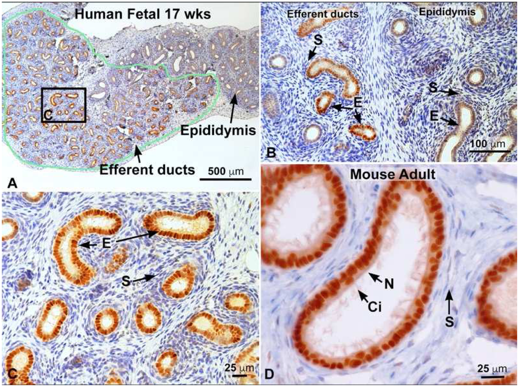
Fig. 3.

Estrogen receptor 1 (ESR1) immunostaining. A) 17 week fetal human efferent ductules in the head of the epididymis outlined in green and staining intensely for ESR1. To the right is the immature epididymal duct, which appears to have far less staining. B) Fetal 17 week human tissue showing efferent ductules to the left and epididymis to the right. Efferent duct stroma (S) is negative, but the epithelial cells (E) are strongly positive for ESR1, while epididymal epithelium is less positive and some stromal cells show slight staining. C) Higher magnification of area outlined in A. Efferent duct epithelial cells (E) are strongly positive for ESR1, but the stroma (S) is negative. D) Mouse adult efferent ductules showing intense staining for ESR1 in both nonciliated (N) resorptive epithelial cells, as well as the motile ciliated cells (Ci). Stromal (S) cells are mostly negative. Human fetal photos provided by Dr. Gerald Cunha, with approval by the Committee on Human Research at UCSF, IRB# 12–08813); Cunha et al. (2020).
