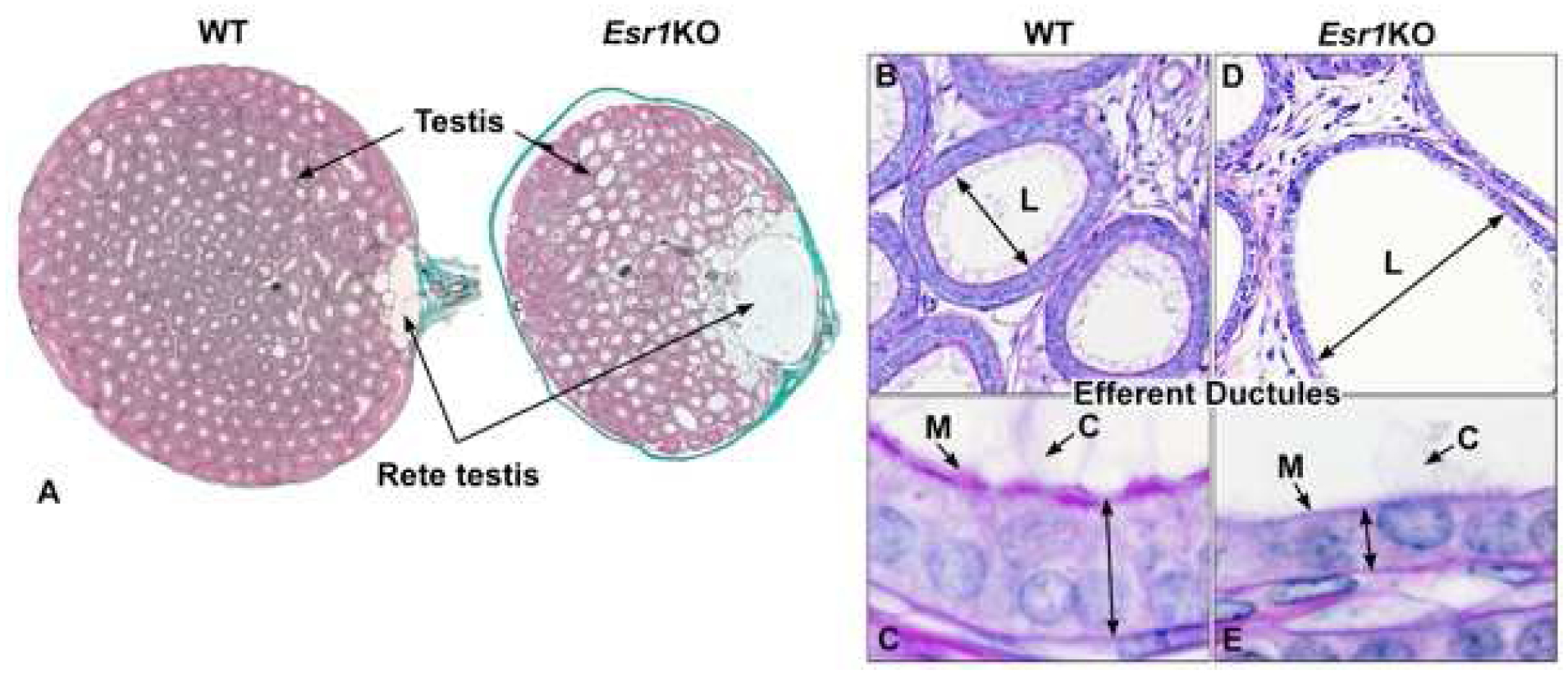
Fig 5.

Testis, rete testis and efferent ductules in the adult wild-type (WT) and Esr1KO mice. A) WT testis shows normal seminiferous tubular lumens with a small flattened rete testis where sperm exit to enter the efferent ductules. In contrast, the rete testis in an Esr1KO mouse shows excessive dilation. B) WT efferent ductules showing a normal luminal (L) diameter. C) Enlargement of the WT epithelium to illustrate normal height, long motile cilia (C) extending into the lumen and adjacent thick microvillus boarder (M) of the nonciliated, resorptive cells that are responsible for reabsorption of nearly 90% of the luminal fluids. D) Esr1KO efferent ductule, showing hyperdilation of the lumen (L). E) Higher magnification of the Esr1KO epithelium, which shows the dramatic decrease in height and loss of apical cytoplasm and microvilli (M) in the resorptive cells. Motile cilia (C) extend into the lumen, but appear to be fewer in number and shorter in length. Adapted from figures in Nanjappa et al. (2016) and Hess et al. (2011) with permission from Oxford University Press and John Wiley and Sons.
