Abstract

SARS-CoV-2 is the etiologic agent of COVID-19, which has led to a dramatic loss of human life and presents an unprecedented challenge to public health worldwide. The gold standard assay for SARS-CoV-2 identification is real-time polymerase chain reaction; however, this assay depends on highly trained personnel and sophisticated equipment and may suffer from false results. Thus, a serological antibody test is a supplement to the diagnosis or screening of SARS-CoV-2. Here, we develop and evaluate the diagnostic performance of an IgM/IgG indirect ELISA method for antibodies against SARS-CoV-2 in COVID-19. The ELISA was constructed by coating with a recombinant nucleocapsid protein of SARS-CoV-2 on an enzyme immunoassay plate, and its sensitivity and specificity for clinical diagnosis of SARS-CoV-2 infection was assessed by detecting the SARS-CoV-2-specific IgM and IgG antibodies in COVID-19 patient’s sera or healthy person’s sera. The SARS-CoV-2 positive serum samples (n = 168) were collected from confirmed COVID-19 patients. A commercial nucleocapsid protein-based chemiluminescent immunoassay (CLIA) kit and a colloidal gold immunochromatography kit were compared with those of the ELISA assay. The specificity, sensitivity, positive predictive value (PPV), and negative predictive value (NPV) of IgM were 100, 95.24, 100, and 91.84%, whereas those of IgG were 100, 97.02, 100, and 94.74%, respectively. We developed a highly sensitive and specific SARS-CoV-2 nucleocapsid protein-based ELISA method for the diagnosis and epidemiologic investigation of COVID-19 by SARS-CoV-2 IgM and IgG antibody detection.
Introduction
The COVID-19 outbreak caused by SARS-CoV-2 was first reported in Wuhan City, China, in December of 2019, which has raised global concern and caused a serious worldwide pandemic. There were 60,074,174 confirmed cases with 1,416,292 deaths by November 27, 2020, worldwide (https://covid19.who.int/). The diagnosis of COVID-19 is dependent mainly on clinical characteristics, CT imaging, and laboratory tests. Laboratory diagnosis of confirmed patients was carried out by detecting viral RNA in throat or nasal swab specimens using real-time reverse transcription polymerase chain reaction (RT-PCR).1 Most reports detected the SARS-CoV-2 viral load peak within the first week of illness.2 However, clinical sensitivity of a RT-PCR is under the influence of the specimen type and the collection time of specimens in relation to the onset of symptoms. The high percentage of false-negative results can lead to missed diagnosis, which limits the role of this assay for epidemic containment. Upon coronavirus infection (3–6 days), the IgM antibodies are produced by short-lived plasma cells during the early phase of the B-cell response, providing the first line of adaptive defense against viral infections, whereas the long-term humoral response is based on high-affinity IgG, which could be detected after 8 days and provide information on the time course of virus infection.3 Therefore, the detection of both IgM and IgG antibodies could provide information for confirming SARS-CoV-2 infection in the suspected patients. The SARS-CoV-2 encodes four structural proteins including spike (S) protein, envelope (E) protein, membrane (M) protein, and nucleocapsid (N) protein.4 Of them, nucleocapsid protein (NP) is not only a major component of the viral replication processes, integral to viral particle assembly, but is also abundantly expressed and is highly immunogenic during infection.4−6 Several serological kits for measuring SARS-CoV-2 IgM and IgG have been approved by the Chinese National Medical Products Administration (CNMPA) with the restriction that they may only be used as companion tests for NAT (blood-related virus’ nucleic acid test) and not to be used for general screening of SARS-CoV-2 infection due to lack of the required specificity and sensitivity.
To explore the accurate and reliable detection for COVID-19 diagnosis, we developed two ELISA assays using recombinant NP of SARS-CoV-2 as the diagnostic target and assessed its performance for the clinical diagnosis of SARS-CoV-2 infections by detecting NP-specific IgM and IgG antibodies in patients. The aim of this study was to critically evaluate the sensitivity and specificity of the ELISA kit.
Materials and Methods
Virus
A confluent monolayer of Vero cells was inoculated with the SARS-COV-2 strain isolated from a patient in 2020 in Wuhan City, China. The virus was harvested 7 days after inoculation and stored at −80 °C for RNA extraction.
Cloning and Expression of SARS-COV-2 N Protein
The RNA of SARS-COV-2 was extracted by TRIzol reagent (Invitrogen, California). An NP-encoding gene was amplified by one-step RT-PCR using specific primers (NP forward: 5′GCTAGCATGTCTGATAATGGACCCCAA3′; NP backward: 5′GGATCCTTAGGCCTGAGTTGAGTCAGCA3′) and cloned into expression vector pET-28(a)+ (Novagen, Wisconsin). pET-28(a)-NP was then transformed chemically into a competent Escherichia coli BL21 for protein expression. The NP was induced in 500 mL LB broth medium with 1 mM isopropyl β-d-1-thiogalactopyranoside at 20 °C for 16 h. The culture pellet containing the recombinant protein was sonicated, and the lysate supernatant was loaded onto a nickel ion affinity column (GE Healthcare, Pennsylvania) for 6HIS-N fusion protein purification. All operations followed the instructions of manufacturers.
Coupling Anti-Human IgG and Anti-Human IgM Monoclonal Antibodies to Horseradish Peroxidase
Monoclonal antibodies (MAbs) to human IgG and human IgM were prepared as previously described and kept in our laboratory and were dissolved in a phosphate-buffered saline (PBS), pH 7.3, at a concentration of 2 mg/mL, respectively. MAbs were coupled to horseradish peroxidase (HRP) by the Lightning-Link HRP conjugation kit (Innova Biosciences, Cambridge, UK).
Sera
Group A: 168 sera from 30 patients with COVID-19 were obtained from hospitals in Wuhan City, China, in 2020 at different times post onset, and all the patients were positive for SARS-CoV-2 with RT-PCR on nasopharyngeal swabs and diagnosed with critical COVID-19. The date of the onset of symptoms, clinical classification (moderate, severe, or critical), and basic demographic information (male/female, age) were recorded for each COVID-19 patient. The patients’ sera were collected on days 1 to 88 after hospitalization. The median day of serum sample collection after disease onset was 40 (ranged from 1 to 88 days).
Group B: 90 sera of healthy humans collected in 2018 in Nanjing City, China, were used as control for specificity test.
Group C: 10 healthy human sera, which were SARS-CoV-2 infection-free, and stored in our laboratory, were used as negative control in ELISA.
This study was approved by the Ethics Committee of Wuhan University (2020YF0051).
Indirect ELISA Procedure for Detection of Human IgG and IgM Antibodies
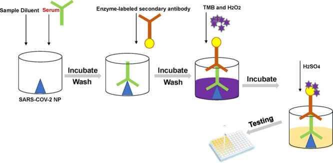
Costar 96-well EIA/RIA Stripwell immunoplates (Corning, New York, USA) were coated with the recombinant NP at a concentration of 10 μg/mL by a carbonate–bicarbonate buffer pH 9.6 (50 μL/well) and incubated overnight at 4 °C. After washing once with a washing buffer consisting of PBS (pH 7.2) and 0.05% Tween-20, the plates were blocked with 300 μL/well of 5% fat-free milk powder in PBS and incubated overnight at 4 °C and then washed as described above. Sera in group A and B were diluted at 1:100 in PBS containing 5% fat-free milk powder and 100 μL of diluted sera was added to the plates. After incubation in a moist chamber for 30 min at 37 °C, the plates were washed five times with washing buffer, and 100 μL per well of anti-human IgG-HRP conjugate diluted 1:4000 (or anti-human IgM–HRP conjugate diluted 1:2500) was added. The plates were incubated for 30 min at 37 °C and washed five times, and 100 μL per well of tetramethylbenzidine and H2O2 substrate (Thermo, Massachusetts) was used for detection. The plates were incubated for 5 min, and 50 μL per well of 0.5 M H2SO4 was added to stop the reaction, and absorbance was read at 450 nm (OD450nm).
Determination of Cut-Off Values and ELISA Diagnostic Accuracy
Cut-off values were determined as the mean value of OD450nm derived from 10 healthy human sera in group C plus two standard deviations. All statistical analyses were performed using SPSS software version 22.0. Estimates of diagnostic sensitivity and specificity, positive predictive value (PPV), and negative predictive value (NPV) were calculated as described previously.7
Nucleocapsid-Based Chemiluminescent Immunoassay Test
We used a commercially available chemiluminescent immunoassay (CLIA) kit and a colloidal gold immunochromatography kit as per the manufacturer’s instruction to test those 168 COVID-19 patients’ sera in group A.
Anti-SARS-CoV-2 Chemiluminescent Immunoassay IgM and IgG (National Medical Products Administration, China; Cat. # C86095M and C86095G, respectively) were performed according to the manufacturer’s instruction. The assay is a CLIA that detects the specific IgM/IgG in the human serum or plasma by binding the SARS-CoV-2 nucleocapsid antigen-coated microparticles. The levels of IgG and IgM antibodies were positively correlated with the relative luminescence unit (RLU) and were calculated as arbitrary units per milliliter (AU/mL). According to the receiver operating characteristic (ROC) curve, the corresponding concentration point of area under the ROC curve greater than 0.9 was defined as the cut-off point, and the level of this point was defined as 10 AU/mL.
Anti-SARS-CoV-2 colloidal gold immunochromatography IgM and IgG (National Medical Products Administration, China) was performed according to the manufacturer’s instruction. The assay detects antibodies against the NP of SARS-CoV-2.
Results
Expression of Recombinant SARS-COV-2 N Protein
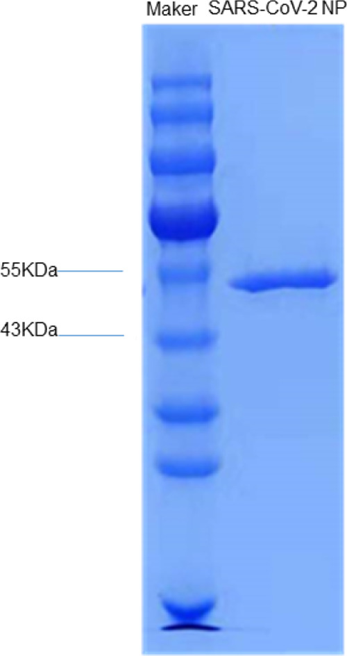
The recombinant NP protein was expressed in E. coli as a soluble protein and purified to homogeneity by the affinity chromatography scheme. The recombinant protein was dialyzed into PBS (pH 7.3) and sodium dodecyl sulfate polyacrylamide gel electrophoresis (SDS-PAGE) and revealed a protein band with a molecular weight of 50 kDa under reducing conditions (Figure 1).
Figure 1.

Purified His-tag recombinant SARS-COV-2 NP at approximately 50 kDa in SDS-PAGE.
Cut-Off Values and Diagnostic Performance of IgM/IgG ELISA
The sensitivity and specificity of SARS-CoV-2 ELISA for IgM and IgG antibodies were tested with confirmed COVID-19 patients’ sera (group A) and healthy persons’ sera (group B). ELISA showed that of 168 confirmed COVID-19 patients’ sera, 160 sera were IgM-positive, 163 sera were IgG-positive, and all sera were IgG- and/or IgM-positive to SARS-CoV-2 (Table 1), demonstrating the good sensitivity of IgM/IgG ELISA. ELISA also showed that none of the 90 healthy persons’ sera were positive for IgM or IgG ELISA. The specificity, sensitivity, PPV, and NPV for IgM are 100, 95.24, 100, and 91.84%, whereas those for IgG are 100, 97.02, 100, and 94.74%, respectively.
Table 1. Specificities, Sensitivities, PPV, and NPV for SARS-CoV-2 Specific IgM and IgG Antibody With ELISAa.
| cut-off | group A sera | group B sera | specificity (%) | sensitivity (%) | PPV (%) | NPV (%) | |
|---|---|---|---|---|---|---|---|
| IgM | 0.233 | 160/168 | 90/90 | 100 | 95.24 | 100 | 91.84 |
| IgG | 0.290 | 163/168 | 90/90 | 100 | 97.02 | 100 | 94.74 |
| IgM and IgG | NA | 168/168 | 90/90 | 100 | 100 | 100 | 100 |
IgM and IgG indicated that a sample was either IgM- or IgG-positive or positive to both IgM and IgG.
Comparison with Two Different Commercial Kits
To evaluate the performance of our ELISA assay, we tested those 168 COVID-19 patients’ sera by using a commercial CLIA kit and a commercial colloidal gold immunochromatography kit, which have been approved by the Chinese National Medical Products Administration (CNMPA) for clinical diagnosis of COVID-19. The commercial CLIA kit identified 78 and 151 to be positive for IgM (46.43%) and IgG (89.88%), respectively, from 168 tested samples. The colloidal gold immunochromatography kit detected 98/168 (57.14%) for IgM and 161/168 (95.83%) for IgG, whereas our ELISA assays showed a sensitivity up to 95.24% for IgM and 97.02% for IgG, respectively. We also compared the analytical characteristics of our assays and other reported detections. The analytical performance of various immunoassay methods for detection is summarized in Table 2.
Table 2. Comparison of Sensitivity and Specificity of ELISA Kits for Diagnosis of COVID-19.
| manufacturer | method | antigen | specificity (%) | sensitivity (%) | class of antibodies | references |
|---|---|---|---|---|---|---|
| Shenzhen YHLO Biotech Co., Ltd., China | chemiluminescence | recombinant NP | 99.49 | 85.96 | IgM | (8) |
| Shenzhen YHLO Biotech Co., Ltd., China | chemiluminescence | recombinant NP | 99.15 | 96.69 | IgG | (8) |
| Zhu Hai Liv Zon Diagnostics Inc., China | LFIA | recombinant NP | 100 | 82.4 | IgG and IgM | (9) |
| Zhu Hai Liv Zon Diagnostics Inc., China | indirect ELISA | recombinant NP | 100 | 87.3 | IgG and IgM | (9) |
| Mikrogen, Germany | indirect sandwich | recombinant NP | 100 | 86.4 | IgG | (9) |
| this study | indirect ELISA | recombinant NP | 100 | 95.24 | IgM | |
| this study | indirect ELISA | recombinant NP | 100 | 97.02 | IgG |
Discussion
The outbreak and rapid spread of SARS-CoV-2, the etiological agent for COVID-19, has posed a huge threat to public health worldwide.10,11 RT-PCR is a popular and routine strategy for laboratory diagnosis of COVID-19,1,12 but it heavily depends on highly trained personnel and sophisticated equipment and is very time-consuming. The sample for RT-PCR testing is mainly derived from oropharyngeal or nasopharyngeal swabs.13,14 Improper sampling time or a site with poor viral loads can result in false-negative results.15 Hence, it is important to develop an accurate, rapid, and cost-effective laboratory SARS-CoV-2 detection method. The serological test including antibodies detection can be used as an alternative to PCR because only a trace of pathogen is enough to induce the human humoral response. Because there is a certain window period for the production of antibodies, the results can be further evaluated through multiple antibody tests combined with clinical manifestations, epidemiological history, and other laboratory-related tests.16 Previous studies have shown that SARS-CoV-2-specific IgM and IgG exhibited positivity within 30 days.16−19 Some studies20−22 showed different times for seroconversion to SARS-CoV-2 depending on the severity of the disease. Qu21 et al. analyzed the profile of IgG and IgM antibodies against SARS-CoV-2 in 347 sera from 41 patients with COVID-19 between 3 and 43 days of their illness and revealed that IgG and IgM antibody responses in the critical patients’ group was delayed compared with noncritical groups. IgM in critical patients group rose on day 10, peaked on day 23, and then began to decline.21 In our cohort, the seropositive rate of IgG and IgM was still observed to maintain at a high level within 80 days after illness onset, which may be due to these 168 sera in group A collected from critical COVID-19 patients.
In this study, two ELISA assays were designed to detect SARS-CoV-2-specific IgM and IgG antibodies, respectively, by using the NP, an abundant and highly immunogenic component in the virions, as a diagnostic target. Of 168 clinical confirmed SARS-CoV-2 sera, 160 were IgM-positive (95.24%), 163 were IgG-positive (97.02%), and IgM and IgG combined sensitivity is up to 100%, which is of great significance for both individual clinical diagnosis and cohort epidemiological investigation. We also evaluated two ELISA assays and compared their performance with two commercial immunoassays, which have been approved by the Chinese National Medical Products Administration (CNMPA) and are widely used in China for the commercial diagnosis of COVID-19. Compared to two commercial kits, our ELISA assay showed a higher sensitivity on the same antigen. For the negative sera in group B, which were taken before the 2019 COVID-19 outbreak, none were IgM-/IgG-positive. The higher sensitivity of our assay may be attributed to two aspects: on the one hand, unlike the traditional indirect ELISA, we used MAbs as enzyme-labeled secondary antibodies instead of polyclonal antibodies, which can make the background noise of ELISA kits lower, so the sensitivity will increase accordingly. On the other hand, addition of excess anti-human IgGFc polyclone antibodies to the sample diluent captured the specific IgG in the serum, leading to the decrease in competition with IgM and the increase in sensitivity of IgM kits.
The study was limited by no determination of potential cross-reactivity with other CoVs. There are 7 human coronaviruses (CoVs) including NL63-CoV and 229E-CoV in genus Alphacoronavirus and SARS-CoV-2, SARS-CoV, MERS-CoV, OC43-CoV, and HKU1-CoV in genus Betacoronavirus. SARS-CoV-2, SARS-CoV, and MERS-CoV may cause severe diseases in humans and other CoVs cause human common cold. Although homology analysis showed that the SARS-CoV-2 nucleocapsid has 90% aminoacid identity to MERS-CoV and SARS-CoV,23 the MERS-CoV had never been introduced into China and SARS-CoV had been eradicated in 2004. Therefore, the positive sera of these viruses were difficult to obtain in China. In addition, the sequence homology (in protein level) between NPs of SARS-CoV-2 and the common cold CoV is very low (from 21 to 30%), which eliminates the possibility of the cross-reaction between them. On the other hand, we only evaluated the diagnostic performance in patients with critical COVID-19 and did not study the antibody response in asymptomatic persons and patients with mild to moderate COVID-19.
In conclusion, we developed a highly sensitive and specific NP-based ELISA for detection of the SARS-CoV-2 IgM and IgG antibodies for diagnosis and epidemiologic investigation of COVID-19.
Acknowledgments
This project was supported by a grant from National Natural Science Funds of China (no. 81971939).
Author Contributions
⊥ Y.-j.J. and X.-j.Y. contributed equally.
The authors declare no competing financial interest.
References
- Pfefferle S.; Reucher S.; Nörz D.; Lütgehetmann M. Evaluation of a quantitative RT-PCR assay for the detection of the emerging coronavirus SARS-CoV-2 using a high throughput system. Eurosurveillance 2020, 25, 2000152. 10.2807/1560-7917.es.2020.25.9.2000152. [DOI] [PMC free article] [PubMed] [Google Scholar]
- Cevik M.; Tate M.; Lloyd O.; Maraolo A. E.; Schafers J.; Ho A. SARS-CoV-2, SARS-CoV, and MERS-CoV viral load dynamics, duration of viral shedding, and infectiousness: a systematic review and meta-analysis. Lancet Microbe 2021, 2, e13–e22. 10.1016/s2666-5247(20)30172-5. [DOI] [PMC free article] [PubMed] [Google Scholar]
- Deeks J. J.; Dinnes J.; Takwoingi Y.; Davenport C.; Spijker R.; Taylor-Phillips S.; Adriano A.; Beese S.; Dretzke J.; Ferrante di Ruffano L.; Harris I. M.; Price M. J.; Dittrich S.; Emperador D.; Hooft L.; Leeflang M. M.; Van den Bruel A. Antibody tests for identification of current and past infection with SARS-CoV-2. Cochrane Database Syst. Rev. 2020, 6, Cd013652. 10.1002/14651858.CD013652. [DOI] [PMC free article] [PubMed] [Google Scholar]
- Lu R.; Zhao X.; Li J.; Niu P.; Yang B.; Wu H.; Wang W.; Song H.; Huang B.; Zhu N.; Bi Y.; Ma X.; Zhan F.; Wang L.; Hu T.; Zhou H.; Hu Z.; Zhou W.; Zhao L.; Chen J.; Meng Y.; Wang J.; Lin Y.; Yuan J.; Xie Z.; Ma J.; Liu W. J.; Wang D.; Xu W.; Holmes E. C.; Gao G. F.; Wu G.; Chen W.; Shi W.; Tan W. Genomic characterisation and epidemiology of 2019 novel coronavirus: implications for virus origins and receptor binding. Lancet 2020, 395, 565–574. 10.1016/s0140-6736(20)30251-8. [DOI] [PMC free article] [PubMed] [Google Scholar]
- Shang B.; Wang X.-Y.; Yuan J.-W.; Vabret A.; Wu X.-D.; Yang R.-F.; Tian L.; Ji Y.-Y.; Deubel V.; Sun B. Characterization and application of monoclonal antibodies against N protein of SARS-coronavirus. Biochem. Biophys. Res. Commun. 2005, 336, 110–117. 10.1016/j.bbrc.2005.08.032. [DOI] [PMC free article] [PubMed] [Google Scholar]
- Liu S.; Leng C.; Lien S.; Chi H.; Huang C.; Lin C.; Lian W.; Chen C.; Hsieh S.; Chong P. Immunological characterizations of the nucleocapsid protein based SARS vaccine candidates. Vaccine 2006, 24, 3100–3108. 10.1016/j.vaccine.2006.01.058. [DOI] [PMC free article] [PubMed] [Google Scholar]
- Jiao Y.; Zeng X.; Guo X.; Qi X.; Zhang X.; Shi Z.; Zhou M.; Bao C.; Zhang W.; Xu Y.; Wang H. Preparation and evaluation of recombinant severe fever with thrombocytopenia syndrome virus nucleocapsid protein for detection of total antibodies in human and animal sera by double-antigen sandwich enzyme-linked immunosorbent assay. J. Clin. Microbiol. 2012, 50, 372–377. 10.1128/jcm.01319-11. [DOI] [PMC free article] [PubMed] [Google Scholar]
- Qian C.; Zhou M.; Cheng F.; Lin X.; Gong Y.; Xie X.; Li P.; Li Z.; Zhang P.; Liu Z.; Hu F.; Wang Y.; Li Q.; Zhu Y.; Duan G.; Xing Y.; Song H.; Xu W.; Liu B.-F.; Xia F. Development and multicenter performance evaluation of fully automated SARS-CoV-2 IgM and IgG immunoassays. Clin. Chem. Lab. Med. 2020, 58, 1601–1607. 10.1515/cclm-2020-0548. [DOI] [PubMed] [Google Scholar]
- Krüttgen A.; Cornelissen C. G.; Dreher M.; Hornef M.; Imöhl M.; Kleines M. Comparison of four new commercial serologic assays for determination of SARS-CoV-2 IgG. J. Clin. Virol. 2020, 128, 104394. 10.1016/j.jcv.2020.104394. [DOI] [PMC free article] [PubMed] [Google Scholar]
- Zhu N.; Zhang D.; Wang W.; Li X.; Yang B.; Song J.; Zhao X.; Huang B.; Shi W.; Lu R.; Niu P.; Zhan F.; Ma X.; Wang D.; Xu W.; Wu G.; Gao G. F.; Tan W. A Novel Coronavirus from Patients with Pneumonia in China, 2019. N. Engl. J. Med. 2020, 382, 727–733. 10.1056/nejmoa2001017. [DOI] [PMC free article] [PubMed] [Google Scholar]
- Guan W.-j.; Ni Z.-y.; Hu Y.; Liang W.-h.; Ou C.-q.; He J.-x.; Liu L.; Shan H.; Lei C.-l.; Hui D. S. C.; Du B.; Li L.-j.; Zeng G.; Yuen K.-Y.; Chen R.-c.; Tang C.-l.; Wang T.; Chen P.-y.; Xiang J.; Li S.-y.; Wang J.-l.; Liang Z.-j.; Peng Y.-x.; Wei L.; Liu Y.; Hu Y.-h.; Peng P.; Wang J.-m.; Liu J.-y.; Chen Z.; Li G.; Zheng Z.-j.; Qiu S.-q.; Luo J.; Ye C.-j.; Zhu S.-y.; Zhong N.-s. Clinical Characteristics of Coronavirus Disease 2019 in China. N. Engl. J. Med. 2020, 382, 1708–1720. 10.1056/nejmoa2002032. [DOI] [PMC free article] [PubMed] [Google Scholar]
- Cheng M. P.; Papenburg J.; Desjardins M.; Kanjilal S.; Quach C.; Libman M.; Dittrich S.; Yansouni C. P. Diagnostic Testing for Severe Acute Respiratory Syndrome-Related Coronavirus 2: A Narrative Review. Ann. Intern. Med. 2020, 172, 726–734. 10.7326/m20-1301. [DOI] [PMC free article] [PubMed] [Google Scholar]
- Wang W.; Xu Y.; Gao R.; Lu R.; Han K.; Wu G.; Tan W. Detection of SARS-CoV-2 in Different Types of Clinical Specimens. JAMA 2020, 323, 1843–1844. 10.1001/jama.2020.3786. [DOI] [PMC free article] [PubMed] [Google Scholar]
- Wang X.; Tan L.; Wang X.; Liu W.; Lu Y.; Cheng L.; Sun Z. Comparison of nasopharyngeal and oropharyngeal swabs for SARS-CoV-2 detection in 353 patients received tests with both specimens simultaneously. Int. J. Infect. Dis. 2020, 94, 107–109. 10.1016/j.ijid.2020.04.023. [DOI] [PMC free article] [PubMed] [Google Scholar]
- Zou L.; Ruan F.; Huang M.; Liang L.; Huang H.; Hong Z.; Yu J.; Kang M.; Song Y.; Xia J.; Guo Q.; Song T.; He J.; Yen H.-L.; Peiris M.; Wu J. SARS-CoV-2 Viral Load in Upper Respiratory Specimens of Infected Patients. N. Engl. J. Med. 2020, 382, 1177–1179. 10.1056/nejmc2001737. [DOI] [PMC free article] [PubMed] [Google Scholar]
- Caturegli G.; Materi J.; Howard B. M.; Caturegli P. Clinical Validity of Serum Antibodies to SARS-CoV-2 : A Case-Control Study. Ann. Intern. Med. 2020, 173, 614–622. 10.7326/m20-2889. [DOI] [PMC free article] [PubMed] [Google Scholar]
- Xiang F.; Wang X.; He X.; Peng Z.; Yang B.; Zhang J.; Zhou Q.; Ye H.; Ma Y.; Li H.; Wei X.; Cai P.; Ma W.-L. Antibody Detection and Dynamic Characteristics in Patients With Coronavirus Disease 2019. Clin. Infect. Dis. 2020, 71, 1930–1934. 10.1093/cid/ciaa461. [DOI] [PMC free article] [PubMed] [Google Scholar]
- Montesinos I.; Gruson D.; Kabamba B.; Dahma H.; Van den Wijngaert S.; Reza S.; Carbone V.; Vandenberg O.; Gulbis B.; Wolff F.; Rodriguez-Villalobos H. Evaluation of two automated and three rapid lateral flow immunoassays for the detection of anti-SARS-CoV-2 antibodies. J. Clin. Virol. 2020, 128, 104413. 10.1016/j.jcv.2020.104413. [DOI] [PMC free article] [PubMed] [Google Scholar]
- Bundschuh C.; Egger M.; Wiesinger K.; Gabriel C.; Clodi M.; Mueller T.; Dieplinger B. Evaluation of the EDI enzyme linked immunosorbent assays for the detection of SARS-CoV-2 IgM and IgG antibodies in human plasma. Clin. Chim. Acta 2020, 509, 79–82. 10.1016/j.cca.2020.05.047. [DOI] [PMC free article] [PubMed] [Google Scholar]
- Long Q.-X.; Liu B.-Z.; Deng H.-J.; Wu G.-C.; Deng K.; Chen Y.-K.; Liao P.; Qiu J.-F.; Lin Y.; Cai X.-F.; Wang D.-Q.; Hu Y.; Ren J.-H.; Tang N.; Xu Y.-Y.; Yu L.-H.; Mo Z.; Gong F.; Zhang X.-L.; Tian W.-G.; Hu L.; Zhang X.-X.; Xiang J.-L.; Du H.-X.; Liu H.-W.; Lang C.-H.; Luo X.-H.; Wu S.-B.; Cui X.-P.; Zhou Z.; Zhu M.-M.; Wang J.; Xue C.-J.; Li X.-F.; Wang L.; Li Z.-J.; Wang K.; Niu C.-C.; Yang Q.-J.; Tang X.-J.; Zhang Y.; Liu X.-M.; Li J.-J.; Zhang D.-C.; Zhang F.; Liu P.; Yuan J.; Li Q.; Hu J.-L.; Chen J.; Huang A.-L. Antibody responses to SARS-CoV-2 in patients with COVID-19. Nat. Med. 2020, 26, 845–848. 10.1038/s41591-020-0897-1. [DOI] [PubMed] [Google Scholar]
- Qu J.; Wu C.; Li X.; Zhang G.; Jiang Z.; Li X.; Zhu Q.; Liu L. Profile of Immunoglobulin G and IgM Antibodies Against Severe Acute Respiratory Syndrome Coronavirus 2 (SARS-CoV-2). Clin. Infect. Dis. 2020, 71, 2255–2258. 10.1093/cid/ciaa489. [DOI] [PMC free article] [PubMed] [Google Scholar]
- Lou B.; Li T.-D.; Zheng S.-F.; Su Y.-Y.; Li Z.-Y.; Liu W.; Yu F.; Ge S.-X.; Zou Q.-D.; Yuan Q.; Lin S.; Hong C.-M.; Yao X.-Y.; Zhang X.-J.; Wu D.-H.; Zhou G.-L.; Hou W.-H.; Li T.-T.; Zhang Y.-L.; Zhang S.-Y.; Fan J.; Zhang J.; Xia N.-S.; Chen Y. Serology characteristics of SARS-CoV-2 infection after exposure and post-symptom onset. Eur. Respir. J. 2020, 56, 2000763. 10.1183/13993003.00763-2020. [DOI] [PMC free article] [PubMed] [Google Scholar]
- Okba N. M. A.; Müller M. A.; Li W.; Wang C.; GeurtsvanKessel C. H.; Corman V. M.; Lamers M. M.; Sikkema R. S.; de Bruin E.; Chandler F. D.; Yazdanpanah Y.; Le Hingrat Q.; Descamps D.; Houhou-Fidouh N.; Reusken C. B. E. M.; Bosch B.-J.; Drosten C.; Koopmans M. P. G.; Haagmans B. L.. SARS-CoV-2 specific antibody responses in COVID-19 patients. 2020, medRxiv 2020.03.18.20038059. [DOI] [PMC free article] [PubMed] [Google Scholar]


