Clinical History
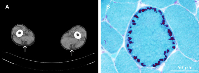
A 31‐year‐old man, with no family history of neuromuscular diseases, received muscle biopsy for slowly progressive muscle weakness. The first symptom he had was difficulty dorsiflexing his feet at age 20 years, which was followed by gradual development of gait disturbance. At age 27 years, he started using a handrail to climb up stairs. At age 28 years, he developed dysphagia for liquid, in addition to difficulty raising arms, which led him to have aspiration pneumonia later in the same year. At age approximately 30 years, he developed dyspnea on exertion. Arterial blood gas analysis revealed hypoxemia (65 mmHg, at room air) and hypercapnia (82 mmHg), with a vital capacity decreased to 780 mL, leading to the diagnosis of chronic type 2 respiratory failure, and non‐invasive ventilation was started. At age 31 years, he was unable to walk without aid and required a wheelchair for long distances. Physical examination revealed moderate muscle weakness and atrophy in an asymmetric limb‐girdle distribution, together with marked muscle atrophy in the tibialis anterior muscles and weakness in ankle dorsiflexion with Medical Research Council grade 1. Mild neck muscle weakness was also noted. Serum creatine kinase level was 375 IU/L (normal: <287 IU/L). Electromyography showed myopathic changes. Skeletal muscle CT demonstrated remarkable fat tissue replacement in the semitendinosus muscles (Figure 1A, arrows) at the thigh level.
Figure 1.

Pathology
Skeletal muscle pathology from the biceps brachii demonstrated myopathic changes with moderate fiber size variation, mildly disorganized myofibrillar networks, some fibers with cytoplasmic bodies, which were often located in line in subsarcolemmal region of a muscle fiber (Figure 1B, modified Gomori trichrome stain), and a few fibers with rimmed vacuoles. What is your diagnosis? What is the next test to confirm it?
Diagnosis
Hereditary myopathy with early respiratory failure (HMERF).
The diagnosis was confirmed by gene analysis showing a heterozygous titin gene (TTN) mutation g.284701T > C (p.Cys31712Arg) in the FN3 119 domain in the A‐band region (ENST00000589042).
Discussion
HMERF is a hereditary myopathy caused by mutations in the A‐band region of TTN 1, 2. Since the causative gene was reported in 2012, an increasing number of patients with HMERF have been identified worldwide 5.
HMERF is clinically characterized by adult‐onset muscle weakness with respiratory insufficiency from an early, typically ambulant stage of the disease. A common initial symptom is gait disturbance because of distal (especially the anterior compartment) and/or proximal lower limb muscle weakness. Weakness of ankle dorsiflexion is prominent, leading to easy tripping and foot drop. There is no or minimal, if any, weakness in the upper limbs at disease onset. Respiratory insufficiency subsequently appears after a median of six years post‐onset (range: 0 − 16 years) 5, although respiratory symptoms can be the initial manifestation in 24%−36% of patients 1, 5. Most patients are ambulant when they need nocturnal ventilation 1. To date, clinically evident primary cardiomyopathy has not been reported in patients with HMERF, although right heart failure secondary to severe respiratory failure is occasionally observed. Nevertheless, a recent study found cardiac conduction abnormalities in 32% (7/22) of patients with the p.Cys31712Arg mutation of TTN and imaging evidence of otherwise unexplained asymptomatic cardiomyopathy in 18% (4/22) (3). The autosomal dominant trait is common, although a few homozygous patients with severe symptoms have been reported. Some patients have no apparent positive family history 5.
Skeletal muscle imaging demonstrates that the semitendinosus muscles and the anterior compartment of the lower legs (preferentially, the peroneus longus muscles) are selectively involved in the early stage 1, 2, 5. Indeed, the selective involvement of the semitendinosus muscles is often observed (93%−100%) 2, 5. The pattern is a radiological clue for diagnosis of HMERF, although it can be observed in other muscle diseases, including desminopathy, αB‐crystallinopathy, and anoctaminopathy 4.
The pathological hallmark of HMERF is necklace cytoplasmic bodies; at cross section, a number of cytoplasmic bodies are often circumferentially aligned in the subsarcolemmal region in muscle fibers, mimicking a necklace 5. The finding of necklace cytoplasmic bodies is highly specific for HMERF (sensitivity: 82%, specificity: 99%) 5. Rimmed vacuoles are also sometimes observed but less specific.
Since TTN encodes the largest known protein consisting of 35,991 amino‐acid polypeptides in 363 exons among those recognized so far, sequence analysis of all the exons requires considerable labor. Even if next generation sequencing is available for the genetic analysis, it would still be difficult to determine which variant is truly pathogenic because TTN is a highly polymorphic gene. Despite this, all HMERF patients reported so far carry a mutation in exon 343 encoding the FN3 119 domain in the A‐band region (ENST00000589042). Therefore, exon 343 should be considered as the primary target for genetic diagnosis of HMERF.
When a patient shows the pathological finding of necklace cytoplasmic bodies, sequencing of exon 343 of TTN should be considered for definite diagnosis of HMERF, even though the patient does not exhibit respiratory problems at that time. If the patient has a mutation in exon 343 of TTN, the patient will eventually develop a severe respiratory insufficiency. Early diagnosis of the disease would enable earlier recognition of asymptomatic respiratory insufficiency and appropriate intervention without delay (eg, nocturnal ventilation).
Acknowledgments
We thank Ms. Kaoru Tatezawa and Ms. Kanako Goto for their technical assistance. This study was supported partly by JSPS KAKENHI Grant Number of 24659437; partly by Research on Rare and Intractable Diseases, Comprehensive Research on Disability Health and Welfare and Applying Health Technology from the Ministry of Health, Labour and Welfare of Japan; and partly by Intramural Research Grant 23‐5 and 26‐8 for Neurological and Psychiatric Disorders of NCNP.
References
- 1. Ohlsson M, Hedberg C, Brådvik B, Lindberg C, Tajsharghi H, Danielsson O et al (2012) Hereditary myopathy with early respiratory failure associated with a mutation in A‐band titin. Brain 135:1682–1694. [DOI] [PubMed] [Google Scholar]
- 2. Pfeffer G, Elliott HR, Griffin H, Barresi R, Miller J, Marsh J et al (2012) Titin mutation segregates with hereditary myopathy with early respiratory failure. Brain 135:1695–1713. [DOI] [PMC free article] [PubMed] [Google Scholar]
- 3. Steele HE, Harris E, Barresi R, Marsh J, Beattie A, Bourke JP et al (2016) Cardiac involvement in hereditary myopathy with early respiratory failure: A cohort study. Neurology 87:1031–1035. [DOI] [PMC free article] [PubMed] [Google Scholar]
- 4. Tasca G, Evilä A, Pane M, Monforte M, Graziano A, Hackman P et al (2014) Isolated semitendinosus involvement in the initial stages of limb‐girdle muscular dystrophy 2L. Neuromuscul Disord 24:1118–1119. [DOI] [PubMed] [Google Scholar]
- 5. Uruha A, Hayashi YK, Oya Y, Mori‐Yoshimura M, Kanai M, Murata M et al (2015) Necklace cytoplasmic bodies in hereditary myopathy with early respiratory failure. J Neurol Neurosurg Psychiatry 86:483–489. [DOI] [PubMed] [Google Scholar]


