Clinical History and Neuroimaging
A previously healthy 42‐year‐old man presented with 2 weeks of confusion, lethargy, headache and malaise. He was diagnosed with AIDS 3 months prior (CD4 count 198 cells/μL; viral load 87 100 copies/mL) and was on trimethoprim/sulfamethoxazole prophylaxis but had not started highly active antiretroviral therapy. Neurological examination was notable for a sleepy man lacking orientation to location or date, perseveration on simple phrases and overall paucity of speech and generalized weakness. He could not stand unassisted. Reflexes were 3+ throughout.
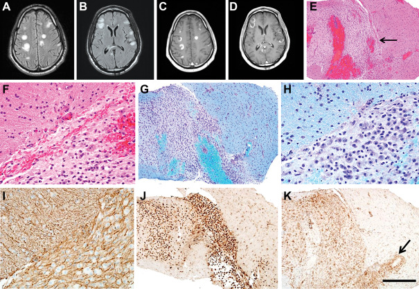
MRI was notable for numerous hyperintense T2/FLAIR lesions in multiple regions within the bilateral cerebral hemispheres (Figure 1A,B, FLAIR sequences) as well as the right cerebellum. These lesions demonstrated T1 hypointensity and contrast‐enhancement, often in incomplete ring‐enhancing patterns (Figure 1C,D).
Figure 1.

Serum antitoxoplasma IgG/IgM antibodies, blastomyces antibody and urine histoplasma antigen were negative. CSF analysis showed 10 red blood cells/mL, 41 nucleated cells/μL (99% lymphocytes), normal glucose, protein 184 mg/dL, 6 oligoclonal bands (<4 normal) and IgG index 0.96 (<0.21 normal). CSF flow cytometry and cytology was negative. CSF was negative for Toxoplasma gondii, Cryptococcus and VZV, HSV, JC, EBV and CMV. CSF cultures including fungal and acid‐fast bacilli cultures were negative. Stereotactic brain biopsy was performed.
Pathological Findings
H&E and LFB‐PAS stains (Figure 1E–H) show a sharply demarcated area of pallor (arrow, Figure 1E) and loss of myelin, with relative preservation of axons by neurofilament immunostain (Figure 1I). The lesion also has numerous CD68‐positive macrophages (Figure 1J). Additionally, the lesion and surrounding an adjacent blood vessel have many CD4‐positive lymphocytes (Figure 1K, arrow, magnification bar: 500 μm). There was no evidence of neoplasia or infection. What is your diagnosis?
Diagnosis
Acute disseminated encephalomyelitis.
Discussion
This case highlights several important principles to both the clinical neurologist as well as the neuropathologist. Initially, highest on the differential was opportunistic infection, lymphoma or demyelination. Due to concern for CNS toxoplasmosis, the patient was empirically treated with pyrimethamine and sulfasalazine. Given the lack of laboratory evidence and clinical improvement to support this treatment strategy, tissue diagnosis was pursued. Just prior to this time, the patient worsened clinically to where he had anarthria, severe dysphagia and left hemiplegia. After the pathology was reviewed and a tissue diagnosis made, the course of treatment was completely changed and he was treated with intravenous methylprednisolone followed by an oral prednisone taper and five rounds of plasma exchange. Follow‐up MRI showed complete resolution of contrast enhancement with stability of the T2/FLAIR signal. Clinically, he became more interactive and verbal with improvement of his left hemiparesis.
There are numerous neurologic manifestations of HIV/AIDS both intrinsic to the infection as well as the immunocompromised state that is seen in other patients, such as transplant recipients who are iatrogenically immunosuppressed. A history of immunodeficiency is salient to the astute clinician and widens the differential considerably. Notably, while this patient did technically meet criteria for AIDS, his CD4 count was on the borderline.
Toxoplasmosis is the most common CNS parasitic opportunistic infection in AIDS patients and the most common AIDS‐defining illness in the CNS. On MRI, it frequently presents with multiple ring‐enhancing lesions, particularly in the grey‐white junction and the basal ganglia. Approximately 95% of these patients will have anti‐toxoplasma IgG antibodies. Primary CNS lymphoma is a form of non‐Hodgkin lymphoma and is second only to toxoplasmosis for mass lesions in the brains of AIDS patients. On neuroimaging, single or multiple contrast enhancing lesions are present and can be ring patterned or homogeneous 2.
ADEM is a disease of acute multifocal demyelination in the CNS that typically follows an infectious illness and presents heterogeneously. Full recovery may occur and treatment consists of therapies such as steroids, IVIg and plasma exchange 3. ADEM may present with ring‐enhancing lesions and can mimic CNS toxoplasmosis or lymphoma 4. There are rare case reports of ADEM in patients with HIV/AIDS but the nature of this relationship needs to be further elucidated 1.
We present a rare case of pathologically confirmed extensive demyelination with multiple incomplete ring‐enhancing lesions in a man with AIDS. This case demonstrates that while immunocompromised patients have an expansive differential including opportunistic infections and malignancies, they are also prone to the diseases that immunocompetent patients are. As the prognosis and management may differ vastly, considering all diagnostic possibilities is important. In this case, brain biopsy was instrumental in making the diagnosis and embarking on a completely different treatment strategy.
References
- 1. Allen SH, Malik O, Lipman MCI, Johnson MA, Wilson LA (2002) Acute demyelinating encephalomyelitis (ADEM) in a patient with HIV infection. J Infect 45:62–64. [DOI] [PubMed] [Google Scholar]
- 2. Bilgrami M, O'Keefe P (2014) Neurologic diseases in HIV‐infected patients. Handbk Clin Neurol 121:1321–1344. [DOI] [PubMed] [Google Scholar]
- 3. Javed A, Khan O (2014) Acute disseminated encephalomyelitis. Handbk Clin Neurol 123:705–716. [DOI] [PMC free article] [PubMed] [Google Scholar]
- 4. Lim KE, Hsu YY, Hsu WC, Chan CY (2003) Multiple complete ring‐shaped enhanced MRI lesions in acute disseminated encephalomyelitis. Clin Imaging 27:281–284. [DOI] [PubMed] [Google Scholar]


