Abstract
At the time of preparing this Perspective, large-scale vaccination for COVID-19 is in progress, aiming to bring the pandemic under control through vaccine-induced herd immunity. Not only does this vaccination effort represent an unprecedented scientific and technological breakthrough, moving us from the rapid analysis of viral genomes to design, manufacture, clinical trial testing and use authorization within the timeframe of less than a year, but also highlights rapid progress in the implementation of nanotechnology to assist vaccine development. These advances enable us to deliver nucleic acid and conformation-stabilized subunit vaccines to regional lymph nodes, with the ability to trigger effective humoral and cellular immunity that prevents viral infection or controls disease severity. In addition to a brief description of the design features of unique cationic lipid and virus-mimicking nanoparticles for accomplishing spike protein delivery and presentation by the cognate immune system, we also discuss the importance of adjuvancy and design features to promote cooperative B- and T-cell interactions in lymph node germinal centers, including through the use of epitope-based vaccines. Although current vaccine efforts have demonstrated short-term efficacy and vaccine safety, key issues are now vaccine durability and adaptability against viral variants. We present a forward-looking perspective of how vaccine design can be adapted to improve durability of the immune response and vaccine adaptation to overcome immune escape by viral variants. Finally, we consider the impact of nano-enabled approaches in the development of COVID-19 vaccines for improve vaccine design against other infectious agents, including for pathogens that may lead to future pandemics.
Keywords: COVID-19, vaccine, antigens, epitopes, lipid nanoparticles, adjuvants, immune escape, durability, viral variants, immunoinformatics
Graphical Abstract
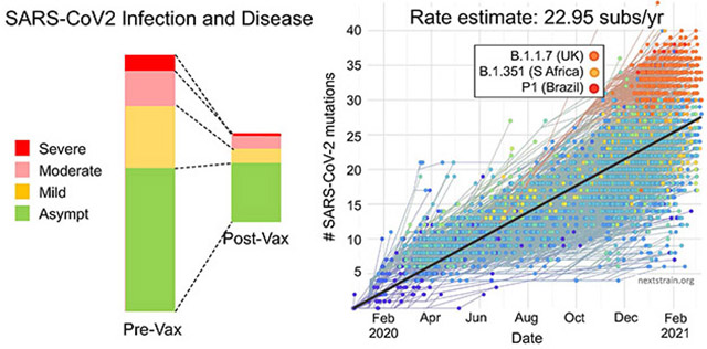
The challenge of developing a SARS-CoV-2 vaccine capable of intervening in the alarming rate of spread and mortality, the likes of which has not been seen since the 1918 influenza contagion, has been a daunting task. Remarkably, the 4–14 year time frame that was required for developing mumps, measles, polio, or human papilloma virus vaccines was condensed into a year to accomplish the same task for COVID-19.1
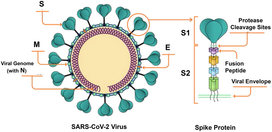
Infectious disease experts have cautioned for years about the pandemic potential of coronaviruses. These concerns were confirmed by the emergence of SARS-CoV-1 in 2003, with a case fatality rate of 15%, and the Middle Eastern Respiratory Syndrome Coronavirus (MERS-CoV) in 2012, with a fatality rate of 36%.2 These short-lived outbreaks stimulated interest in understanding coronavirus pathogenesis and immunity, leading to the development of experimental vaccines in animal models.3-8 Unfortunately, due to the finite duration of these disease episodes, none of the efforts resulted in vaccine development for human use. Nonetheless, these efforts provided critical information about the role of the trimeric spike (S) glycoprotein, which is responsible for SARS-CoV uptake into host cells through a binding interaction with angiotensin-converting enzyme 2 (ACE2) receptors (Figures 1 and 2).3,6,7,9-11 In particular, it was revealed that the development of neutralizing antibodies against the receptor-binding domain (RBD) of the SARS-CoV-1 or MERS-CoV spike were effective in blocking viral uptake. This finding was instrumental in earmarking the generation of neutralizing antibodies against the spike protein as a viable vaccine strategy against coronaviruses.3,6,7,9-11 Moreover, research on experimental SARS-CoV-1 and respiratory syncytial virus (RSV) vaccines helped to refine a structural vaccinology approach in which the spike or fusion proteins were engineered to obtain a stabilized antigen conformation that optimizes the generation of neutralizing antibodies.12-14 These efforts subsequently became a blueprint for expedited SARS-CoV-2 vaccine development.
Figure 1. SARS-CoV-2 components for generating protective antiviral immune responses.
These include the spike or S glycoprotein, membrane or M protein, envelope or E protein, and nucleocapsid or N protein (associates with viral RNA genome; not shown). The current choice for vaccine generation is the S glycoprotein, which is capable of generating neutralizing antibody responses in addition to eliciting CD8+ and CD4+ T-cells. The spike protein exhibits a screw-like shape, composed of a larger head and a long, thin stalk.206 Three spike proteins interact to form a trimer that is held in place by a stalk (composed of S1 and S2 regions), which stands away from the viral surface and exhibits a host protease (furin) cleavage site, the role of which is explained in Figure 2. Adapted with permission from ref 206. Copyright 2020 CAS.
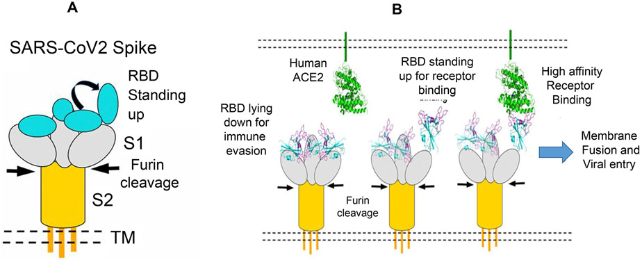
Figure 2. SARS-CoV-2 spike (S) glycoprotein.
A. The S protein includes (i) the trimeric S1 subunit, which contains 3 receptor-binding domains (RBDs) (two of which are lying down, with one standing up); (ii) the membrane-associated S2 subunit, which includes a fusion peptide; (iii) a transmembrane (TM) anchor and (iv) an intracellular tail.60 B. Schematic to show the early stage of viral uptake.60 Viral uptake commences with proteolytic cleavage by furin, which separates the S1 and S2 subunits, enabling one RBD to stand up. Next, the 2nd and then the 3rd RBD domains stand up. The achievement of a pre-fusion complex (with 3 RBDs standing up) leads to two important outcomes: (i) exposure and immune recognition of S1 epitopes that were covered up by the RBDs in the lying down conformation; (ii) high affinity binding of RBDs to the host hACE2 receptor to enable viral docking. Once docked onto the host cell membrane, the contraction of the S2 fusion peptide blends the viral envelope with the host cell membrane. Adapted with permission from ref 60 under a Creative Commons Attribution License 4.0 (CC BY). Copyright 2020 The Authors.
As the explosive spread of SARS-CoV-2 infections and accompanying morbidity and mortality became apparent, vaccine producers recognized that curtailing the pandemic would require technologies that support rapid development, scalable manufacturing, and rapid deployment. An attractive approach, albeit one that had never been tested at large scale in humans, was the delivery of mRNA for in situ production of genetically optimized antigens.1,15-19 Although the preclinical feasibility of mRNA-based therapeutics and vaccines was reported in the 1990s, practical implementation of this approach only became apparent over the past decade. Early studies encountered major obstacles, including high innate immune reactivity to foreign RNA, rapid degradation by RNases in body fluids, and inefficient in vivo delivery to the translational machinery of host target cells.15-19
Pioneering work by Katalin Karikó and colleagues showed that exogenous RNA stimulates innate immunity in part through activation of endosomally localized Toll-like receptors (TLRs, e.g., TLR3, TLR7, and TLR8), and that the immune reactivity of RNA can be systematically down-modulated by incorporation of modified nucleosides (e.g., m5C, m6A, m5U, s2U, or Ψ).17,20 Modified nucleosides increase both the safety profile and the expression efficiency of mRNA delivered in vivo; understanding this relationship was a critical step in advancing the technology to the clinic.21,22 The development of high-yield in vitro transcription systems for RNA production, methods for increasing translation using synthetic 5' cap analogs and capping enzymes, and advances in template design and purification now facilitate rapid, robust, and scalable manufacturing of mRNA.16,23-25
Solutions to the remaining challenges of in vivo stability and delivery emerged with the advent of nanotechnologies for encapsulating mRNA into virus-sized (~100 nm) cationic lipid nanoparticles (LNPs) that provide protection from extracellular RNases while also facilitating uptake and endomosomal release of mRNA in target cells.16,26 Intradermal, intramuscular, or subcutaneous delivery of mRNA, packaged in cationic LNPs, results in prolonged antigen expression and the induction of B-cell and CD4+ T follicular helper (TFH) cell responses that originate in the germinal centers of secondary lymphoid organs. This outcome results in the production of potent and long-lived antibody responses.27, 28 In addition to CD4+ T-cells, mRNA vaccines also induce CD8+ T-cells that recognize and kill antigen-expressing cells, including SARS-CoV-2 infected cells.29-36
The design, implementation, efficacy, and safety profiles of mRNA-based nanoparticle vaccines expressing SARS-CoV-2 antigens, as exemplified by the COVID-19 vaccines recently developed by Moderna Inc. and Pfizer/BioNTech, are discussed below. We also review nano-enabled approaches for direct delivery of protein subunits, such as the Novavax vaccine, as well as in silico designed, self-assembling nanoparticles that mimic the virus display of the receptor-binding domain (RBD). Table 1 provides a short list of the large number of contemporary and next-generation vaccines that are currently entering clinical trials according to the World Health Organization (WHO) COVID-19 vaccine database.37 In addition to discussing nano-enabled platforms that either have received or are poised to obtain U.S. Food and Drug Administration (FDA) approval (vide infra), we briefly discuss future refinements, such as the development of nanoparticles that are capable of delivering multiple epitopes or of promoting B- and T-cell cooperation in lymph node germinal centers. Finally, challenges associated with vaccine durability, avoidance of vaccine side effects, and adapting vaccine coverage to include SARS-CoV-2 escape mutants are also discussed.
Table 1:
CoV-2 Vaccine Technologies Advancing to Clinical Trials or Approval
| Contemporary Vaccine Technology | |||
|---|---|---|---|
| Category | Developer | Clinical development stage |
Phase 3 Efficacy* |
| Live-attenuated vaccines | Codagenix (COVI-VAC) India | Phase 1 | |
| Inactivated viral vaccines | Sinovac Research (CoronaVac) China39, 194, 195 | Phase 3 Approved# | 50% |
| Sinopharm (BBIP-CorV)39, 196 China and other countries | Phase 3 Approved# | 79% | |
| Viral-vectored vaccines | |||
| • Adenovirus (nonreplicating) | CanSino [Convidicea (Ad5-nCoV)] China 39, 186, 197 | Phase 3 | |
| Gamaleya Res Institute (Sputnik V) Russia198 | Phase 3 Approved# | 91.6% | |
| Johnson & Johnson (JNJ-78436735) USA183,199 | Phase 3 FDA EUA | 66% | |
| • Chimp Adenovirus (nonreplicating) | AstraZeneca/Oxford (AZD1222) UK200-202 | Phase 3, FDA EUA pending | |
| Next-Generation Vaccine Technology, Including Nano-Enabled Vaccines | |||
| mRNA vaccines | Pfizer/BioNTech (BNT162b2)42-48 Multinational | Phase 3 FDA-EUA Granted | 95% |
| Moderna (mRNA-1273) 38-44 USA | Phase 3, FDA-EUA granted | 94.5% | |
| DNA vaccines | Inovio Pharmaceuticals DNA plasmid (INO-4800) USA39,84 | Phase 2/3 | |
| Zydus Cadila DNA plasmid (ZyCoV-D) India39 | Phase 3 | ||
| Entos Proteolipid vesicle | Preclinical | ||
| Protein subunit vaccines | Novavax (Nvx-CoV2373) USA39,61 | Phase 3 | 89% |
| Peptide subunit vaccines | Vektor State Research Center of Virology and Biotechnology (EpiVacCorona) Russia | Phase 1/2 Approved# | |
| Virus-like particles | Medicago; GSK: Dynavax Plant-based vaccine DNA delivery39,85 | Phase 2/3 | |
Efficacy data provided by the manufacturers upon completion of phase 3 clinical trials, using heterogeneous criteria that included, in most instances, looking at symptomatic infections, but occasionally also considered serious infections and mortality.183 Efficacy was also observed be impacted by viral variants.
Approval denotes permissible use in some countries (which are not specified here). Vaccines receiving emergency use authorization (EUA) in the United States are labeled separately.
Development of the mRNA Template for the SARS-CoV-2 Antigen
Antigen selection and composition are of key importance for vaccine development, in addition to the choice of adjuvant and ensuring efficacious vaccine delivery to the host’s innate and adaptive immune systems. The first vaccines to receive emergency use authorization (EUA) from the FDA following successful completion of phase 3 clinical studies are mRNA-delivering nanoparticles developed by Moderna, Inc. and Pfizer/BioNTech.38-51 The selection and inclusion of the COVID-19 spike protein as the preferred immunogen is premised on the observation that neutralizing antibodies to the S glycoprotein of SARS-CoV-1 can prevent virus uptake by host cells by interfering in ACE-2 binding.4,9,52,53 The decision to use an mRNA source to express S protein in host cells is based on the scalability of nucleic acid technology, enabling billions of vaccine doses to be produced rapidly. The employment of nucleic acids, in turn, necessitated nanoparticle construction to protect RNA from being degraded, while also improving vaccine delivery to regional lymph nodes, where the expressed antigen is presented to the cognate immune system by antigen presenting cells (APCs).16 Nanoparticles also enable multicargo loading (e.g., antigens, epitopes, adjuvants), with the added potential to include surface modifications (e.g., targeting ligands or surface coatings) to facilitate lymph node and germinal center access.21,28 These advantages contribute to the current trend of using nanoparticles for vaccine delivery (e.g., the human papilloma virus vaccine), premised on the preference of APCs for particle encapsulated compared to soluble antigens.13,19,54-57 A wide variety of materials such as lipids, liposomes, polymers, dendrimers, and self-assembled proteins can be used for nanoparticle construction.25,39,58
In addition to guiding the choice of antigen selection, the development of experimental SARS-CoV-1 and MERS vaccines informed engineering of the RNA template to ensure high spike protein expression and to obtain an antigen conformation that provides maximum immunogenicity.5,6,59 Figures 1 and 2 illustrate the structural composition of the homotrimeric S protein, with two subunits (S1 and S2) separated by a protease cleavage site.60 A key characteristic of the S1 subunits is that their receptor-binding domains (RBDs) can either assume an “up” or a “down” conformation, with impact on spike immunogenicity.5-7,53,60-63 Upon release of virus particles from cells, the S1/S2 junction is cleaved by a host protease, furin (Figures 1 and 2).60 This cleavage causes a conformational change that sequentially impacts one, two, and then all three RBDs to assume a “stand up” conformation. ACE2 binding enables virus docking and fusion of the virus with the mammalian cell membrane, a process requiring contraction of a fusion peptide in the S2 subunit (Figure 2). The three-dimensional (3D) conformation of the trimeric S protein when all RBD subunits are standing up, also called a “pre-fusion” complex, is critical for viral uptake as well as for exposing linear and conformational S1 epitopes required to generate neutralizing antibodies.64-67 Not only is the spatial distribution of the B-cell epitopes critical for spike protein crosslinking by neutralizing antibodies, but mutational alteration of these binding sites could also play a role in decreased vaccine efficacy and the possibility of immune escape by viral variants.68,69 We discuss this aspect further below.
Based on structural vaccinology considerations, a key design feature of SARS-CoV-2 vaccines has been genetic engineering of the RNA constructs to enable S protein expression as a prefusion complex.6,59 Mutational engineering was accomplished by eliminating the furin cleavage site plus introducing two proline substitutions (referred to as “2-P”) in the S2 peptide loop that is involved in the RBD stand-up conformation.6,59,70 Figure 3 demonstrates attainment of a pre-fusion conformation for the MERS S-2P trimer, which was significantly more immunogenic than the monomeric S1 subunit in animal vaccination studies.6,20,71 Similar engineering approaches for preserving neutralization-sensitive epitopes have also been accomplished for other class I viral fusion proteins, such as RSV, HIV-1, and influenza. In addition to the 2-P mutational approach of Moderna Inc. and Pfizer/BioNTech, other approaches for achieving antigen stabilization are possible, including substituting additional proline residues as well as developing self-assembling nanoparticles with improved RBD displays.70,72 Arcturus/Duke-NUS and Imperial College London/Acuitas24 have also developed mRNA-delivering nanoparticles that have progressed to clinical trials (Table 2). Several other companies are pursuing similar strategies.
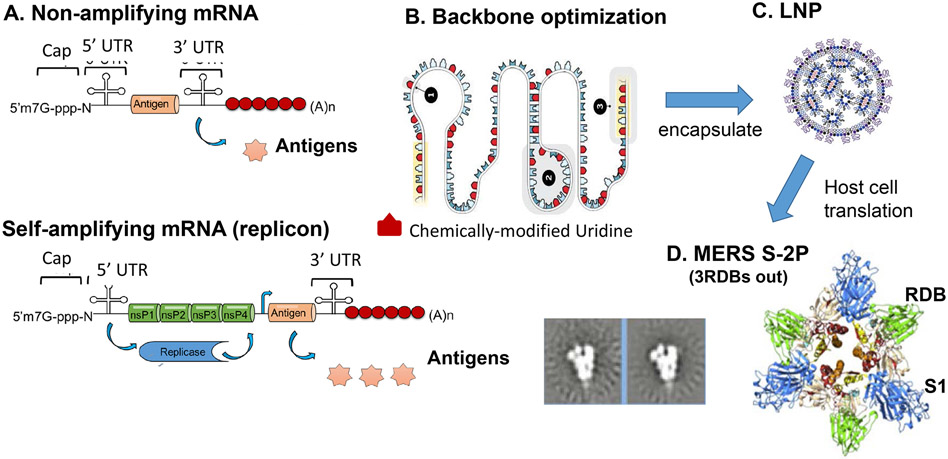
Figure 3. Use of non-amplifying and self-amplifying mRNA (SAM) for expression of stabilized pre-fusion S1 complexes for MERS, SARS-CoV-1 or SARS-CoV-2 in host cells.
A. mRNA vaccines utilize non-amplifying or self-amplifying RNA constructs. Non-amplifying mRNA contains the basic RNA structure as it appears in the viral open reading frame (ORF) for S1.71 The major characteristics of non-replicating mRNA vaccines are: (i) relatively small mRNA size (~2-3 kb); (ii) absence of additional potentially immunogenic viral proteins; (iii) ease of manufacturing; and (iv) facile genetic engineering to accomplish stable antigen expression.71 In contrast, SAM RNA encodes alphavirus replication machinery and 5' capping functions in addition to spike protein sequences. SAM vaccines increase antigen expression for a duration of up to ~2 months.71 On the downside, the replicon is less amenable to tolerating RNA stabilizing synthetic nucleotide modifications and also expresses other viral proteins that may be immunogenic. Reprinted with permission from ref 71. Copyright 2019 Elsevier. B. RNA stability and gene expression are enhanced by modifications of the mRNA cap, poly (A) tail, 5’ and 3’ untranslated regions (UTRs), and the nucleoside bases.207 Multiple sequence elements are also engineered in the 5' and 3' UTRs to impact mRNA stability and expression. In addition, nucleoside substitution of uridine with pseudouridine or 1-methylpseudouridine impacts the stability and adjuvant properties of the RNA. Reprinted with permission from ref 207. "Image by V. ALTOUNIAN/SCIENCE. Copyright 2020 AAAS. C. Lipid nanoparticle (LNP) construction and encapsulation are discussed in Figures 4 and 5.23 Reprinted with permission from ref 23. Copyright 2018 John Wiley and Sons. D. The encapsulated RNA is also engineered to include two proline (2-P) substitutions plus elimination of the furin cleavage site to allow expression of a stabilized pre-fusion complex. This feature allows the exposure of hidden epitopes, which are required for a robust neutralizing antibody response. The schematic shows the crystal structure and negative EM staining of a MERS S-2P complex.6 Reprinted with permission from ref 6. Copyright 2017 The Authors.
Table 2:
Leading mRNA Vaccine Nanoparticles
| Developer | Vaccine name |
Nanoparticle formulation |
Antigen/adjuvancy | Clinical advancement |
|---|---|---|---|---|
| Moderna38-44 | mRNA-1273 (100 μg/dose) |
Lipid nanoparticle, 80–100 nm, composed of the ionizable cationic lipid (designated “H”), PC, cholesterol and PEG (molar ratio 50:10:38.5:1.5) | Nonreplicating RNA, encoding full length S protein in its pre-fusion formation (2P mutation plus intact S1/S2 cleavage site). Uridine-modified RNA provides adjuvancy. | Phases 1–3 completed FDA–EUA |
| Pfizer/BioNtech42-48 | BNT162b2 (plus other experimental variations) (30 μg/dose) |
Lipid nanoparticle, 80 nm, composed of ionizable cationic lipid, ALC-0315 (Acuitas), PC, cholesterol and PEG | Self-replicating RNA coding for full length S protein in its pre-fusion formation (additional variants with non-replicating RNA, expressing RBD that contains a T4 fibritin-derived trimerization domain were also developed and tested). Uridine-modified RNA provides adjuvancy. | Phases 1–3 completed FDA–EUA |
| Imperial College London24 | LNP-nCoV-saRNA (1 μg/dose) |
Lipid nanoparticle (LPNP100), composed of ionizable cationic Acuitas lipid (designated A9), PC, cholesterol and a PEG-lipid | Self-replicating RNA, encoding for full length S protein in its prefusion formation. The plasmid vector for synthesizing the self-amplifying replicon was derived from the Trinidad donkey Venezuelan equine encephalitis virus strain (VEEV) alphavirus | Phase 1/2 |
| Arcturus (Duke/NUS)118 | ARCT-021 (1-10 μg/dose) |
Lipid nanoparticle LUNAR®, composed of 50% ionizable amino lipids (Lipid2.2), 7% PC, 40% cholesterol, 3% dimyristoyl-sn-glycerol and methoxy-polyethylene glycol | STARR™ self-replicating mRNA technology, full length spike protein | Phase 1/2 |
Abbreviations: PC, phosphatidylcholine; PEG, polyethylene glycol; RBD, receptor-binding domain.
The RNA vaccines in Table 2 include the use of nonamplifying or self-amplifying mRNA constructs.15,24,26,71,73 Whereas the Moderna vaccine delivers conventional single-stranded mRNA for spike protein expression, the vaccines developed by Pfizer/Biontech, Arcturus, and Imperial College include self-amplifying positive-sense (RNA+) constructs that borrow expression elements from an alpha virus genome (Figure 3).71 The Imperial College vaccine uses a gene sequence from the equine encephalitis alphavirus that encodes nonstructural proteins assisting RNA capping and replication, and the Arcturas vaccine uses a self-transcribing and replicating RNA (STARR) platform.
The upscale production of RNA constructs for nanoparticle encapsulation begins with large-scale production of linearized plasmid DNA, using E. coli fermentation or enzymatic processes such as doggybone™.23,74 mRNA is produced from DNA templates in bioreactors optimized for in vitro transcription by T7 RNA polymerase and 5' RNA capping with 7-methylguanosine to ensure optimal antigen expression.74,75 Following RNA synthesis, template DNA is digested by DNAse I and RNA is purified by tangential flow filtration (TFF) plus ion exchange chromatography to remove enzymes and impurities. A second TFF processes is used to provide a buffered aqueous RNA solution for sterile filtration. In addition to engineering antigen stabilization, modified nucleosides are used to increase construct stability and to tune immunological “danger signals” (Figure 3). 20-22,76 A frequently used approach is uridine substitution by pseudouridine or 1-methyl pseudouridine.20,22 The delivery of danger signals plays an important role in the self-adjuvanting properties of RNA vaccines, including generation of pro-inflammatory responses.77 Modifications of 5' and 3' untranslated regions of mRNA constructs are also used to control stability and expression.
Nanoparticle Construction to Deliver RNA Vaccines
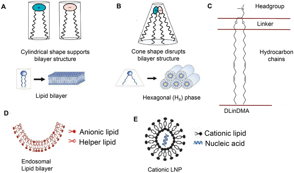
To prevent RNA degradation before delivery to the host translational machinery, an effective packaging system is required. This prerequisite can be accomplished by making use of cationic lipid nanoparticles (LNPs), which were originally developed for nucleic acid delivery for cancer immunotherapy and other vaccine applications (Figure 4).16,19,23,73 The use of cationic lipids effectively condenses mRNA into solid lipid nanoparticles that can be taken up by host APCs through endocytic or phagosomal pathways.71 These processing pathways also facilitate nucleic acid release into the cytoplasm, where the mRNA is expressed.78 The packaging and encapsulation of mRNA was originally developed by the use of ionizable, cationic lipids (such as dioleoyl-3-dimethyl ammoniumpropane, DODAP) which exhibit a high binding affinity for negatively charged RNA (Figure 5).23,78,79 In addition to DODAP, other lipids have been developed using a more flexible hinge region to link amino-lipid head groups to hydrophobic lipid tails.16,73,79-81 Examples include 1,2-dilinoleyloxy-3-dimethylaminopropane (DLinDMA) and dilinoleylmethyl-4 dimethyl aminobutyrate (DLin-MC3-DMA), which, in the presence of helper/structural lipids (e.g., distearoyl-sn-glycerol-3 phosphocholine, DSPC) and cholesterol, lead to the formation of micellar structures in the presence of RNA.79-81 Finally, coating with poly(ethylene glycol) (PEG)-conjugated lipids leads to the formation of nanoparticles with colloidal stability.82
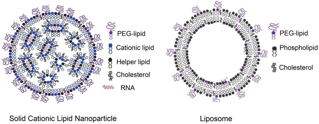
Figure 4. Lipid nanoparticles (LNP) for the encapsulation and delivery of mRNA.
RNA has theoretical advantages over DNA delivery because the RNA payload only has to reach the cytosol instead of making it to the nucleus. Moreover, RNA does not pose the danger of integration into host genomes, is easier to manufacture, and only transiently expressed compared to DNA. Lipid nanoparticles differ from liposomes by the presence of an electron dense core that forms during complexation of cationic lipids to a nucleic acid. Instead, liposomes are composed of a lipid bilayer with an aqueous interior.71 LNP formulations are typically made up of: (i) an ionizable amino-lipid (discussed in Figure 5) for electrostatic complexing to RNA (in red), leading to the formation of hydrophobic inverted micelles; (ii) helper lipids providing structural components that stabilize cell membranes, including zwitterionic lipids (e.g., DOPE) or DSPC; (iii) cholesterol, promoting tight packaging of lipid components; and (iv) a glycol (PEG)-lipid, which provides a surface hydrating layer that improves colloidal stability with reduced protein adsorption.71 Reprinted with permission from ref 23. Copyright 2018 John Wiley and Sons.
Figure 5. The role of cationic lipids in nucleic acid delivery nanoparticles.
A. Membrane lipids normally adopt a cylindrical molecular shape, which accommodates their incorporation into lipid bilayers of endosomal membranes.79 B. However, when cationic and anionic lipids are mixed together, ion pairs form a cross-sectional area in which the head groups occupy a volume that is less than that of the lipid side-chains, which are splayed in cone-shaped fashion.79 This conformation is also known as a hexagonal (HII) lipid phase, which has the capacity to interfere with the lipid bilayer in endosomes upon contact with the LNP (Figure 6).78 C. Ionizable amino-lipids are characterized by a cationic headgroup, a linker, and hydrocarbon side-chains. These cationic lipids have acid dissociation constants (pKas) of less than 7.0, conferring neutral charge at physiological pH (7.4), but converting to a positive charge in acidified (pH <6.0) endosomal compartments.23 Ionizable amino-lipids with a linker group (such as in DLinDMA) serve a number of purposes: (i) nucleic acid entrapment with high encapsulation efficiency; (ii) maintenance of a neutral particle surface charge under physiological pH conditions, such as in the interstitial and lymphatic fluids; (iii) endosomal escape under acidic, intracellular conditions, as explained in Figure 6.23 D and E. The planar lipid bilayer of the endosomal membrane is composed mostly of anionic and helper lipids plus cholesterol, which differs from the hexagonal lipid phase, displayed by the cationic lipid nanoparticles (also see Figure 6). A, B, and C reprinted with permission from ref 79. Copyright 2019 Springer Nature. D and E adapted with permission from ref 78 under the terms of the Creative Commons Attribution 3.0 License. Copyright 2012 The Authors.
In addition to their role in RNA complexation, cationic lipids also contribute to the assembly of a hexagonal (HII) lipid phase in the nanoparticles (Figure 5).79,81 The hexagonal phase is the result of binding interactions between cationic head groups and anionic bystander lipids. This phase leads to the formation of cone-shaped lipid structures, in contrast to the more cylindrical lipid assembly found in the planar lipid bilayers that compose endosomal membranes.79 Under acidifying endosomal conditions, lipid exchanges take place between the particle HII lipid phase and the endosomal membrane (Figure 6).78 The consequence of mixing particle and endosomal membrane lipids is the release of mRNA into the cytosol. This release may be assisted by an increase in hydrostatic pressure in the endosomal compartment as a result of the cationic lipids turning on the proton pump, leading to an influx of H+, Cl− and H2O.78 Thus, the efficacy of mRNA release is directly related to the pKa of the cationic head group.79,83 Cationic head groups may also contribute to the generation of immune danger signals by TLRs that are sensitive to surface charge.
Figure 6. RNA delivery to the cytosol.
The prevailing hypothesis is that the hexagonal lipid phase in nanoparticles within endosomes catalyzes lipid exchange that destabilizes the composition of the anionic endosomal membrane.78 The suggested flow of events is that lipid nanoparticle (LNP) entry into endosomes is followed by electrostatic interaction of cationic, ionizable lipids in the LNP with the anionic lipids in the endosomal membrane. This interaction leads to lipid exchange and fusion of the particle with the endosomal lipid phase, followed by nucleic acid escape to the cytoplasm, possibly assisted by the endosomal proton pump, which is activated by cationic lipids. Every proton leads to the importation of a chloride and water molecule, leading to increased hydrostatic pressure. Reprinted with permission from ref 78 under the terms of the Creative Commons Attribution 3.0 License. Copyright 2012 The Authors.
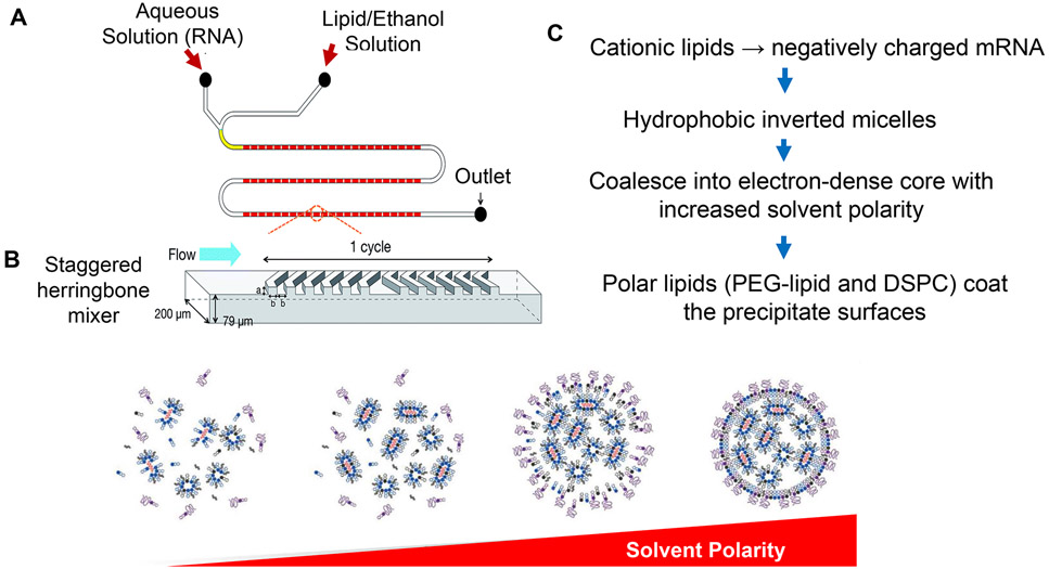
Although LNP synthesis can be accomplished by sonicating lipid suspensions in a buffered aqueous solution, efficient industrial-level synthesis of RNA nanoparticles is based on a scaled-up process involving microfluidics mixing, as outlined in Figure 7.23 Mixing of the RNA-containing aqueous phase with the ethanol-suspended lipid phase is accompanied by a progressive increase in solvent polarity, which acts as a driver for nanoparticle assembly. Assembly commences with the formation of hydrophobic inverted micelles when the cationic lipids are complexed to negatively charged RNA. A further increase in solvent polarity leads to the coalescence of lipid particle cores, followed by coating with PEGylated lipids and DSPC.23 The ultimate outcome is the formation of nanoparticles with electron-dense cores, surrounded by a PEGylated lipid monolayer (Figure 4).
Figure 7. Upscale synthesis process for manufacturing lipid nanoparticles (LNPs).
A. The microfluidic process is premised on an ethanol injection approach, which leads to precipitation of dissolved lipids when mixed with an aqueous solution in a microfluidic device. In this example, the device is equipped with a staggered herringbone micromixer.208 Reprinted with permission from ref 208 under a Creative Commons Attribution-Non Commercial 3.0 Unported License. Copyright 2015 The Royal Society of Chemistry. B and C. The assembly of the nanoparticle components is driven by the increase in solvent polarity with introduction of the aqueous phase.23 The first interaction during the mixing of ethanol with the aqueous stream is binding of the cationic lipid with the negatively charged nucleic acid to generate hydrophobic, inverted micelles. With further increase in solvent polarity, the hydrophobic micelles coalesce into nano-precipitates that generate the LNP core. With continuous mixing, more polar lipids (such as PEG-lipid and DSPC) coat the surface of the precipitates, resulting in particles with electron-dense cores surrounded by a lipid monolayer.23 Reprinted with permission from ref 23. Copyright 2018 John Wiley and Sons.
DNA Delivery Platforms for COVID-19 Vaccination
The premier delivery platforms for DNA leading to the expression of SARS-CoV-2 spike protein are adenovirus-vectored vaccines, which fall outside the scope of this review (Table 1). Other examples of DNA delivery are the vaccines being developed by Entos Pharmaceuticals, Mediphage Bioceuticals, Zydus Cadila, and Inovio Pharmaceuticals.39,84,85 Entos Pharmaceuticals developed a proteolipid vesicle (PLV) for nucleic acid delivery, premised on Fusogenix nucleic acid transfer technology.86 These neutral lipid vesicles display a proprietary fusion-associated small transmembrane (FAST) protein that catalyzes lipid exchange between the PLV and the host cell plasma membrane. This highly efficient fusion of the vesicle with the lipid membrane results in delivery of the nucleic acid payload by a process that bypasses the endocytic route. To date, leading CoV-2 vaccine candidates have shown robust efficacy and safety in preclinical animal studies and have progressed to the clinical trial stage in humans. Utilizing a Ministring platform comprising mini-linear DNA vectors that encode the gene of interest plus eukaryotic expression elements (devoid of unwanted bacterial sequences), Mediphage Bioceuticals has developed virus-like particles (VLPs) for delivery of SARS-CoV-2 DNA sequences.87 This vaccine platform is in preclinical development.
In addition to nanoparticles, some companies have developed plasmid-based technologies for electroporation-driven DNA delivery, with the prospect of providing SARS-CoV-2 vaccination through the expression of spike protein. This includes the “plug-and-play” plasmid delivery platform (INO-4800) from Inovio Pharmaceuticals, which is injected intradermally, followed by electroporation that involve the company’s CELLECTRA® device.84 To date, this vaccine effort has demonstrated efficacy and safety in preclinical studies and has advanced to a phase 2 clinical trial in humans. Zydus Cadila has also developed a DNA plasmid, ZyCoV-D, that has performed well in preclinical studies and recently advanced to a phase 3 vaccine trial in humans.
Nano-Enabled Protein Subunit/Peptide Vaccines and Virus-Like Particles
An alternative approach to employing nanoparticles for CoV-2 vaccine development is to use full length spike protein or viral subunits for encapsulation or self-assembly into nanoparticles. The value proposition of subunit vaccines is that nanoparticles enhance the efficacy of cargo delivery to endosomal APC compartments, from which the proteolytically cleaved peptides are transported to (i) the surface of APCs by vesicles containing type II major histocompatibility complexes (MHC-II); and (ii) the cytosol, from where the peptides are imported to vesicles that transport MHC-I complexes to the surface of most body cells.88 As a result, MHC-I-mediated peptide presentation to CD8+ T-cells is enabled, with the possibility of triggering the development of cytotoxic T-cells that kill virus-infected cells. In contrast, antigen presentation by MHC-II on APC surfaces leads to activation of different CD4+ lineages that will be discussed later. The efficacy of subunit vaccines is boosted by the inclusion of independent adjuvants into the nanoparticle.
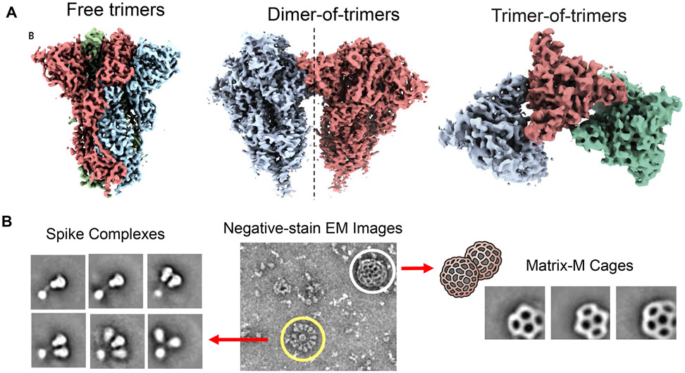
The current front-runner for protein-based antigen presentation efforts is the Novavax vaccine, which recently entered phase 3 clinical trials.39,61,89 NVX-CoV2373 is a self-assembled nanoparticle vaccine derived from the recombinant expression of full-length S protein by a Baclovirus expression system in moth cells (Figure 8).61 The expressed 2-P full-length S protein spontaneously assembles as pre-fusion complexes in the presence of Sorbitol 80 to form free trimers, dimers-of-trimers, trimers-of-trimers, or multitrimer rosettes (up to 14 trimers, Figure 8). Tight clustering of the spike proteins increases immunogenicity as demonstrated for other type I fusion proteins, e.g., influenza hemagglutinin. The Novavax vaccine includes a proprietary adjuvant, MatrixM™,90 which consists of 40 nm honeycomb-like nanoparticles derived from plant saponins, mixed with cholesterol and a phospholipid (Figure 8). A significant advantage of the NVX-CoV2373 formulation is transportability and above-freezing storage temperatures (35–46 °F), compared to the deep freeze requirements of the mRNA LNP vaccines.
Figure 8. COVID-19 vaccine development through self-assembly of stabilized, full-length SARS-CoV-2 S subunits into nanoparticles.
A. Novavax developed a full-length S protein subunit vaccine that is expressed as a stabilized protein with mutational deletion of the furin cleavage site plus 2 proline (2-P) substitutions (K986P and V987P) that confer an "RBD-up" conformation.61 This leads to expression of SARS-CoV-2 3Q-2P spike protein as a pre-fusion complex. When reconstituted in Polysorbate 80 (PS 80), the protein adopts tertiary structures that include free trimers, dimers-of-trimers, trimers-of-trimers or multi-trimer rosettes (with as many as 14 trimer transmembrane domains being enclosed in micellar PS 80 cores).61 Moreover, the vaccine is further reconstituted with the Matrix-M adjuvant. Reprinted with permission from ref 61. Copyright 2020 AAAS. B. Negative stain electron microscopy of the full-length spike (reconstituted in PS 80), admixed with the cage-like Matrix-M component (from plant origin).61,209 The spike rosettes are circled in yellow and Matrix-M adjuvant cages are circled in white.90 The imaging confirms the presence of trimeric spike as free trimers or as multitrimer rosettes. Matrix-M does not appear to interact with the spike nanoparticles. Reprinted with permission from ref 209. Copyright 2020 AAAS.
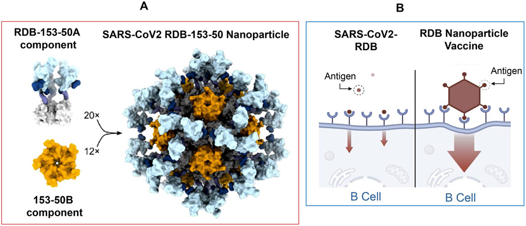
An elegant demonstration of the use of protein self-assembly to generate a SARS-CoV-2 vaccine was provided by Walls et al., who constructed a self-assembling nanoparticle that mimics RBD expression on the viral surface (Figure 9).72 Using a structure-based in silico design, a two-component nanoparticle was derived through the self-assembly of 60 RBD fusion protein subunits on the exterior surface of an icosahedral subunit, obtained by self-assembly of 120 computer designed components (panel B). The multivalent display of RBD subunits enabled generation of neutralizing antibody levels that were 10 times greater than antibody titers obtained with stabilized spike complexes, delivered at a 5-fold higher dose to animals.72 It is tempting to speculate that multivalent display of RBD subunits instructs the development of highly effective neutralizing antibodies capable of crosslinking spike proteins on the SARS-CoV-2 surface. The scalability of this platform introduces a further variation of structure-based vaccine design to be considered in future vaccine efforts.
Figure 9. Self-assembly of an in silico-designed nanoparticle protein vaccine that displays the receptor-binding domain (RBD) as a highly immunogenic array that mimics natural virus structure.
A. Utilizing a structure-based in silico vaccine design approach, a self-assembling protein nanoparticle vaccine with resemblance of viral morphology was developed.72 Each particle is composed of two components, an icosahedral 120 subunit core (153-50B) that supports binding to 60 SARS-CoV-2 RBD domains (RBD-153-50A), conjugated to a 153-50B-interactive domain. B. The nanoparticles roughly resemble a virus, which may account for their enhanced ability to provoke a diverse, highly potent, and protective antibody responses during animal testing.72 The lead nanoparticle vaccine candidate is being manufactured for clinical trials. Reprinted with permission from ref 72 under a Creative Commons CC-BY license. Copyright 2020 Elsevier.
In addition to Novavax, Sanofi Pasteur/GSK, Vaxine, and Clover Biopharmaceuticals/GSK/Dynavax are engaged in SARS-CoV-2 subunit vaccine efforts that have progressed to phase 1 clinical trials. These vaccines make use of adjuvants developed by GSK, Dynavax, and Vaxine. The GSK adjuvant (AS03) is composed of α-squalene and polysorbate 80 in an oil-in-water emulsion,91 while the Dynavax adjuvant (CpG 1018)92 consists of a 22-mer oligonucleotide that interacts with TLR9. The Vaxine adjuvant (Advax) is a microparticle composed of delta-inulin polysaccharides.93 Adjuvant choice is important in instructing the type of helper T-cell responses that are key to the attainment of vaccine efficacy and safety, as discussed below.
An alternative approach for developing subunit vaccines is to use VLPs, which are constructed by the self-assembly of capsid proteins from mammalian, insect or plant viruses.39,58,94,95 The production of VLPs is accomplished by fermentation processes or the use of molecular farming techniques. Medicago96 and iBio/CC-Pharming97 produce tobacco plant virus particles that include the CoV-2 S protein, and AdaptVac uses their ExpreS2ion vector for expressing the S2 subunit in insect cells.98 The Medicago vaccine, which utilizes the GSK adjuvant, has entered clinical trials, while the iBio/CC-Pharming and AdaptVac VLPs are in preclinical development (Table 1).
Another vaccine category making progress is epitope-based vaccines, which could rely on nanoparticle delivery of individual or epitope combinations. Currently, the primary goals of these particles are to enhance T-cell responses and to obtain improved cooperation between T- and B-cells for boosting the production of neutralizing antibodies.3,30,85,99-105 Peptide-based vaccines hold several advantages over conventional subunit vaccines, including low production cost, no need for microbial cultures, high specificity, high stability, and easy scalability. On the downside, the genetic variation of the human leukocyte antigen (HLA) allele gene pool in the population could mean that individual T-cell epitopes are not equally effective in every person, necessitating selection tools to identify promiscuous epitopes for MHC-II presentation to CD4+ T-cells or for MHC-I presentation to CD8+ T-cells.3,30,64,99,103 The ability to design new peptide-based vaccines has received a major boost from the availability of epitope-mapping tools such as the Immune Epitope Database (IEDB) for predicting B- and T-cell epitope sequences (Table 3).106,107 In addition, a number of immuno-informatics approaches have emerged to facilitate the prediction-making process for multi-epitope vaccine design,30,64,65,94,100,102,104,108 including computational tools that predict epitope binding to diverse HLA alleles (e.g., the IEDB Population Coverage Tool).107 It is also possible to use HLA ligandome analysis for calculating the probability of peptide interactions with consensus motifs, including 3D predictions for viral peptide docking to cytokine-inducing receptors (e.g., TLR2 and TLR4).65,109 Molecular dynamics simulations are used for molecular in silico predictions of vaccine antigenicity (Table 3).109 Collectively, these tools now enable multi-epitope vaccine design to test the feasibility of obtaining cooperative B- and T-cell interactions, as well as possibly soliciting specific CD4+ and CD8+ T-cell responses (discussed in in a later section).30,64,65,102,104,108,110
Table 3:
Epitope Identification and Selection for Vaccine Development
| Epitopes Selection Tools | Refs |
|---|---|
- Prediction of linear and conformational B-cell epitopes - Population coverage (consideration of HLA allele heterogeneity) |
203, 106, 107, 3, 30, 140, 30, 66, 67, 204 3, 30, 64, 99, 101, 103, 109 |
| - Conservancy analysis - Epitope distribution analysis |
65, 101 |
| Immunoinformatics Tools to Design Epitope-Based Vaccines | 30, 64, 65, 94, 100, 102 |
|
205 |
| Rationale for Epitope Selection | |
|
32 |
| 99, 100, 155 | |
| 103 | |
| 109 | |
| 102, 154 |
Abbreviations: HLA, human leukocyte antigen; TLR, Toll-like receptor
Although several peptide vaccines have been constructed and are in preclinical development, comparatively few CoV-2 peptide vaccines have advanced to the clinical stage. One example is EpiVacCorona, which was developed in Russia and is composed of synthesized SARS-CoV-2 peptide antigens, conjugated to a protein carrier that is adsorbed onto an aluminum-containing adjuvant.111 This vaccine has advanced to phase 1 and 2 clinical trials, and has demonstrated the ability to induce protective immunity following intramuscular injection. The second example is a peptide vaccine (UB-612) developed by COVAXX plus United Biomedical, that has entered phase 2 and 3 clinical trials.112 UB-612 is composed of 8 components and was designed to induce a combination of neutralizing antibodies plus T-cell responsivity through the inclusion of a S1-RBD-sFc fusion protein, 6 synthetic peptides (one universal plus 5 SARS-CoV-2-derived peptides), a proprietary CpG oligonucleotide (TLR-9 binding agonist), and an aluminum phosphate adjuvant. Vaccination studies in guinea pigs and rats demonstrated the generation of high titers of neutralizing antibodies against S1-RBD, robust cellular immunity, and TH1 skewing of the immune response. Subsequent challenge studies in a nonhuman primate animal model confirmed disease prevention and reduction of viral load. The third example is the development of the IMP-CoVac-1 vaccine by IMV Inc., which has entered a Phase 1 clinical trial.113 This vaccine is based on the use of a lipid-based DPX platform that can be formulated with peptide antigens, which are capable of activating B- and T-cell responses. The vaccine can be stored for extended time periods in a dry form and is easy to reconstitute for injection. In addition to these examples, new vaccine products that are in preclinical development include a vaccine by Oncogen that incorporates synthetic peptides that mimic S and M protein epitopes,25,114 a vaccine by Vaxil Corporation that utilizes a signal peptide technology, an adjuvanted microspheric peptide platform by FlowVax, and the Ii-Key hybrid peptide platform developed by EpiVax Inc. in collaboration with Generex Biotechnology Corp.113
Short-Term Efficacy Studies Confirm the Immunogenicity of Vaccinating Nanoparticles.
Despite being the first of their kind, the RNA vaccines developed by Pfizer/BioNTech and Moderna represent the fastest vaccine development attempts ever, replacing the previous record of 4 years held by the mumps vaccine. In addition to the authorization of these vaccines by the U.S. Food and Drug Administration (FDA), the Arcturas Therapeutics and Imperial College vaccines are poised to advance to phase 3 clinical trials at the time of writing of this Perspective (Table 2).
Prior to commencing human clinical trials, the antigen-specificity and immunogenicity of mRNA vaccine candidates were confirmed by animal studies, which demonstrated that dose-dependent neutralizing antibody responses to the S protein are capable of reducing lung infection and viral loads of mouse-adapted SARS-CoV-2 strains.38,40,44,47,115 Moreover, antibody subclasses recognizing the S protein showed dominance of the IgG2a subclass over IgG1, which is an indirect reflection of the differential triggering of T-helper 1 cell (TH1) versus T-helper 2 cell (TH2) immune responses.44 Whereas TH1 cells facilitate IgG2a class switching, TH2 cells favor IgG1 class switching. This notion was corroborated by the dominance of IFN-γ versus IL-4 production by splenocytes from immunized animals; IFN-γ is produced by TH1 cells, while IL-4 is produced by TH2 cells.44 The importance of obtaining TH1 dominance is important for the prevention of side effects, as observed for MERS and SARS-CoV1 vaccines. Additional preclinical studies in nonhuman primates confirmed the generation of high titers of neutralizing antibodies capable of preventing SARS-CoV-2 infection in the upper and lower respiratory tracts.38,47 These studies also assisted in dosimetry development and demonstrating that the intramuscular route of administration generates sufficient inflammatory effects to assist the recruitment of APCs and antigen presentation in regional lymph nodes.21,116,117
In addition to the success of the FDA-approved vaccines, Arcturus (ARCT-021) demonstrated effective induction of protective antibody levels in primates after a single injection of the mRNA-delivering LNP.118 Interestingly, this vaccine also provided protection to immune-deficient animals that were depleted of B-cells, but not to mice depleted of CD8+ T-cells, demonstrating the independent contribution of cellular immunity in preventing SARS-CoV-2 infection.118
The success of first-generation vaccines in preclinical studies was duplicated by observations of protective immunity in human clinical trials, which have been extensively reviewed elsewhere (Table 2).38,40-43,45-47,50,119 In brief, all mRNA vaccines subjected to large-scale clinical trials have proven to be effective in generating high protective antibody titers against the S protein and its RBD in humans. In fact, the IgG titers to RBD or the S protein are frequently higher than antibody titers in convalescent sera of subjects recovering from natural COVID-19 infections. In addition, humoral immune responses were generally accompanied by evidence of CD4+ and CD8+ mediated T-cell immunity, including evidence of a TH1 skewed immune response (e.g., IFN-γ production). An unprecedented milestone was the demonstration that two rounds of intramuscular injection with the Moderna and Pfizer/BioNTech vaccines were ~95% effective in preventing symptomatic disease (Table 2). A recent non-peer-reviewed update from Arcturus also demonstrated favorable immunogenicity and tolerability in phase 1 and 2 human studies, with all vaccinated subjects showing high antibody titers.118
Preclinical studies with the Novavax vaccine in mice and nonhuman primates also demonstrated the generation of high SARS-CoV-2 neutralizing antibodies, along with strong B- and T-cell responses.61,89 Subsequent phase 1 and 2 clinical trials confirmed the generation of vaccine-induced antibody titers that exceed the immunoglobulin titers after natural infections.120 The vaccine has recently entered phase 3 clinical trials.
Among VLPs, the Medicago vaccine has entered phase 2/3 clinical trials, while the iBio/CC-Pharming and AdaptVac VLPs are still in preclinical development (Table 1).96,97 Medicago recently reported unpublished data indicating that 100% of human test subjects receiving the adjuvanted vaccine develop protective humoral and cellular immunity after two doses.96
All considered, the above data are indicative of a high success rate for CoV-2 RNA and subunit vaccines, now allowing comparisons of vaccine-induced versus natural immunity. We can now test the hypothesis that the generation of immunity against the spike protein may suffice in achieving herd immunity and be capable of bringing the pandemic under control. To succeed, the current vaccine drive has to overcome the logistic challenges of vaccinating a sufficient number of people to reach this level of protection. It is also urgent to complete this task in as short a time interval as possible to control the global viral burden, which contributes to the generation of potential immune escape viral variants.
Critical Safety Considerations for the Development of SARS-CoV-2 Vaccines
A key consideration for COVID-19 vaccine development is safety, with an emphasis on avoiding adverse outcomes encountered during experimental MERS and SARS-CoV-1 vaccination studies.47,50,95,115,121-124 These studies demonstrated two major adverse response categories, namely, (i) antibody-mediated disease enhancement (ADE), and (ii) the occurrence of a skewed cellular immune response in which eosinophil recruitment resulted in severe lung damage at the time of viral challenge in vaccinated animals.122 Eosinophil lung damage was also observed during development of the RSV vaccine (which includes a type I fusion protein) in humans.95,115,121,125,126 The basis for ADE was ascribed to a non-neutralizing antibody response to MERS and SARS-CoV-1, which, instead of interfering in viral uptake, was accompanied by accelerated Fcγ-mediated uptake of antibody-bound viral particles by host phagocytic cells.122,127 As a result, the internalized virus triggered innate immune responses that led to pathological levels of cytokine and chemokine production. In contrast, eosinophil-mediated lung damage was ascribed to the development of an unbalanced TH2-mediated immune response, which is characterized by IL-4, IL-5, and IL-13 production, leading to eosinophil recruitment and lung damage.95,128,129 TH2 skewing of the immune response also favors IgGi class switching by IL-4. Postulated reasons for the TH2 skewing of immune responses by some MERS or SARS-CoV-1 vaccines include antigen selection and the use of TH2 adjuvants (e.g., alum).123,129
The take-home message from prior vaccine efforts was to focus on the use of the spike protein or subunits as the major immunogenic target, plus the use of TH1-skewing adjuvants.93,123 In addition to relying on the intrinsic adjuvant properties of the RNA in nucleic acid vaccines, independent adjuvant use was introduced during development of VLP and subunit vaccines (e.g., MatrixM, Advax, the GSK adjuvants, STING agonists).124 The TH1 skewing effects of mRNA vaccines were confirmed by phase 1 (safety) clinical trials in humans.40,45 Clinical trials demonstrated that leukocytes from vaccinated human subjects predominantly produce TH1 (e.g., INF-γ, IL-2, TNF) compared to TH2 (e.g., IL-4, IL-13) cytokines. Moreover, no evidence for ADE or eosinophilic immunopathology were observed in phase 3 studies. When side effects did occur following administration of the Moderna or Pfizer vaccines, they were predominantly characterized by mild or moderate symptoms.29,40 The most frequently experienced side effects were localized pain at the injection site, occasional low-grade muscle and joint pain, fatigue, headaches, fever and chills. Ultimately, their combined safety features plus the demonstration of ~95% efficacy in the clinical trials led to the Moderna and Pfizer/BioNTech vaccines obtaining EUA from the FDA.46
Although mild to moderate side effects continue to be the predominant experience during population vaccination efforts, a handful of subjects have reported more serious allergic side effects,130-133 such as the development of anaphylaxis, characterized by a severe drop in blood pressure, breathing problems, wheezing, and tissue swelling. This condition was infrequently observed in earlier clinical safety studies, which excluded people with serious allergic disorders from participating. Although the exact cause of the anaphylaxis is still a matter of debate, the Centers for Disease Control and Prevention (CDC) advises people with a severe allergic reaction to ingredients in the RNA vaccine not to take the injection.134 This recommendation includes not taking a second vaccine dose if there was a severe allergic reaction to the first dose. People who experience immediate allergic reactions to any other vaccine or injectable therapeutics are advised to seek medical attention before considering vaccine administration. In contrast, the CDC recommends that people who experience severe allergic reactions not related to vaccines or injectable medications (e.g., environmental, food, pet, or latex allergies) to get vaccinated. The same advice applies to people with a history of allergies to oral medications or a family history of severe allergies.130
In considering possible ingredients that may contribute to anaphylactoid RNA vaccine responses, a potential role for PEG has been noted.130 Poly(ethylene glycol) is often used for coating nanoparticle surfaces to provide colloidal stability. Different molecular weight PEGs are also used as softeners or moisture carriers in consumer products such as toothpaste and shampoo, and may also be included in biopharmaceuticals and laxatives. Although it has been documented that PEG is capable of generating IgM or IgG antibody responses that may lead to complement activation at the particle surface, an alternative opinion is that PEGylated nanoparticles (e.g., Doxil) may trigger a nonspecific complement activation-related pseudoallergic (CARPA) disorder.131 Yet another twist to the story is the discovery of IgE antibodies to PEG or PEGylated drugs that may play roles in anaphylaxis. However, there is also a group of experts who doubt that PEG is involved in anaphylaxis because of its low content in RNA vaccines.133
The best practical approach for at-risk people is to follow the CDC guidelines. It is also recommended that all subjects receiving the vaccine wait 30 min after administration to monitor severe side effects, which usually occurs within the first 15 min. Although it is possible to develop serological screening assays to detect PEG antibodies, the best advice for at-risk people not able to take RNA vaccines is to consider an alternative vaccine formulation that excludes PEG. If no alternative is available, it is theoretically possible to use a prophylactic cocktail composed of injectable H1 and H2 histamine receptor blockers plus dexamethasone, to reduce the severity of the adverse responses, such as used for people with severe reactions to radiocontrast media.132 However, this strategy would require consultation with an allergist and performance of the procedure in an appropriate healthcare setting.
Duration of the Immune Response to COVID-19 and Use of Nano-Enabled Approaches to Enhance Vaccine Durability
How long does the protective immune response to COVID-19 last? The current expectation is that neutralizing antibody responses to spike will be of sufficient duration to bring the pandemic under control by providing herd immunity. Although there is good evidence that front-runner nucleic acid, subunit, and viral-vectored vaccines provide protective immunity against COVID-19 that lasts for at least 8 months, we are uncertain about the longer-term duration and completeness of the neutralizing antibody response.41,43 To address this question, we briefly review what is known about the development of protective immunity after coronavirus infections. The neutralizing antibody response to seasonal (“cold”) coronaviruses is of transient duration, allowing the occurrence of reinfections.33 In contrast, the protective antibody responses to SARS-CoV-1 and MERS lasted a minimum of 2–3 years after recovery. The current observation period has been insufficient for the evaluation of vaccine durability against SARS-CoV-2, but early studies performed on convalescent patient sera provide some clues. One study calculated the half-life of anti-RBD antibody decline to be ~ 36 days;135 however, other studies failed to observe a decline over observation periods of 4–7 months.136,137 These variations could reflect differences in the severity of infection, because it has been demonstrated that people with milder infections generate lower antibody titers that decline more rapidly.138 Severity of infection may also explain why the more virulent SARS-CoV-1 and MERS viruses generated more durable immunity.
Longitudinal studies to determine the duration of the protective response after natural infection or vaccination are ongoing and of great importance for establishing future public health policies. An early predictor of what may happen comes from a comprehensive study looking at SARS-CoV-2 specific CD4+ and CD8+ T-cell responses as well as antibody levels in a cohort of 188 individuals over a time period of 8 months.139 This cohort includes people with a variety of disease severities, ranging from mild to severe, as typically observed across the United States. Neutralizing IgG antibody titers against the Spike protein and RBD remained relatively stable, with only a modest decline over 6–8 months. Spike-specific memory B-cells increased during this time span, whereas memory CD4+ and CD8+ T-cells declined with estimated half-lives of 3–5 months. The decay kinetics of memory T-cell responses after COVID-19 are similar to the vaccination response to the yellow fever virus, which is known to confer long-lasting immunity.29 Although precise correlates of protection against secondary SARS-CoV-2 infection and disease are unknown, the presence of durable cellular and humoral responses in the majority of subjects being studied over 8 months suggests that most individuals recovering from COVID-19 will have significant protection against further disease. These findings are further strengthened by the observations of Peng et al., who recently noted that disease severity may determine the type of T-cell response.31 Thus, whereas activated CD8+ T-cells tended to dominate in mild infections, CD4+ T-cells dominated after severe infections. Moreover, Grifoni et al. demonstrated good correlation between the appearance of spike-specific T-cells and antibody responses.140
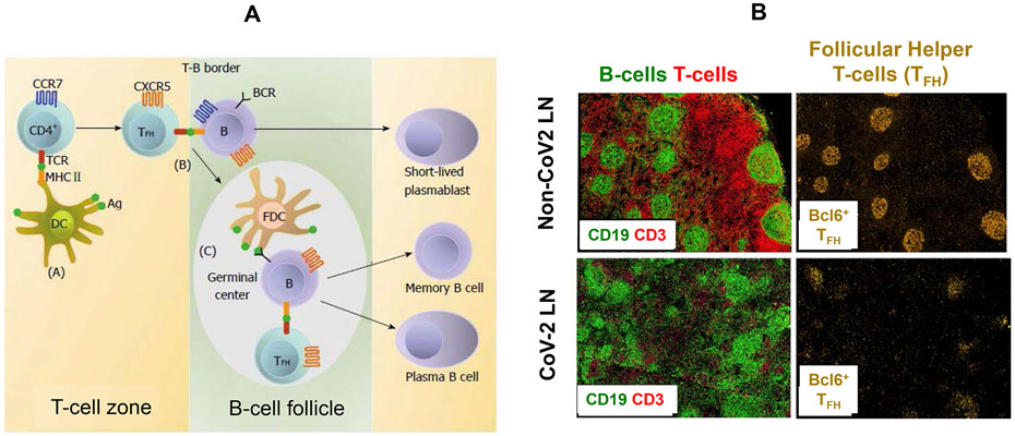
How do CD4+ T-cells contribute to the generation of durable humoral immunity and how can this cooperation be exploited during vaccine development? To get to the bottom of this question, it is important to understand how B-cells are activated in a micro-anatomical lymph node compartment known as the germinal center. Here, a memory CD4+ subset, known as follicular helper T-cells (TFH) cells, cooperate with germinal center B-cells to improve their survival, proliferation, immunoglobulin-class-switching ability, and somatic hypermutation (Figure 10A).21,28,141 This improvement culminates in the production of antibodies with high diversity and affinity by B-cell precursors that ultimately differentiate into plasma cells and long-lived memory B-cells. Moreover, it has been demonstrated that that there is a critical requirement for TFH cooperation with germinal center B-cells in the development of durable immunity to polio, smallpox, and other viral vaccines. Similar cooperativity is likely required for effective COVID-19 vaccination, as indicated by a postmortem study conducted by Kaneko et al. in 15 SARS-CoV-2 infected subjects succumbing to serious disease with high viral loads.27 Of particular significance, there was a severe disruption of thoracic lymph node architecture, compared to 29 age-matched controls succumbing to non-COVID-19 related causes (Figure 10). Not only did the COVID cases demonstrate a ~70% decline in circulating B- and T-cells, but histological analysis backed by immunohistochemistry staining demonstrated comparable reduction in the number of germinal center B-cells and accompanying Bcl-6+ TFH cells.27 These findings are in agreement with the low levels of somatic hypermutation in 403 monoclonal antibodies that were developed from B-cells recovered from the blood of convalescent subjects by Brouwer et al.142
Figure 10. Lymph node germinal centers—key to memory B-cell development—are disrupted by COVID-19.
A. Naïve CD4+ T-cells are activated in the T-cell zone by antigen-presenting dendritic cells (DC).141 This includes the generation of follicular helper T lymphocytes (TFH), which upregulate CXC-chemokine receptor 5 (CXCR5) expression before activating B-cells at the T-B border. Both cell types migrate to B-cell-follicles to form germinal centers.141 Active cooperation of TFH with germinal center B-cells promotes immunoglobulin hypermutation, class switching and affinity maturation, during which B-cells differentiate into memory cells and long-lived plasma cells. (FDC: Follicle dendritic cell; BCR: B-cell receptor; MHC: major histocompatibility complex; TCR: T-cell receptor). Reprinted with permission from ref 141 under a Creative Commons Attribution-Noncommercial (CC BY-NC 4.0) License. Copyright 2014 The Authors. B. Acutely ill COVID-19 patients carrying high viral loads exhibit a striking absence of germinal centers, with a marked reduction of germinal center B-cells and TFH cells.27,210 Although there is robust activation of nongerminal center B-cells, this does not give rise to long-lived memory B-cells or the production of high affinity antibodies. Reprinted with permission from ref 27. Copyright 2020 Elsevier.
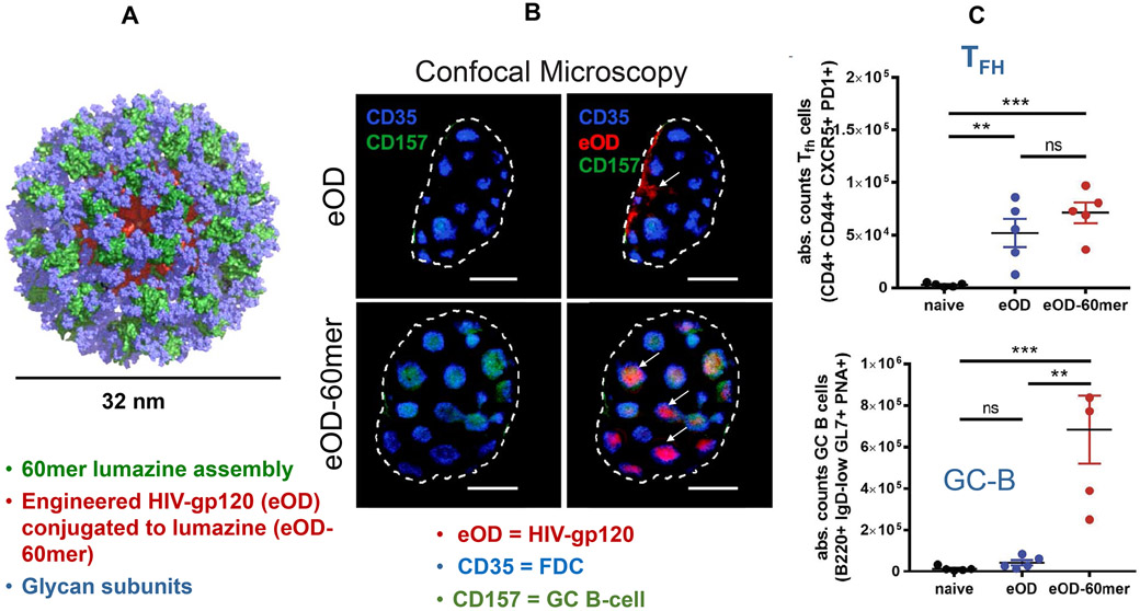
Are germinal centers involved in the development of antibody responses induced by SARS-CoV-2 vaccination? During the use of mRNA delivering vaccines against Zika virus, influenza hemagglutinin, and HIV-Env in mice and primates, Parodi et al. demonstrated that administered lipid nanoparticles induce robust cooperation of TFH cells with germinal center B-cells.21,143,144 Similar effects were observed during the vaccination of rhesus macaques against influenza by Lindgren et al., who also used nucleic-acid-carrying nanoparticles that could improve antibody production in lymph node germinal centers.145 These findings suggest that the same impact may be achieved by mRNA-delivering LNPs during SARS-CoV-2 vaccination. Should that not be the case, a promising approach for augmenting memory B-cell responses in COVID-19 could be to develop vaccinating nanoparticles that improve antibody production in germinal centers. An example of how that might be accomplished is depicted in Figure 11, in which HIV-gp120 was encapsulated in self-assembling nanoparticles that form during the polymerization of a highly glycosylated bacterial protein, conjugated to a gp120 fusion protein.146 Following intravenous (IV) injection of these glycan-rich nanoparticles, the polysaccharide ligands on the particle surface were shown to bind to manose-binding protein present in host serum, which led to complement activation and, subsequently, particle recruitment to germinal centers. As a consequence, a high titer of neutralizing antibodies was generated to the encapsulated, compared to the monomeric, antigen. Similar success was achieved by glycosylated nanoparticles delivering an HIV-gp160 (Env) trimer as well as influenza hemagglutinin.146 These findings suggest that the level of particle or antigen glycosylation could be a means of boosting the durability of neutralizing antibody responses by vaccines. Additional approaches for improving humoral immunity through the creative design of lymph node targeting nanoparticles were recently reviewed by Irvine and Read.147
Figure 11. Use of nanoparticle glycosylation to target lymph node germinal centers.
Particulate HIV immunogens are more capable of activating low-affinity germline precursor B-cells than monomeric antigens, in addition to promoting TFH cooperation with germinal center B-cells.146 This capability can be used for nano-enabled enhancement of neutralizing antibody responses. A. The engineered outer domain (eOD) of the soluble HIV-gp120 monomer was formulated into ~32-nm nanoparticles by fusion to a glycan-rich bacterial protein, lumazine synthase, which self-assembles into a 60-mer (eOD-60mer).146 B. Confocal microscopy of a lymph node showing that soluble monomers enter the subcapsular sinus of the lymph node but do not gain access to germinal centers (darker blue). During challenge with monomers, the centers exhibit sparse follicular dendritic cells (FDC) (lighter blue) and B-cells (green). However, administration of the eOD-60mer demonstrates access to the germinal center, loaded with FDC and B-cells.146 The mechanism of germinal center recruitment has been ascribed to glycans on the particle surface binding to mannose-binding protein. This signal triggers complement activation, which supports polymerized antigen access to the germinal center.146 C. Quantitative expression of the number of TFH and B cells in the germinal centers. Reprinted with permission from ref 146. Copyright 2019 AAAS.
Vaccine Development to Boost T-Cell Contributions to Neutralizing Antibody Production and Cell-Mediated Immune Defense
Although initial efforts focused on achieving protection by generating neutralizing antibodies, a challenge for COVID-19 vaccine developers is uncertainty about the immunological correlates of vaccine efficiency.148 Do we evaluate immunological biomarkers (e.g., antibody titers), absence of symptomatic infections, decline in hospitalization, or decreased mortality rates? Although contemporary vaccine trials suggest that IgG levels provide a good proxy for vaccine efficacy, we know that neutralizing antibodies do not provide viral clearance from infected sites. Viral clearance requires the participation of cytotoxic CD8+ T-cells as well as the cooperation of CD4+ T-cells. A key question from a vaccination perspective, therefore, becomes to what extent do we rely on the sterilizing effects of antibodies as compared to the additional contribution of T-cells, including providing long-term efficacy? Not only do activated CD4+ and cytotoxic CD8+ T-cells play critical roles in defense against acute viral infections, but evidence has been collected during mild or asymptomatic infections that show the appearance of T-cells in the absence of significant antibody production (e.g., the appearance of T-cells in the circulation of sero-negative family members exposed to COVID-19).149,150 In addition, epitope-mapping studies have shown the appearance of cross-reactive T-cell responses to the spike or membrane (M) proteins in a total of 28% of healthy blood donors before the onset of the pandemic.149 This result highlights the possibility that non-spike proteins could play an important role in cross-reactive immunity to multiple coronaviruses, a finding that is further corroborated by the detection of cross-reactive T-cells in 20–50% of uninfected people in high-impact COVID-19 communities.151
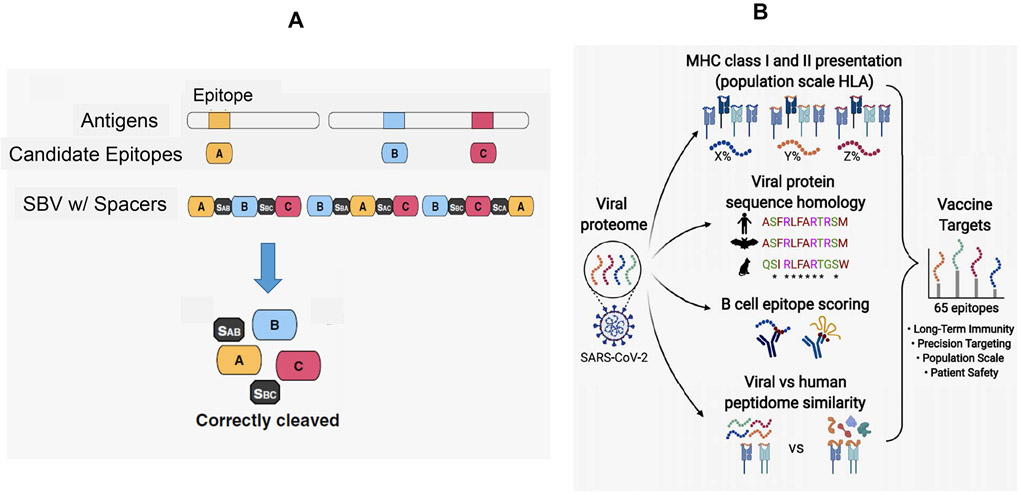
Although there is good evidence for T-cell involvement in the vaccine response to mRNA, protein subunit, and viral-vectored CoV-2 vaccines, it is becoming clear that the roles of T-cells require additional consideration for future vaccine development. Of particular interest is determining if there are differences in T-cell phenotypes stimulated by different vaccine types, with the possibility that there could be complementarity of action by combining current vaccines (e.g., adenovirus-vectored with mRNA vaccines).152 A more deliberate future attempt would be to exploit nano-enabled design features to improve T-B cooperation. In addition to the principles explained in Figures 10 and 11, another approach would be to develop a multi-epitope vaccine strategy, such as the “string-of-beads vaccines” (SBV) concept, previously used for cytomegalovirus and influenza (Figure 12A).110,153,154 This strategy could entail the selection and combination of epitopes from diverse SARS-CoV-2 antigens (M, N, E, and S proteins) that are spliced together with the assistance of cleavable spacer sequences.110 Similar outcomes can also be achieved by the design of DNA or RNA minigenes, as demonstrated by Fomsgaard et al. for influenza.154 The selection of potentially synergistic epitope combinations will benefit from the use immunoinformatics tools, as described above.65,94,99,100,102,104,109 This approach could include the use of tools that enable the design of appropriate linker and codon adjustment strategies for minigene design.64,102 Ultimately, it should be possible to use suitably designed nanocarriers to deliver multi-epitope peptide sequences or minigene nucleic acid constructs to the host immune system for antigen presentation. Vaccine efficiency will depend on epitope selection as well as the correct choice of spacers to enable epitope release by the immunoproteosome.110
Figure 12. String-of-beads vaccines (SBVs) for multi-epitope delivery.
A. The concept of SBVs for infectious disease agents (e.g., influenza and cytomegalovirus) is based on expressing multiple epitopes from pathogen antigens separated by cleavable spacers.110 It is envisaged that the design tools for the selection of multiple epitopes and their encapsulation in suitable nanocarriers will facilitate SARS-CoV-2 epitope delivery to regional lymph nodes, where individual epitopes will be released by proteolytic processing and presented by dendritic cells. Vaccine efficiency would depend on the optimal combination of B and T-cell epitopes as well as the use of appropriate spacers to allow efficient epitope release.110 Reprinted with permission from ref 110 under the Creative Commons Attribution 4.0 International License. Copyright 2016 Springer Nature. B. Multi-epitope vaccines have received a boost from new immunoinformatic tools for vaccine design, using a series prediction tools as outlined in Table 3. In this example, Yarmarkovich et al. report the design of multi-epitope vaccines that deliver 65 x 33-mer SARS-CoV-2 peptides, making use of the following guidelines: (i) selection of peptide sequences from 15 related coronaviruses; (ii) epitope ability to activate CD4+ and CD8+ T-cells by enabling interactions with diverse HLA gene sequences; (iii) B-cell activation by linear and conformational epitope sequences; (iv) high immunogenicity through sequence selection that is significantly dissimilar to the self-proteome; (v) vaccine safety.109 Reprinted with permission from ref 109 under the Creative Commons License CC-BY 4.0. Copyright 2020 The Authors.
An example of multi-epitope vaccine design, aiming for durable immunity through a combination of B-and T-cell epitopes, was recently demonstrated by Yarmarkovich et al.109 These investigators developed 65 x 33mer-peptides using the design principles outlined in Figure 12B. Epitope selections included evolutionarily conserved coronavirus sequences, including T-cell epitopes for population-based HLA coverage, in addition to the inclusion of linear and conformational B-cell epitopes. To maximize immunogenicity, only viral regions with the highest degree of dissimilarity to the human immunopeptidome were chosen. This vaccine design also enables conjugated TH1 adjuvants to be included with the peptides. Although the efficacy of this design still awaits animal experimentation, we have outlined the success that was achieved by COVAXX using their 8-component multi-epitope vaccine (UB-612) to induce protective immune responses in rats, guinea pigs, and a nonhuman primate model.112
Multi-epitope vaccine design could also enable the generation of cross-reactive immunity that deliberately expands the contribution of different types of memory T-cells.155 The feasibility of this approach is supported by a number of studies showing broad-based T-cell reactivity in 20–50% of people with no known exposure to SARS-CoV-2.151 Memory T-cells can be classified as central memory, effector memory, and tissue-resident (TRM) memory T-cells, which can be expressed as CD4+ and CD8+ phenotypes.156,157 Lipsitch et al. recently conducted a thought experiment that considers multiple scenarios of how cross-reactive CD4+ memory T-cells may impact the development of SARS-CoV-2 transmission and herd immunity.155 In one hypothetical scenario, cross-reactive memory TFH cells were projected to participate in triggering more robust and rapid neutralizing antibody responses that reduce the magnitude and duration of symptomatic SARS-CoV-2 infections. This outcome was projected to enhance the durability of the immune response, but to exert little effect on viral loads in the respiratory tract. This scenario was also postulated to lead to fewer hospitalizations and deaths, with a moderate impact on viral spread.155 A second scenario envisages a role for cross-reactive TRM cells that have the inherent capacity to induce rapid control of virus spread in the respiratory tract through their ability to recruit cytotoxic T-cells, resulting in rapid clearance from virally infected cells as a result of the failure of the sterilizing antibody defense.155 Thus, this form of T-cell memory could allay the development of severe disease at the time of re-exposure, leading to short asymptomatic infectious episodes with low viral loads. However, this immune response would be launched at the expense of generating durable memory responses. Although this thought experiment still needs to be experimentally tested, a key question now becomes whether it is possible through epitope selections and use of appropriate adjuvants to engage and activate specific memory phenotypes selectively in vaccine development. Although there is currently no answer to this question, it is tempting to speculate that future vaccine development efforts will be able to exploit the early surveillance role of TRM cells to boost neutralizing antibody defenses against SARS-CoV-2.156,157 Along similar lines, it may also be possible to select cross-reactive coronavirus regions to generate long-lived memory T-cells to develop a pan-coronavirus vaccine.
Adaptability of Nano-Enabled Vaccines to Counter SARS-CoV-2 Evolution
There is presently great concern about the ability of COVID-19 vaccines to protect against viral variants that have emerged in the United Kingdom (UK), South Africa, and Brazil.158 Replication of the 29.9 kb single-stranded positive-sense RNA genome of SARS-CoV-2 requires the production of negative strand complementary RNA templates, which are copied into positive stranded viral genomes. In all cases, RNA synthesis is catalyzed by the viral replication–transcription complex that includes the RNA-dependent RNA polymerase subunits that confer processivity (nsp7, nsp8), and a 3'-5' exonuclease (nsp14) that proofreads nascent RNA and excises misincorporated nucleotides—a rare activity among viral RdRp complexes.76 Coronaviruses are endowed with genomes that are among the largest of all RNA viruses, and the proofreading function of nsp14 is highly conserved in the Coronaviridae family. Proofreading helps maintain the integrity of coronavirus genomes and is likely essential for the evolution of their complexity and adaptability.76,159 Despite this relative stability, mutations do occur and their cumulative effects are a source of global concern.
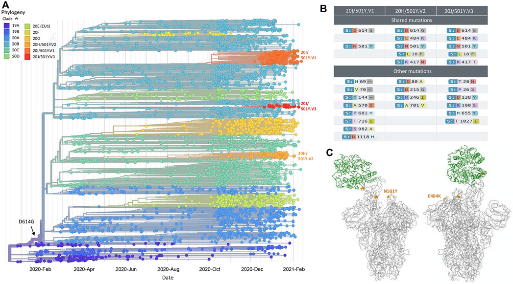
The mean rate of evolutionary change in SARS-CoV-2, or, more accurately, the rate at which alleles are fixed in the population, can be estimated using viral sequence information and sampling dates (Figure 13A).160,161 Current data from a curated global subsample of SARS-CoV-2 sequences suggests the virus is evolving at a rate of approximately 7.5 x 10⁻⁴ substitutions per site per year, which corresponds to about one substitution every 16 days.161,162 Nucleotide insertions and deletions are often observed, but synonymous substitutions are the primary source of genetic variation. Given its recent spillover from a zoonotic reservoir, estimated to have occurred in late November 2019, further adaptation toward increased fitness in humans is to be expected.162 Indeed, the first “mutation of concern”, an Asp to Gly substitution at amino acid position 614 in the SARS-CoV-2 spike protein (D614G), was first identified early in the pandemic, becoming the globally dominant form of the virus by June 2020.163 The D614G substitution increases the efficiency of cell entry and replication in vitro,164 and enhances transmission in animal models165 and in human populations.166 The D614G mutation lies outside the RBD, and recent cryoEM studies suggest that it facilitates the transition of spike protein to the open conformation, thereby promoting receptor binding, membrane fusion and viral infection.167
Figure 13. Global phylogeny of SARS-CoV-2 and the evolution of variant strains.
A. Phylogenetic tree showing evolutionary relationships between SARS-CoV-2 viruses from the ongoing COVID-19 pandemic, from 3913 genomes sampled between Dec 2019 and Feb 2021. The spike protein D614G substitution (arrow), which is carried by virtually all currently circulating viruses, emerged early in the course of the pandemic. Clade 20I/501Y.V1 (UK variant B.1.1.7); 20C/501Y.V2 (South African variant B.1.351); 20J/501Y.V3 (Brazil variant P1) are indicated. Figure is adapted from Nextstrain, under a C-BY-4.0 license.160,161 B. Amino acid substitutions in the SARS-CoV-2 spike locus shared by variants from the UK (20I/501Y.V1; B.1.1.7), South Africa (20C/501Y.V2; B.1.351), and Brazil (20J/501Y.V3; P1) are shown along with other lineage-defining mutations. C. Locations of the N501Y (left) and E484K (right) substitutions in SARS-CoV-2 spike trimers (gray), showing the RBD of one subunit bound to the human ACE2 receptor (green). B. and C. are adapted from ref 211, CoVariants: SARS-CoV-2 Mutations and Variants of Interest, licensed under a GNU Affero General Public License (AGPL).211 Copyright 2020–2021 Emma Hodcroft.
At least two waves of adaptation can be expected when a newly evolved pathogen acquires the ability to infect new host populations. While selection for increased transmission between immunologically naive hosts initially prevails, as the immune status of the host population increases due to natural infection or vaccination, further adaptations take place in response to selective pressures to evade immunity. Substantial evidence indicates that both selective forces are now shaping the global phylodynamics of SARS-CoV-2, resulting in the emergence of new viral strains that display partially overlapping collections of mutations, presumably due to convergent evolution (Figure 13B).162,168,169 Three newly emerged variant strains, all decedents of the D614 lineage, are now attracting global attention.
The most widely distributed variant SARS-CoV-2 strain, B.1.1.7 (also 201/501Y.V1, Figure 13), first emerged in southeast England in December, 2020170 and rapidly became the predominant strain throughout the UK. As of February 23, 2021, its global distribution has expanded to include 93 countries.171 The strain was initially defined by a collection of 14 nonsynonymous substitutions and 3 deletions, with 8 of 17 mutations located in the spike protein. The extent of divergence of B.1.1.7 was surprising, as most branches in the SARS-CoV-2 phylogenetic tree show only a few defining mutations that accumulate slowly over time. Several alterations in spike protein had been observed in different lineages, suggesting they arose independently in response to similar selective pressures. These include a 2 amino acid deletion (H69-) in the N-terminal domain that had previously been associated with immune evasion,172 a substitution (P681H) located adjacent to the S1/S2 furin cleavage site that is essential for entry into human lung cells,173 and the N501Y mutation in the spike RBD which increases binding affinity for ACE2 receptors (Figure 13C).174 Although observations on the properties of individual mutations may be informative, the critical question is how they may act together to affect virus behavior. Presently, B.1.1.7 has a substantial fitness advantage over other circulating lineages, with an estimated increase in person to person transmission of 50–70%.175 Fortunately, there is little evidence for a clinically significant decline in antibody neutralization titers in convalescent sera,176 including sera from individuals previously vaccinated with the Moderna177 or Pfizer mRNA products.178 Whether B.1.1.7 causes more severe illness remains controversial, but a preliminary study reports an estimated 35% (CI 12–64%) higher hazard of death associated with infection by this strain.179 As the number of infections due to this hyper-transmissible lineage increases worldwide, so does the likelihood that further mutations will evolve.
Two more variant strains have recently emerged that are also of major concern, one discovered in South Africa and designated B.1.351 (also 20H/501Y.V2), and the other first identified in Brazil (P.1, also 20J/501Y.V3). Both variants carry multiple mutations which include the N501Y allele found in B.1.1.7 as well as additional mutations that are shared with each other (Figure 13B). Most notable is the E484K substitution in the RBD (Figure 13C), which increases ACE2 binding in a manner that is further augmented by N501Y.180 Most concerning is mounting evidence that the E484K allele emerged independently on multiple occasions as a result of immune-driven selection.181 E484K facilitates measurable escape from neutralizing antibodies present in convalescent serum as well as a modest decrease in neutralization by serum from individuals that were vaccinated with the Moderna or Pfizer mRNA vaccines.182 Although the initial Phase 3 clinical trials conducted by Moderna and Pfizer preceded the discovery of these strains, subsequent Phase 3 trials with newer vaccines included populations with high rates of infection by variants of concern. In a recent trial by Johnson & Johnson involving 45,000 volunteers, the efficacy of their single-dose, adenoviral-vectored, Ad26.COV2.S vaccine in conferring protection against moderate to severe COVID-19 was 72% in the United States, 66% in Latin America and 57% in South Africa, 28 days post-vaccination.183 The lower efficacy in South Africa is suspected to be due to the rise of the B.1.351 variant. Despite this result, the protective efficacy against severe disease was 85% across all regions studied, with complete protection observed against hospitalization and death.53 A preliminary analysis of Phase 3 clinical trial data by Novavax indicated that their two-dose nanoparticle vaccine, NVX-CoV2373, conferred nearly 90% protection against COVID-19 in the UK, but just under 50% protection in South Africa.184 In all cases that were analyzed, vaccine failures were caused by the B.1.351 variant, and even more troubling was the finding that several trial participants became infected with B.1.351 despite having previously suffered from COVID-19.
We are now at a critical point where advances in vaccine technology and deployment must be strategically aligned with viral evolution. This task could become increasingly challenging as the number of SARS-CoV-2 infections continues to expand. The recent discovery of B.1.1.7 variants that have acquired the E484K substitution vividly illustrates this challenge, raising concern that hypertransmissive strains with increased capacity for immune evasion may have already emerged. Gaining control of the situation will require a number of parallel efforts. The first is to vaccinate as many people as possible on a global scale and to do it as quickly as we can to decrease the global viral pool participating in the formation of mutants. The second is to exploit the inherent adaptability of newly developed vaccine platforms. mRNA vaccines, for example, are rapidly modifiable to encode newly evolved antigens, and both Moderna and Pfizer have announced plans to build second-generation vaccines expressing B.1.351 variant spike proteins to provide variant-specific boosting.158,185 Similarly, Novavax has announced plans to incorporate B.1.351 spike sequences in their self-assembling protein nanoparticles. Similar strategies could be used to issue immunological updates to vaccinated hosts over time to keep pace with the evolution of SARS-CoV-2.
Should the above strategies fail to control immune escape variants, a longer-term strategy would be to boost T-cell contributions and to target CoV-2 antigenic sites that are not subject to the same rate of mutation as the spike protein.186 One possible approach could be to develop multivalent vaccines that present multiple B-cell and T-cell epitopes, as described above.
Looking Forward
The global COVID-19 vaccine drive has introduced a new era in structure-based vaccine design, in addition to drawing on nano-enabled approaches to deliver engineered vaccines to the immune system.1,4,10,13,14,55,70,187,188 These efforts have advanced the structural vaccinology concept that was initially implemented to obtain stable expression of type I fusion proteins from RSV (F protein), HIV-1 gp160 (env), influenza hemagglutinin, and the spike proteins from MERS and SARS-CoV1. Moderna and Pfizer took this discovery forward by developing engineered nucleic acid constructs for the expression of the SARS-CoV-2 spike protein in a pre-fusion conformation, thereby enabling these vaccines to generate strong neutralizing antibody responses. A parallel development was the self-assembly of engineered full length S protein and computerized design of a viral-like particle with multivalent RBD display.72 Not only are the strategies coming from current SARS-CoV-2 vaccination efforts reinvigorating vaccine design for RSV, influenza, HIV-1, and other pathogens, but they are also highlighting principles for continuously updating vaccine composition using rapidly reprogrammable platforms. This provides a new approach to vaccinology. With the current rate of technological improvement, we can expect that COVID-19 vaccines will emerge that are capable of generating high titers of neutralizing antibodies, eliciting robust TH1-mediated immune responses, inducing long-term memory, and providing broader protection against SARS-CoV-2 variants and other cross-reactive coronaviruses. Another important lesson from current vaccine efforts is the necessity of developing comprehensive and real-time surveillance, data analysis, high-throughput approaches for genotyping and phenotyping viral isolates, as well as efficient methods for altering, testing, approving, manufacturing, and deploying newly synthesized CoV-2 vaccines.160,163 Just as software updates are a fact of life, vaccine updates should be similarly expected and expedited.
Although it is too early to make firm predictions about the outcomes of the current vaccine drive, it is reasonable to expect major impacts on symptomatic as well as severe disease, hospitalization, and mortality, even prior to reaching the herd immunity threshold, estimated to occur when 70–85% of the population is protected. There are already indications from the phase 3 clinical trials conducted by Johnson & Johnson that their adenoviral-vectored vaccine is 66% effective at preventing moderate to severe COVID-19, with 85% efficacy against the most serious illness.183 The vaccine also prevented mortality among the 43,000 participants in that trial. In addition, it has recently been demonstrated that Pfizer’s vaccine trial data holds up in the real-world vaccination drive in Israel.189 This study compared almost 600,000 people receiving the Pfizer vaccine between December 20, 2020, and February 1, 2021, against a cohort of similar size that was not vaccinated. After two vaccine doses, the vaccinated subjects were 94% less likely to develop symptomatic illness and 87% less likely to be hospitalized. It is also of interest that this outcome was achieved in a study cohort where a large number of the infections could be ascribed to the UK CoV-2 variant (B.1.1.7). It is important to take into consideration that the Israeli vaccination campaign is far ahead of most other countries, and we need to keep an eye on what will happen in other sectors of the world where vaccination may proceed less rapidly due to inadequate supply lines, ineffective vaccine distribution, lack of societal acceptance, and the rapid evolution of new viral variants.
If and when herd immunity can be achieved remain unanswered questions. There are an increasing number of predictions that COVID-19 will become an endemic disease.190,191,192 A Nature survey among immunologists, infectious disease experts, and virologists (who are active in the coronavirus field), recently found that 90% of the respondents were of the opinion that the coronavirus will become endemic, with the implication being that the virus may continue to circulate for a number of years in small cohorts in the global community.190 Although this circulation could lead to the continuation of infection, similar to disease occurrence related to common cold coronaviruses, the failure to eradicate SARS-CoV-2 completely does not mean that disease severity, mortality, or social isolation will continue at current levels, particularly if the vaccination campaign can push populations close to herd immunity. Interestingly, the factors that were identified as possible drivers for endemic SARS-CoV-2 circulation in the survey were listed, in decreasing order of importance, as immune escape, waning immunity, uneven vaccine distribution, vaccine hesitancy, lack of political will, and the presence of animal reservoirs.193
Suggested pull quotes.
Antigen selection and composition are of key importance for vaccine development, in addition to the choice of adjuvant and ensuring efficacious vaccine delivery to the host’s innate and adaptive immune systems.
An alternative approach to employing nanoparticles for CoV-2 vaccine development is to use full length spike protein or viral subunits for encapsulation or self-assembly into nanoparticles.
Another vaccine category making progress is epitope-based vaccines, which could rely on nanoparticles to deliver individual or epitope combinations.
Despite being the first of their kind, the RNA vaccines developed by Pfizer/BioNTech and Moderna represent the fastest vaccine development attempts ever, replacing the previous record of 4 years held by the mumps vaccine.
All mRNA vaccines subjected to large-scale clinical trials have proven to be effective in generating high protective antibody titers against the S protein and its receptor-binding domains in humans.
Longitudinal studies to determine the duration of the protective response after natural infection or vaccination are ongoing and of great importance for establishing future public health policies.
Multi-epitope vaccine design could also enable the generation of cross-reactive immunity that deliberately expands the contribution of different types of memory T-cells.
We are now at a critical point where advances in vaccine technology and deployment must be strategically aligned with viral evolution.
The global COVID-19 vaccine drive has introduced a new era in structure-based vaccine design, in addition to drawing on nano-enabled approaches to deliver engineered vaccines to the immune system.
Acknowledgments:
We thank Holly Bunje for assistance in obtaining permission to use published material for the figures and proofreading the manuscript, as well as updating the references. Research in Dr. Nel’s lab is supported by the National Cancer Institute of the National Institutes of Health under Award Number, 1R01CA247666-01A1. Research in Dr. Miller's lab is supported by the Kavli Foundation and the Shaffer Family Foundation.
Footnotes
Competing Financial Interests: Andre E. Nel is a co-founder and equity holder in Westwood Biosciences Inc. and NAMMI Therapeutics. He also serves on the Board for Westwood Biosciences Inc. Jeff F. Miller is a co-founder, equity holder and board member of Pylum Biosciences, Xyphos Inc, and Dermolytica Inc., and SAB member of Notitia Biosciences.
References
- 1.Mascola JR; Fauci AS Novel Vaccine Technologies for the 21st Century. Nat. Rev. Immunol 2020, 20, 87–88. [DOI] [PMC free article] [PubMed] [Google Scholar]
- 2.de Wit E; van Doremalen N; Falzarano D; Munster VJ SARS and MERS: Recent Insights Into Emerging Coronaviruses. Nat. Rev. Microbiol 2016,14, 523–534. [DOI] [PMC free article] [PubMed] [Google Scholar]
- 3.Ahmed SF; Quadeer AA; McKay MR Preliminary Identification of Potential Vaccine Targets for the COVID-19 Coronavirus (SARS-CoV-2) Based on SARS-CoV Immunological Studies. Viruses 2020,12, 254. [DOI] [PMC free article] [PubMed] [Google Scholar]
- 4.Dai L; Zheng T; Xu K; Han Y; Xu L; Huang E; An Y; Cheng Y; Li S; Liu M; Yang M; Li Y; Cheng H; Yuan Y; Zhang W; Ke C; Wong G; Qi J; Qin C; Yan J; Gao GF A Universal Design of Betacoronavirus Vaccines against COVID-19, MERS, and SARS. Cell 2020, 182, 722–733.e11. [DOI] [PMC free article] [PubMed] [Google Scholar]
- 5.Kirchdoerfer RN; Cottrell CA; Wang N; Pallesen J; Yassine HM; Turner HL; Corbett K; Graham BS; McLellan JS; Ward AB Pre-Fusion Structure of a Human Coronavirus Spike Protein. Nature 2016, 531, 118–121. [DOI] [PMC free article] [PubMed] [Google Scholar]
- 6.Pallesen J; Wang N; Corbett KS; Wrapp D; Kirchdoerfer RN; Turner HL; Cottrell CA; Becker MM; Wang L; Shi W; Kong WP; Andres EL; Kettenbach AN; Denison MR; Chappell JD; Graham BS; Ward AB; McLellan JS Immunogenicity and Structures of a Rationally Designed Prefusion MERS-CoV Spike Antigen. Proc. Natl. Acad. Sci. U. S. A 2017, 114, E7348–e7357. [DOI] [PMC free article] [PubMed] [Google Scholar]
- 7.Yuan Y; Cao D; Zhang Y; Ma J; Qi J; Wang Q; Lu G; Wu Y; Yan J; Shi Y; Zhang X; Gao GF Cryo-EM Structures of MERS-CoV and SARS-CoV Spike Glycoproteins Reveal the Dynamic Receptor Binding Domains. Nat Commun 2017, 8, 15092. [DOI] [PMC free article] [PubMed] [Google Scholar]
- 8.Zhou H; Chen Y; Zhang S; Niu P; Qin K; Jia W; Huang B; Zhang S; Lan J; Zhang L; Tan W; Wang X Structural Definition of a Neutralization Epitope on the N-Terminal Domain of MERS-CoV Spike Glycoprotein. Nat. Commun 2019, 10, 3068. [DOI] [PMC free article] [PubMed] [Google Scholar]
- 9.Du L; He Y; Zhou Y; Liu S; Zheng BJ; Jiang S The Spike Protein of SARS-CoV--A Target for Vaccine and Therapeutic Development. Nat. Rev. Microbiol 2009, 7, 226–236. [DOI] [PMC free article] [PubMed] [Google Scholar]
- 10.Walls AC; Park YJ; Tortorici MA; Wall A; McGuire AT; Veesler D Structure, Function, and Antigenicity of the SARS-CoV-2 Spike Glycoprotein. Cell 2020, 181, 281–292.e6. [DOI] [PMC free article] [PubMed] [Google Scholar]
- 11.Yuan M; Wu NC; Zhu X; Lee CD; So RTY; Lv H; Mok CKP; Wilson IA A Highly Conserved Cryptic Epitope in the Receptor Binding Domains of SARS-CoV-2 and SARS-CoV. Science 2020, 368, 630–633. [DOI] [PMC free article] [PubMed] [Google Scholar]
- 12.Espeseth AS; Cejas PJ; Citron MP; Wang D; DiStefano DJ; Callahan C; Donnell GO; Galli JD; Swoyer R; Touch S; Wen Z; Antonello J; Zhang L; Flynn JA; Cox KS; Freed DC; Vora KA; Bahl K; Latham AH; Smith JS; et al. Modified mRNA/lipid Nanoparticle-Based Vaccines Expressing Respiratory Syncytial Virus F Protein Variants are Immunogenic and Protective in Rodent Models of RSV Infection. NPJ Vaccines 2020, 5, 16. [DOI] [PMC free article] [PubMed] [Google Scholar]
- 13.Graham BS; Gilman MSA; McLellan JS Structure-Based Vaccine Antigen Design. Annu. Rev. Med 2019, 70, 91–104. [DOI] [PMC free article] [PubMed] [Google Scholar]
- 14.McLellan JS; Chen M; Joyce MG; Sastry M; Stewart-Jones GB; Yang Y; Zhang B; Chen L; Srivatsan S; Zheng A; Zhou T; Graepel KW; Kumar A; Moin S; Boyington JC; Chuang GY; Soto C; Baxa U; Bakker AQ; Spits H; et al. Structure-Based Design of a Fusion Glycoprotein Vaccine for Respiratory Syncytial Virus. Science 2013, 342, 592–598. [DOI] [PMC free article] [PubMed] [Google Scholar]
- 15.Iavarone C; O'Hagan D T; Yu D; Delahaye NF; Ulmer JB Mechanism of Action of mRNA-Based Vaccines. Expert Rev. Vaccines 2017, 16, 871–881. [DOI] [PubMed] [Google Scholar]
- 16.Reichmuth AM; Oberli MA; Jaklenec A; Langer R; Blankschtein D mRNA Vaccine Delivery Using Lipid Nanoparticles. Ther. Deliv 2016, 7, 319–334. [DOI] [PMC free article] [PubMed] [Google Scholar]
- 17.Sahin U; Karikó K; Türeci Ö mRNA-Based Therapeutics--Developing a New Class of Drugs. Nat. Rev. Drug Discov 2014, 13, 759–780. [DOI] [PubMed] [Google Scholar]
- 18.Scorza FB; Pardi N New Kids on the Block: RNA-Based Influenza Virus Vaccines. Vaccines (Basel) 2018, 6, 20. [DOI] [PMC free article] [PubMed] [Google Scholar]
- 19.Zhuang X; Qi Y; Wang M; Yu N; Nan F; Zhang H; Tian M; Li C; Lu H; Jin N mRNA Vaccines Encoding the HA Protein of Influenza A H1N1 Virus Delivered by Cationic Lipid Nanoparticles Induce Protective Immune Responses in Mice. Vaccines (Basel) 2020, 8, 123. [DOI] [PMC free article] [PubMed] [Google Scholar]
- 20.Karikó K; Muramatsu H; Welsh FA; Ludwig J; Kato H; Akira S; Weissman D, Incorporation of Pseudouridine into mRNA Yields Superior Nonimmunogenic Vector with Increased Translational Capacity and Biological Stability. Mol. Ther 2008, 16, 1833–1840. [DOI] [PMC free article] [PubMed] [Google Scholar]
- 21.Pardi N; Hogan MJ; Naradikian MS; Parkhouse K; Cain DW; Jones L; Moody MA; Verkerke HP; Myles A; Willis E; LaBranche CC; Montefiori DC; Lobby JL; Saunders KO; Liao HX; Korber BT; Sutherland LL; Scearce RM; Hraber PT; Tombácz I; et al. Nucleoside-Modified mRNA Vaccines Induce Potent T Follicular Helper and Germinal Center B Cell Responses. J. Exp. Med 2018, 215, 1571–1588. [DOI] [PMC free article] [PubMed] [Google Scholar]
- 22.Pardi N; Weissman D Nucleoside Modified mRNA Vaccines for Infectious Diseases. Methods Mol. Biol 2017, 1499, 109–121. [DOI] [PubMed] [Google Scholar]
- 23.Evers MJW; Kulkarni JA; van der Meel R; Cullis PR; Vader P; Schiffelers RM State-of-the-Art Design and Rapid-Mixing Production Techniques of Lipid Nanoparticles for Nucleic Acid Delivery. Small Methods 2018, 2, 1700375. [Google Scholar]
- 24.McKay PF; Hu K; Blakney AK; Samnuan K; Brown JC; Penn R; Zhou J; Bouton CR; Rogers P; Polra K; Lin PJC; Barbosa C; Tam YK; Barclay WS; Shattock RJ Self-Amplifying RNA SARS-CoV-2 Lipid Nanoparticle Vaccine Candidate Induces High Neutralizing Antibody Titers in Mice. Nat. Commun 2020, 11, 3523. [DOI] [PMC free article] [PubMed] [Google Scholar]
- 25.Shin MD; Shukla S; Chung YH; Beiss V; Chan SK; Ortega-Rivera OA; Wirth DM; Chen A; Sack M; Pokorski JK; Steinmetz NF COVID-19 Vaccine Development and a Potential Nanomaterial Path Forward. Nat. Nanotechnol 2020, 15, 646–655. [DOI] [PubMed] [Google Scholar]
- 26.Brito LA; Kommareddy S; Maione D; Uematsu Y; Giovani C; Berlanda Scorza F; Otten GR; Yu D; Mandl CW; Mason PW; Dormitzer PR; Ulmer JB; Geall AJ Self-Amplifying mRNA Vaccines. Adv. Genet 2015, 89, 179–233. [DOI] [PubMed] [Google Scholar]
- 27.Kaneko N; Kuo HH; Boucau J; Farmer JR; Allard-Chamard H; Mahajan VS; Piechocka-Trocha A; Lefteri K; Osborn M; Bals J; Bartsch YC; Bonheur N; Caradonna TM; Chevalier J; Chowdhury F; Diefenbach TJ; Einkauf K; Fallon J; Feldman J; Finn KK; Garcia-Broncano P; Hartana CA; Hauser BM; Jiang C; Kaplonek P; Karpell M; Koscher EC; Lian X; et al. Loss of Bcl-6-Expressing T Follicular Helper Cells and Germinal Centers in COVID-19. Cell 2020, 183, 143–157.e13. [DOI] [PMC free article] [PubMed] [Google Scholar]
- 28.Nutt SL; Tarlinton DM Germinal Center B and Follicular Helper T Cells: Siblings, Cousins or Just Good Friends? Nat. Immunol 2011, 12, 472–477. [DOI] [PubMed] [Google Scholar]
- 29.Akondy RS; Fitch M; Edupuganti S; Yang S; Kissick HT; Li KW; Youngblood BA; Abdelsamed HA; McGuire DJ; Cohen KW; Alexe G; Nagar S; McCausland MM; Gupta S; Tata P; Haining WN; McElrath MJ; Zhang D; Hu B; Greenleaf WJ; et al. Origin and Differentiation of Human Memory CD8 T Cells After Vaccination. Nature 2017, 552, 362–367. [DOI] [PMC free article] [PubMed] [Google Scholar]
- 30.Fatoba AJ; Maharaj L; Adeleke VT; Okpeku M; Adeniyi AA; Adeleke MA Immunoinformatics Prediction of Overlapping CD8(+) T-Cell, IFN-γ and IL-4 Inducer CD4(+) T-Cell and Linear B-Cell Epitopes Based Vaccines Against COVID-19 (SARS-CoV-2). Vaccine 2021, 39, 1111–1121. [DOI] [PMC free article] [PubMed] [Google Scholar]
- 31.Peng Y; Mentzer AJ; Liu G; Yao X; Yin Z; Dong D; Dejnirattisai W; Rostron T; Supasa P; Liu C; López-Camacho C; Slon-Campos J; Zhao Y; Stuart DI; Paesen GC; Grimes JM; Antson AA; Bayfield OW; Hawkins D; Ker DS; et al. Broad and Strong Memory CD4(+) and CD8(+) T Cells Induced by SARS-CoV-2 in UK Convalescent Individuals Following COVID-19. Nat. Immunol 2020, 21, 1336–1345. [DOI] [PMC free article] [PubMed] [Google Scholar]
- 32.Schulien I; Kemming J; Oberhardt V; Wild K; Seidel LM; Killmer S; Sagar; Daul F; Salvat Lago M; Decker A; Luxenburger H; Binder B; Bettinger D; Sogukpinar O; Rieg S; Panning M; Huzly D; Schwemmle M; Kochs G; Waller CF; et al. Characterization of Pre-Existing and Induced SARS-CoV-2-Specific CD8(+) T Cells. Nat Med 2021, 27, 78–85. [DOI] [PubMed] [Google Scholar]
- 33.Sette A; Crotty S Adaptive Immunity to SARS-CoV-2 and COVID-19. Cell 2021, 184, 861–880. [DOI] [PMC free article] [PubMed] [Google Scholar]
- 34.Swadling L; Maini MK T Cells in COVID-19 – United in Diversity. Nat. Immunol 2020, 21, 1307–1308. [DOI] [PubMed] [Google Scholar]
- 35.Tarke A; Sidney J; Kidd CK; Dan JM; Ramirez SI; Yu ED; Mateus J; da Silva Antunes R; Moore E; Rubiro P; Methot N; Phillips E; Mallal S; Frazier A; Rawlings SA; Greenbaum JA; Peters B; Smith DM; Crotty S; Weiskopf D; Grifoni A; Sette A Comprehensive Analysis of T Cell Immunodominance and Immunoprevalence of SARS-CoV-2 Epitopes in COVID-19 Cases. Cell Rep. Med 2021, 2, 100204. [DOI] [PMC free article] [PubMed] [Google Scholar]
- 36.Bialkowski L; van Weijnen A; Van der Jeught K; Renmans D; Daszkiewicz L; Heirman C; Stangé G; K Breckpot K; Aerts JL; Thieleman K Intralymphatic mRNA Vaccine Induces CD8 T-Cell Responses that Inhibit the Growth of Mucosally Located Tumours. Scl. Rep 2016, 6, 22509. doi: 10.1038/srep22509. [DOI] [PMC free article] [PubMed] [Google Scholar]
- 37.WHO Draft Landscape and Tracker of COVID-19 Candidate Vaccine. https://www.who.int/publications/m/item/draft-landscape-of-covid-19-candidate-vaccines (accessed February 23, 2021).
- 38.Baden LR; El Sahly HM; Essink B; Kotloff K; Frey S; Novak R; Diemert D; Spector SA; Rouphael N; Creech CB; McGettigan J; Khetan S; Segall N; Solis J; Brosz A; Fierro C; Schwartz H; Neuzil K; Corey L; Gilbert P; et al. Efficacy and Safety of the mRNA-1273 SARS-CoV-2 Vaccine. N. Engl. J. Med 2021, 384, 403–416. [DOI] [PMC free article] [PubMed] [Google Scholar]
- 39.Chung YH; Beiss V; Fiering SN; Steinmetz NF COVID-19 Vaccine Frontrunners and Their Nanotechnology Design. ACS Nano 2020, 14, 12522–12537. [DOI] [PubMed] [Google Scholar]
- 40.Jackson LA; Anderson EJ; Rouphael NG; Roberts PC; Makhene M; Coler RN; McCullough MP; Chappell JD; Denison MR; Stevens LJ; Pruijssers AJ; McDermott A; Flach B; Doria-Rose NA; Corbett KS; Morabito KM; O'Dell S; Schmidt SD; Swanson PA 2nd; Padilla M; et al. An mRNA Vaccine Against SARS-CoV-2 – Preliminary Report. N. Engl. J. Med 2020, 383, 1920–1931. [DOI] [PMC free article] [PubMed] [Google Scholar]
- 41.Widge AT; Rouphael NG; Jackson LA; Anderson EJ; Roberts PC; Makhene M; Chappell JD; Denison MR; Stevens LJ; Pruijssers AJ; McDermott AB; Flach B; Lin BC; Doria-Rose NA; O'Dell S; Schmidt SD; Neuzil KM; Bennett H; Leav B; Makowski M; et al. Durability of Responses after SARS-CoV-2 mRNA-1273 Vaccination. N. Engl. J. Med 2021, 384, 80–82. [DOI] [PMC free article] [PubMed] [Google Scholar]
- 42.Anderson EJ; Rouphael NG; Widge AT; Jackson LA; Roberts PC; Makhene M; Chappell JD; Denison MR; Stevens LJ; Pruijssers AJ; McDermott AB; Flach B; Lin BC; Doria-Rose NA; O'Dell S; Schmidt SD; Corbett KS; Swanson PA 2nd; Padilla M; Neuzil KM; et al. Safety and Immunogenicity of SARS-CoV-2 mRNA-1273 Vaccine in Older Adults. N. Engl. J. Med 2020, 383, 2427–2438. [DOI] [PMC free article] [PubMed] [Google Scholar]
- 43.Bradley T; Grundberg E; Selvarangan R Antibody Responses Boosted in Seropositive Healthcare Workers After Single Dose of SARS-CoV-2 mRNA Vaccine. medRxiv Preprint (03/01/21) 2021. https://www.medrxiv.org/content/10.1101/2021.02.03.21251078v1 [Google Scholar]
- 44.Corbett KS; Edwards DK; Leist SR; Abiona OM; Boyoglu-Barnum S; Gillespie RA; Himansu S; Schäfer A; Ziwawo CT; DiPiazza AT; Dinnon KH; Elbashir SM; Shaw CA; Woods A; Fritch EJ; Martinez DR; Bock KW; Minai M; Nagata BM; Hutchinson GB; et al. SARS-CoV-2 mRNA Vaccine Design Enabled by Prototype Pathogen Preparedness. Nature 2020, 586, 56–571. [DOI] [PMC free article] [PubMed] [Google Scholar]
- 45.Mulligan MJ; Lyke KE; Kitchin N; Absalon J; Gurtman A; Lockhart S; Neuzil K; Raabe V; Bailey R; Swanson KA; Li P; Koury K; Kalina W; Cooper D; Fontes-Garfias C; Shi PY; Türeci Ö; Tompkins KR; Walsh EE; Frenck R; et al. Phase I/II study of COVID-19 RNA Vaccine BNT162b1 in Adults. Nature 2020, 586, 589–593. [DOI] [PubMed] [Google Scholar]
- 46.Oliver SE; Gargano JW; Marin M; Wallace M; Curran KG; Chamberland M; McClung N; Campos-Outcalt D; Morgan RL; Mbaeyi S; Romero JR; Talbot HK; Lee GM; Bell BP; Dooling K The Advisory Committee on Immunization Practices' Interim Recommendation for Use of Pfizer-BioNTech COVID-19 Vaccine - United States, December 2020. MMWR Morb. Mortal. Wkly. Rep 2020, 69, 1922–1924. [DOI] [PMC free article] [PubMed] [Google Scholar]
- 47.Polack FP; Thomas SJ; Kitchin N; Absalon J; Gurtman A; Lockhart S; Perez JL; Pérez Marc G; Moreira ED; Zerbini C; Bailey R; Swanson KA; Roychoudhury S; Koury K; Li P; Kalina WV; Cooper D; Frenck RW Jr.; Hammitt LL; et al. Safety and Efficacy of the BNT162b2 mRNA Covid-19 Vaccine. N. Engl. J. Med 2020, 383, 2603–2615. [DOI] [PMC free article] [PubMed] [Google Scholar]
- 48.Sahin U; Muik A; Derhovanessian E; Vogler I; Kranz LM; Vormehr M; Baum A; Pascal K; Quandt J; Maurus D; Brachtendorf S; Lörks V; Sikorski J; Hilker R; Becker D; Eller AK; Grützner J; Boesler C; Rosenbaum C; Kühnle MC; Luxemburger U; Kemmer-Brück A; Langer D; Bexon M; Bolte S; Karikó K; Palanche T; Fischer B; Schultz A; et al. COVID-19 Vaccine BNT162b1 Elicits Human Antibody and T(H)1 T Cell Responses. Nature 2020, 586, 594–599. [DOI] [PubMed] [Google Scholar]
- 49.Shi PY; Xie X; Zou J; Fontes-Garfias C; Xia H; Swanson K; Cutler M; Cooper D; Menachery V; Weaver S; Dormitzer P Neutralization of N501Y Mutant SARS-CoV-2 by BNT162b2 Vaccine-Elicited Sera. Res. Sq 2021, DOI: 10.21203/rs.3.rs-143532/v1. [DOI] [PubMed] [Google Scholar]
- 50.Walsh EE; Frenck RW Jr.; Falsey AR; Kitchin N; Absalon J; Gurtman A; Lockhart S; Neuzil K; Mulligan MJ; Bailey R; Swanson KA; Li P; Koury K; Kalina W; Cooper D; Fontes-Garfias C; Shi PY; Türeci Ö; Tompkins KR; Lyke KE; et al. Safety and Immunogenicity of Two RNA-Based Covid-19 Vaccine Candidates. N. Engl. J. Med 2020, 383, 2439–2450. [DOI] [PMC free article] [PubMed] [Google Scholar]
- 51.Xie X; Liu Y; Liu J; Zhang X; Zou J; Fontes-Garfias CR; Xia H; Swanson KA; Cutler M; Cooper D; Menachery VD; Weaver SC; Dormitzer PR; Shi PY Neutralization of SARS-CoV-2 Spike 69/70 Deletion, E484K and N501Y Variants by BNT162b2 Vaccine-Elicited Sera. Nat. Med 2021, DOI: 10.1038/s41591-021-01270-4. [DOI] [PubMed] [Google Scholar]
- 52.Barnes CO; Jette CA; Abernathy ME; Dam KA; Esswein SR; Gristick HB; Malyutin AG; Sharaf NG; Huey-Tubman KE; Lee YE; Robbiani DF; Nussenzweig MC; West AP Jr.; Bjorkman PJ SARS-CoV-2 Neutralizing Antibody Structures Inform Therapeutic Strategies. Nature 2020, 588, 682–687. [DOI] [PMC free article] [PubMed] [Google Scholar]
- 53.Yang J; Wang W; Chen Z; Lu S; Yang F; Bi Z; Bao L; Mo F; Li X; Huang Y; Hong W; Yang Y; Zhao Y; Ye F; Lin S; Deng W; Chen H; Lei H; Zhang Z; Luo M; et al. A Vaccine Targeting the RBD of the S protein of SARS-CoV-2 Induces Protective Immunity. Nature 2020, 586, 572–577. [DOI] [PubMed] [Google Scholar]
- 54.Bharali DJ; Mousa SA; Thanavala Y Micro- and Nanoparticle-Based Vaccines for Hepatitis B. Adv. Exp. Med. Biol 2007, 601, 415–421. [DOI] [PubMed] [Google Scholar]
- 55.Butkovich N; Li E; Ramirez A; Burkhardt AM; Wang SW Advancements in Protein Nanoparticle Vaccine Platforms to Combat Infectious Disease. Wiley Interdiscip. Rev. Nanomed. Nanobiotechnol 2020, e1681. [DOI] [PMC free article] [PubMed] [Google Scholar]
- 56.Pati R; Shevtsov M; Sonawane A Nanoparticle Vaccines Against Infectious Diseases. Front Immunol 2018, 9, 2224. [DOI] [PMC free article] [PubMed] [Google Scholar]
- 57.Saunders KO; Pardi N; Parks R; Santra S; Mu Z; Sutherland L; Scearce R; Barr M; Eaton A; Hernandez G; Goodman D; Hogan MJ; Tombacz I; Gordon DN; Rountree RW; Wang Y; Lewis MG; Pierson TC; Barbosa C; Tam Y; et al. Lipid Nanoparticle Encapsulated Nucleoside-Modified mRNA Vaccines Elicit Polyfunctional HIV-1 Antibodies Comparable to Proteins in Nonhuman Primates. bioRxiv Preprint 2020, DOI: 10.1101/2020.12.30.424745. [DOI] [PMC free article] [PubMed] [Google Scholar]
- 58.Chan SK; Du P; Ignacio C; Mehta S; Newton IG; Steinmetz NF Biomimetic Virus-Like Particles as Severe Acute Respiratory Syndrome Coronavirus 2 Diagnostic Tools. ACS Nano 2021, 15,1259–1272. [DOI] [PubMed] [Google Scholar]
- 59.Kirchdoerfer RN; Wang N; Pallesen J; Wrapp D; Turner HL; Cottrell CA; Corbett KS; Graham BS; McLellan JS; Ward AB Stabilized Coronavirus Spikes are Resistant to Conformational Changes Induced by Receptor Recognition or Proteolysis. Sci. Rep 2018, 8, 15701. [DOI] [PMC free article] [PubMed] [Google Scholar]
- 60.Shang J; Wan Y; Luo C; Ye G; Geng Q; Auerbach A; Li F Cell Entry Mechanisms of SARS-CoV-2. Proc. Natl. Acad. Sci. U. S. A 2020, 117, 11727–11734. [DOI] [PMC free article] [PubMed] [Google Scholar]
- 61.Bangaru S; Ozorowski G; Turner HL; Antanasijevic A; Huang D; Wang X; Torres JL; Diedrich JK; Tian JH; Portnoff AD; Patel N; Massare MJ; Yates JR 3rd; Nemazee D; Paulson JC; Glenn G; Smith G; Ward AB Structural Analysis of Full-Length SARS-CoV-2 Spike Protein from an Advanced Vaccine Candidate. Science 2020, 370, 1089–1094. [DOI] [PMC free article] [PubMed] [Google Scholar]
- 62.Juraszek J; Rutten L; Blokland S; Bouchier P; Voorzaat R; Ritschel T; Bakkers MJG; Renault LLR; Langedijk JPM Stabilizing the Closed SARS-CoV-2 Spike Trimer. Nat. Commun 2021, 12, 244. [DOI] [PMC free article] [PubMed] [Google Scholar]
- 63.Wrapp D; Wang N; Corbett KS; Goldsmith JA; Hsieh CL; Abiona O; Graham BS; McLellan JS Cryo-EM Structure of the 2019-nCoV Spike in the Prefusion Conformation. Science 2020, 367, 1260–1263. [DOI] [PMC free article] [PubMed] [Google Scholar]
- 64.Ahammad I; Lira SS Designing a Novel mRNA Vaccine Against SARS-CoV-2: An Immunoinformatics Approach. Int. J. Biol. Macromol 2020, 162, 820–837. [DOI] [PMC free article] [PubMed] [Google Scholar]
- 65.Behmard E; Soleymani B; Najafi A; Barzegari E Immunoinformatic Design of a COVID-19 Subunit Vaccine Using Entire Structural Immunogenic Epitopes of SARS-CoV-2. Sci. Rep 2020, 10, 20864. [DOI] [PMC free article] [PubMed] [Google Scholar]
- 66.Poh CM; Carissimo G; Wang B; Amrun SN; Lee CY; Chee RS; Fong SW; Yeo NK; Lee WH; Torres-Ruesta A; Leo YS; Chen MI; Tan SY; Chai LYA; Kalimuddin S; Kheng SSG; Thien SY; Young BE; Lye DC; Hanson BJ; et al. Two Linear Epitopes on the SARS-CoV-2 Spike Protein that Elicit Neutralising Antibodies in COVID-19 Patients. Nat. Commun 2020, 11, 2806. [DOI] [PMC free article] [PubMed] [Google Scholar]
- 67.Yi Z; Ling Y; Zhang X; Chen J; Hu K; Wang Y; Song W; Ying T; Zhang R; Lu H; Yuan Z Functional Mapping of B-Cell Linear Epitopes of SARS-CoV-2 in COVID-19 Convalescent Population. Emerg. Microbes Infect 2020, 9, 1988–1996. [DOI] [PMC free article] [PubMed] [Google Scholar]
- 68.Gupta AM; Chakrabarti J; Mandal S Non-Synonymous Mutations of SARS-CoV-2 Leads Epitope Loss and Segregates its Variants. Microbes Infect. 2020, 22, 598–607. [DOI] [PMC free article] [PubMed] [Google Scholar]
- 69.Yurkovetskiy L; Wang X; Pascal KE; Tomkins-Tinch C; Nyalile T; Wang Y; Baum A; Diehl WE; Dauphin A; Carbone C; Veinotte K; Egri SB; Schaffner SF; Lemieux JE; Munro J; Rafique A; Barve A; Sabeti PC; Kyratsous CA; Dudkina N; Shen K; Luban J Structural and Functional Analysis of the D614G SARS-CoV-2 Spike Protein Variant. Cell 2020, 183, 739–751. [DOI] [PMC free article] [PubMed] [Google Scholar]
- 70.Henderson R; Edwards RJ; Mansouri K; Janowska K; Stalls V; Gobeil SMC; Kopp M; Li D; Parks R; Hsu AL; Borgnia MJ; Haynes BF; Acharya P Controlling the SARS-CoV-2 Spike Glycoprotein Conformation. Nat. Struct. Mol. Biol 2020, 27, 925–933. [DOI] [PMC free article] [PubMed] [Google Scholar]
- 71.Kowalski PS; Rudra A; Miao L; Anderson DG Delivering the Messenger: Advances in Technologies for Therapeutic mRNA Delivery. Mol. Ther 2019, 27, 710–728. [DOI] [PMC free article] [PubMed] [Google Scholar]
- 72.Walls AC; Fiala B; Schäfer A; Wrenn S; Pham MN; Murphy M; Tse LV; Shehata L; O'Connor MA; Chen C; Navarro MJ; Miranda MC; Pettie D; Ravichandran R; Kraft JC; Ogohara C; Palser A; Chalk S; Lee EC; Guerriero K; et al. Elicitation of Potent Neutralizing Antibody Responses by Designed Protein Nanoparticle Vaccines for SARS-CoV-2. Cell 2020, 183, 1367–1382.e17. [DOI] [PMC free article] [PubMed] [Google Scholar]
- 73.Lou G; Anderluzzi G; Schmidt ST; Woods S; Gallorini S; Brazzoli M; Giusti F; Ferlenghi I; Johnson RN; Roberts CW; O'Hagan DT; Baudner BC; Perrie Y Delivery of Self-Amplifying mRNA Vaccines by Cationic Lipid Nanoparticles: The Impact of Cationic Lipid Selection. J. Control. Release 2020, 325, 370–379. [DOI] [PubMed] [Google Scholar]
- 74.Kis Z; Kontoravdi C; Shattock R; Shah N Resources, Production Scales and Time Required for Producing RNA Vaccines for the Global Pandemic Demand. Vaccines (Basel) 2020, 9, 3. [DOI] [PMC free article] [PubMed] [Google Scholar]
- 75.Baronti L; Karlsson H; Marušič M; Petzold K A Guide to Large-Scale RNA Sample Preparation. Anal. Bioanal. Chem 2018, 410, 3239–3252. [DOI] [PMC free article] [PubMed] [Google Scholar]
- 76.Robson F; Khan KS; Le TK; Paris C; Demirbag S; Barfuss P; Rocchi P; Ng WL, Coronavirus RNA Proofreading: Molecular Basis and Therapeutic Targeting. Mol. Cell 2020, 79, 710–727. [DOI] [PMC free article] [PubMed] [Google Scholar]
- 77.Edwards DK; Jasny E; Yoon H; Horscroft N; Schanen B; Geter T; Fotin-Mleczek M; Petsch B; Wittman V Adjuvant Effects of a Sequence-Engineered mRNA Vaccine: Translational Profiling Demonstrates Similar Human and Murine Innate Response. J. Transl. Med 2017, 15, 1. [DOI] [PMC free article] [PubMed] [Google Scholar]
- 78.Liang W; Lam KW Endosomal Escape Pathways for Non-Viral Nucleic Acid Delivery Systems. In Molecular Regulation of Endocytosis. Ceresa B, Ed. INTECH Open Science; [Online], 2012, DOI: 10.5772/46006. [DOI] [Google Scholar]
- 79.Semple SC; Akinc A; Chen J; Sandhu AP; Mui BL; Cho CK; Sah DW; Stebbing D; Crosley EJ; Yaworski E; Hafez IM; Dorkin JR; Qin J; Lam K; Rajeev KG; Wong KF; Jeffs LB; Nechev L; Eisenhardt ML; Jayaraman M; et al. Rational Design of Cationic Lipids for siRNA Delivery. Nat. Biotechnol 2010, 28, 172–176. [DOI] [PubMed] [Google Scholar]
- 80.Zhi D; Zhang S; Cui S; Zhao Y; Wang Y; Zhao D The Headgroup Evolution of Cationic Lipids for Gene Delivery. Bioconjug. Chem 2013, 24, 487–519. [DOI] [PubMed] [Google Scholar]
- 81.Zuhorn IS; Bakowsky U; Polushkin E; Visser WH; Stuart MC; Engberts JB; Hoekstra D Nonbilayer Phase of Lipoplex-Membrane Mixture Determines Endosomal Escape of Genetic Cargo and Transfection Efficiency. Mol. Ther 2005, 11, 801–810. [DOI] [PubMed] [Google Scholar]
- 82.Suk JS; Xu Q; Kim N; Hanes J; Ensign LM PEGylation as a Strategy for Improving Nanoparticle-Based Drug and Gene Delivery. Drug Deliv. Rev 2016, 1, 28–51. [DOI] [PMC free article] [PubMed] [Google Scholar]
- 83.Cullis PR; Hope MJ Lipid Nanoparticle Systems for Enabling Gene Therapies. Mol. Ther 2017, 25, 1467–1475. [DOI] [PMC free article] [PubMed] [Google Scholar]
- 84.Smith TRF; Patel A; Ramos S; Elwood D; Zhu X; Yan J; Gary EN; Walker SN; Schultheis K; Purwar M; Xu Z; Walters J; Bhojnagarwala P; Yang M; Chokkalingam N; Pezzoli P; Parzych E; Reuschel EL; Doan A; Tursi N; et al. Immunogenicity of a DNA Vaccine Candidate for COVID-19. Nat. Commun 2020, 11, 2601. [DOI] [PMC free article] [PubMed] [Google Scholar]
- 85.Crooke SN; Ovsyannikova IG; Kennedy RB; Poland GA Immunoinformatic Identification of B Cell and T Cell Epitopes in the SARS-CoV-2 Proteome. Sci. Rep 2020, 10, 14179. [DOI] [PMC free article] [PubMed] [Google Scholar]
- 86.Entos Pharmaceuticals. The Challenge: Effective Nucleic Acid Delivery. https://www.entospharma.com/fusogenix (Accessed February 23, 2021).
- 87.Mediphage Bioceuticals. The Next-Generation Non-Viral Gene Therapy Platform: Mediphage’s Response to COVID-19. https://www.mediphage.ca/ (Accessed February 23, 2021).
- 88.Blum JS; Wearsch PA; Cresswell P Pathways of Antigen Processing. Annu. Rev. Immunol 2013, 31, 443–473. [DOI] [PMC free article] [PubMed] [Google Scholar]
- 89.Guebre-Xabier M; Patel N; Tian JH; Zhou B; Maciejewski S; Lam K; Portnoff AD; Massare MJ; Frieman MB; Piedra PA; Ellingsworth L; Glenn G; Smith G NVX-CoV2373 Vaccine Protects Cynomolgus Macaque Upper and Lower Airways Against SARS-CoV-2 Challenge. Vaccine 2020, 38, 7892–7896. [DOI] [PMC free article] [PubMed] [Google Scholar]
- 90.Magnusson SE; Altenburg AF; Bengtsson KL; Bosman F; de Vries RD; Rimmelzwaan GF; Stertman L Matrix-M™ Adjuvant Enhances Immunogenicity of Both Protein- and Modified Vaccinia Virus Ankara-Based Influenza Vaccines in Mice. Immunol. Res 2018, 66, 224–233. [DOI] [PMC free article] [PubMed] [Google Scholar]
- 91.Cohet C; van der Most R; Bauchau V; Bekkat-Berkani R; Doherty TM; Schuind A; Tavares Da Silva F; Rappuoli R; Garçon N; Innis BL Safety of AS03-Adjuvanted Influenza Vaccines: A Review of the Evidence. Vaccine 2019, 37, 3006–3021. [DOI] [PubMed] [Google Scholar]
- 92.Campbell JD Development of the CpG Adjuvant 1018: A Case Study. Methods Mol. Biol 2017, 1494, 15–27. [DOI] [PubMed] [Google Scholar]
- 93.Hayashi M; Aoshi T; Haseda Y; Kobiyama K; Wijaya E; Nakatsu N; Igarashi Y; Standley DM; Yamada H; Honda-Okubo Y; Hara H; Saito T; Takai T; Coban C; Petrovsky N; Ishii KJ Advax, a Delta Inulin Microparticle, Potentiates In-Built Adjuvant Property of Co-Administered Vaccines. EBioMedicine 2017, 15, 127–136. [DOI] [PMC free article] [PubMed] [Google Scholar]
- 94.Ghorbani A; Zare F; Sazegari S; Afsharifar A; Eskandari MH; Pormohammad A Development of a Novel Platform of Virus-Like Particle (VLP)-Based Vaccine Against COVID-19 by Exposing Epitopes: An Immunoinformatics Approach. New Microbes New Infect. 2020, 38, 100786. [DOI] [PMC free article] [PubMed] [Google Scholar]
- 95.Tseng CT; Sbrana E; Iwata-Yoshikawa N; Newman PC; Garron T; Atmar RL; Peters CJ; Couch RB Immunization with SARS Coronavirus Vaccines Leads to Pulmonary Immunopathology on Challenge with the SARS Virus. PLoS One 2012, 7, e35421. [DOI] [PMC free article] [PubMed] [Google Scholar]
- 96.Medicago COVID-19 Vaccine Development Program. https://www.medicago.com/en/covid-19-programs/ (Accessed February 23, 2021).
- 97.iBio. iBio and CC-Pharming Initiate Joint Development of Coronavirus Vaccine. https://www.globenewswire.com/news-release/2020/02/03/1979068/0/en/iBio-and-CC-Pharming-Initiate-Joint-Development-of-Coronavirus-Vaccine.html (Accessed February 23, 2021).
- 98.ExpreS2ion Biotechnologies. ExpreS2ion Provides Update on its Expectations to the COVID-19 Vaccine Program Prior to the Q2 2020 report. https://news.cision.com/expres2ion-biotechnologies/r/expres2ion-provides-update-on-its-expectations-to-the-covid-19-vaccine-program-prior-to-the-q2-2020-,c3170279 (Accessed February 23, 2021).
- 99.Abdelmageed MI; Abdelmoneim AH; Mustafa MI; Elfadol NM; Murshed NS; Shantier SW; Makhawi AM Design of a Multiepitope-Based Peptide Vaccine against the E Protein of Human COVID-19: An Immunoinformatics Approach. Biomed. Res. Int 2020, 2020, 2683286. [DOI] [PMC free article] [PubMed] [Google Scholar]
- 100.Bhattacharya M; Sharma AR; Patra P; Ghosh P; Sharma G; Patra BC; Lee SS; Chakraborty C Development of Epitope-Based Peptide Vaccine against Novel Coronavirus 2019 (SARS-COV-2): Immunoinformatics Approach. J. Med. Virol 2020, 92, 618–631. [DOI] [PMC free article] [PubMed] [Google Scholar]
- 101.Cun Y; Li C; Shi L; Sun M; Dai S; Sun L; Shi L; Yao Y COVID-19 Coronavirus Vaccine T Cell Epitope Prediction Analysis Based on Distributions of HLA Class I Loci (HLA-A, -B, -C) Across Global Populations. Hum. Vaccin. Immunother 2020, 1–12, DOI: 10.1080/21645515.2020.1823777. [DOI] [PMC free article] [PubMed] [Google Scholar]
- 102.Dong R; Chu Z; Yu F; Zha Y Contriving Multi-Epitope Subunit of Vaccine for COVID-19: Immunoinformatics Approaches. Front. Immunol 2020, 11, 1784. [DOI] [PMC free article] [PubMed] [Google Scholar]
- 103.Kiyotani K; Toyoshima Y; Nemoto K; Nakamura Y Bioinformatic Prediction of Potential T Cell Epitopes for SARS-Cov-2. J. Hum. Genet 2020, 65, 569–575. [DOI] [PMC free article] [PubMed] [Google Scholar]
- 104.Naz A; Shahid F; Butt TT; Awan FM; Ali A; Malik A Designing Multi-Epitope Vaccines to Combat Emerging Coronavirus Disease 2019 (COVID-19) by Employing Immuno-Informatics Approach. Front. Immunol 2020, 11, 1663. [DOI] [PMC free article] [PubMed] [Google Scholar]
- 105.Shrock E; Fujimura E; Kula T; Timms RT; Lee IH; Leng Y; Robinson ML; Sie BM; Li MZ; Chen Y; Logue J; Zuiani A; McCulloch D; Lelis FJN; Henson S; Monaco DR; Travers M; Habibi S; Clarke WA; Caturegli P; Laeyendecker O; et al. Viral Epitope Profiling of COVID-19 Patients Reveals Cross-Reactivity and Correlates of Severity. Science 2020, 370, eabd4250. [DOI] [PMC free article] [PubMed] [Google Scholar]
- 106.Dhanda SK; Mahajan S; Paul S; Yan Z; Kim H; Jespersen MC; Jurtz V; Andreatta M; Greenbaum JA; Marcatili P; Sette A; Nielsen M; Peters B IEDB-AR: Immune Epitope Database-Analysis Resource in 2019. Nucleic Acids Res. 2019, 47, W502–w506. [DOI] [PMC free article] [PubMed] [Google Scholar]
- 107.Vita R; Overton JA; Greenbaum JA; Ponomarenko J; Clark JD; Cantrell JR; Wheeler DK; Gabbard JL; Hix D; Sette A; Peters B The Immune Epitope Database (IEDB) 3.0. Nucleic Acids Res. 2015, 43, D405–412. [DOI] [PMC free article] [PubMed] [Google Scholar]
- 108.Enayatkhani M; Hasaniazad M; Faezi S; Guklani H; Davoodian P; Ahmadi N; Einakian MA; Karmostaji A; Ahmadi K Reverse Vaccinology Approach to Design a Novel Multi-Epitope Vaccine Candidate against COVID-19: An in Silico Study. J. Biomol. Struct. Dyn 2020, 1–16, DOI: 10.1080/07391102.2020.1756411. [DOI] [PMC free article] [PubMed] [Google Scholar]
- 109.Yarmarkovich M; Warrington JM; Farrel A; Maris JM Identification of SARS-CoV-2 Vaccine Epitopes Predicted to Induce Long-Term Population-Scale Immunity. Cell Rep. Med 2020, 1, 100036. [DOI] [PMC free article] [PubMed] [Google Scholar]
- 110.Schubert B; Kohlbacher O Designing String-of-Beads Vaccines with Optimal Spacers. Genome Med. 2016, 8, 9. [DOI] [PMC free article] [PubMed] [Google Scholar]
- 111.Precision Vaccinations. EpiVacCorona Vaccine. https://www.precisionvaccinations.com/vaccines/epivaccorona-vaccine (Acessed February 23, 2021).
- 112.Guirakhoo F, Kuo L, Peng J, Huang JH; Kuo B; Lin F; Liu K; Liu Z; Wu G; Ding S; Hou L-L; Cheng J; Yang V; Jiang H; Wang J; Chen T; Xia W; Lin E; Hung CH; Chen K; et al. A Novel SARS-CoV-2 Multitope Protein/Peptide Vaccine Candidate is Highly Immunogenic and Prevents Lung Infection in an Adeno Associated Virus Human Angiotensin-Converting Enzyme 2 (AAV hACE2) Mouse Model. bioRxiv Preprint 2021, DOI: 10.1101/2020.11.30.399154. [DOI] [Google Scholar]
- 113.Di Natale C; La Manna S; De Benedictis I; Brandi P; Marasco D Perspectives in Peptide-Based Vaccination Strategies for Syndrome Coronavirus 2 Pandemic. Front. Pharmacol 2020, 11, 578382. [DOI] [PMC free article] [PubMed] [Google Scholar]
- 114.Wang N; Shang J; Jiang S; Du L Subunit Vaccines Against Emerging Pathogenic Human Coronaviruses. Front. Microbiol 2020, 11, 298. [DOI] [PMC free article] [PubMed] [Google Scholar]
- 115.Agrawal AS; Tao X; Algaissi A; Garron T; Narayanan K; Peng BH; Couch RB; Tseng CT Immunization with Inactivated Middle East Respiratory Syndrome Coronavirus Vaccine Leads to Lung Immunopathology on Challenge with Live Virus. Hum. Vaccin. Immunother 2016, 12, 2351–2356. [DOI] [PMC free article] [PubMed] [Google Scholar]
- 116.Nakamura T; Harashima H Dawn of Lipid Nanoparticles in Lymph Node Targeting: Potential in Cancer Immunotherapy. Adv. Drug Deliv. Rev 2020, 167, 78–88. [DOI] [PubMed] [Google Scholar]
- 117.Zukancic D; Suys EJA; Pilkington EH; Algarni A; Al-Wassiti H; Truong NP The Importance of Poly(ethylene glycol) and Lipid Structure in Targeted Gene Delivery to Lymph Nodes by Lipid Nanoparticles. Pharmaceutics 2020, 12, 1068. [DOI] [PMC free article] [PubMed] [Google Scholar]
- 118.Businesswire. Arcturus Therapeutics Received Approval from Singapore Health Sciences Authority to Proceed with Phase 2 Study of ARCT-021 (LUNAR-COV19) Vaccine Candidate and Provides New and Updated Clinical and Preclinical Data. https://www.businesswire.com/news/home/20201228005277/en/ (Accessed February 23, 2021).
- 119.Ura T; Yamashita A; Mizuki N; Okuda K; Shimada M New Vaccine Production Platforms Used in Developing SARS-CoV-2 Vaccine Candidates. Vaccine 2021, 39, 197–201. [DOI] [PMC free article] [PubMed] [Google Scholar]
- 120.Keech C; Albert G; Cho I; Robertson A; Reed P; Neal S; Plested JS; Zhu M; Cloney-Clark S; Zhou H; Smith G; Patel N; Frieman MB; Haupt RE; Logue J; McGrath M; Weston S; Piedra PA; Desai C; Callahan K; et al. Phase 1-2 Trial of a SARS-CoV-2 Recombinant Spike Protein Nanoparticle Vaccine. N. Engl. J. Med 2020, 383, 2320–2332. [DOI] [PMC free article] [PubMed] [Google Scholar]
- 121.Lindsley AW; Schwartz JT; Rothenberg ME Eosinophil Responses During COVID-19 Infections and Coronavirus Vaccination. J. Allergy Clin. Immunol 2020, 146, 1–7. [DOI] [PMC free article] [PubMed] [Google Scholar]
- 122.Speiser DE; Bachmann MF COVID-19: Mechanisms of Vaccination and Immunity. Vaccines (Basel) 2020, 8, 404. [DOI] [PMC free article] [PubMed] [Google Scholar]
- 123.Honda-Okubo Y; Barnard D; Ong CH; Peng BH; Tseng CT; Petrovsky N Severe Acute Respiratory Syndrome-Associated Coronavirus Vaccines Formulated with Delta Inulin Adjuvants Provide Enhanced Protection While Ameliorating Lung Eosinophilic Immunopathology. J. Virol 2015, 89, 2995–3007. [DOI] [PMC free article] [PubMed] [Google Scholar]
- 124.Lin LC; Huang CY; Yao BY; Lin JC; Agrawal A; Algaissi A; Peng BH; Liu YH; Huang PH; Juang RH; Chang YC; Tseng CT; Chen HW; Hu CJ Viromimetic STING Agonist-Loaded Hollow Polymeric Nanoparticles for Safe and Effective Vaccination against Middle East Respiratory Syndrome Coronavirus. Adv. Funct. Mater 2019, 29, 1807616. [DOI] [PMC free article] [PubMed] [Google Scholar]
- 125.Azkur AK; Akdis M; Azkur D; Sokolowska M; van de Veen W; Brüggen MC; O'Mahony L; Gao Y; Nadeau K; Akdis CA Immune Response to SARS-CoV-2 and Mechanisms of Immunopathological Changes in COVID-19. Allergy 2020, 75, 1564–1581. [DOI] [PMC free article] [PubMed] [Google Scholar]
- 126.Hotez PJ; Bottazzi ME; Corry DB The Potential Role of Th17 Immune Responses in Coronavirus Immunopathology and Vaccine-Induced Immune Enhancement. Microbes Infect. 2020, 22, 165–167. [DOI] [PMC free article] [PubMed] [Google Scholar]
- 127.Arvin AM; Fink K; Schmid MA; Cathcart A; Spreafico R; Havenar-Daughton C; Lanzavecchia A; Corti D; Virgin HW A Perspective on Potential Antibody-Dependent Enhancement of SARS-CoV-2. Nature 2020, 584, 353–363. [DOI] [PubMed] [Google Scholar]
- 128.van Doremalen N; Lambe T; Spencer A; Belij-Rammerstorfer S; Purushotham JN; Port JR; Avanzato V; Bushmaker T; Flaxman A; Ulaszewska M; Feldmann F; Allen ER; Sharpe H; Schulz J; Holbrook M; Okumura A; Meade-White K; Pérez-Pérez L; Bissett C; Gilbride C; et al. ChAdOx1 nCoV-19 Vaccine Prevents SARS-CoV-2 Pneumonia in Rhesus Macaques. Naure 2020, 586, 578–582. [DOI] [PMC free article] [PubMed] [Google Scholar]
- 129.Alhetheel A; Albarrag A; Shakoor Z; Somily A; Barry M; Altalhi H; Bakhrebah M; Nassar M; Alfageeh MB; Assiri A; Alfaraj SH; Memish ZA Assessment of Th1/Th2 Cytokines Among Patients with Middle East Respiratory Syndrome Coronavirus Infection. Int. Immunol 2020, 32, 799–804. [DOI] [PMC free article] [PubMed] [Google Scholar]
- 130.CDC COVID-19 Response Team; Food and Drug Administration. Allergic Reactions Including Anaphylaxis After Receipt of the First Dose of Pfizer–BioNTech COVID-19 Vaccine – United States, December 14–23, 2020. MMWR Morb. Mortal. Wkly. Rep 2021, 70, 46–51. [DOI] [PMC free article] [PubMed] [Google Scholar]
- 131.Szebeni J Complement Activation-Related Pseudoallergy: A Stress Reaction in Blood Triggered by Nanomedicines and Biologicals. Mol. Immunol 2014, 61, 163–173. [DOI] [PubMed] [Google Scholar]
- 132.Liccardi G; Lobefalo G; Di Florio E; Di Iorio C; Occhiochiuso L; Romano L; Savoia G; Massa RM; D'Amato G Strategies for the Prevention of Asthmatic, Anaphylactic and Anaphylactoid Reactions During the Administration of Anesthetics and/or Contrast Media. J. Investig. Allergol. Clin. Immunol 2008, 18, 1–11. [PubMed] [Google Scholar]
- 133.de Vrieze J Pfizer's Vaccine Raises Allergy Concerns. Science 2021, 371, 10–11. [DOI] [PubMed] [Google Scholar]
- 134.Patel A; Jernigan DB Initial Public Health Response and Interim Clinical Guidance for the 2019 Novel Coronavirus Outbreak – United States, December 31, 2019–February 4, 2020. MMWR Morb. Mortal. Wkly. Rep 2020, 69, 140–146. [DOI] [PMC free article] [PubMed] [Google Scholar]
- 135.Ibarrondo FJ; Fulcher JA; Goodman-Meza D; Elliott J; Hofmann C; Hausner MA; Ferbas KG; Tobin NH; Aldrovandi GM; Yang OO Rapid Decay of Anti-SARS-CoV-2 Antibodies in Persons with Mild Covid-19. N. Engl. J. Med 2020, 383, 1085–1087. [DOI] [PMC free article] [PubMed] [Google Scholar]
- 136.Jiang XL; Wang GL; Zhao XN; Yan FH; Yao L; Kou ZQ; Ji SX; Zhang XL; Li CB; Duan LJ; Li Y; Zhang YW; Duan Q; Wang TC; Li ET; Wei X; Wang QY; Wang XF; Sun WY; Gao YW; et al. Lasting Antibody and T Cell Responses to SARS-CoV-2 in COVID-19 Patients Three Months After Infection. Nat. Commun 2021, 12, 897. [DOI] [PMC free article] [PubMed] [Google Scholar]
- 137.Snyder TM; Gittelman RM; Klinger M; May DH; Osborne EJ; Taniguchi R; Zahid HJ; Kaplan IM; Dines JN; Noakes MN; Pandya R; Chen X; Elasady S; Svejnoha E; Ebert P; Pesesky MW; De Almeida P; O'Donnell H; DeGottardi Q; Keitany G; et al. Magnitude and Dynamics of the T-Cell Response to SARS-CoV-2 Infection at Both Individual and Population Levels. medRxiv 2020, DOI: 10.1101/2020.07.31.20165647. [DOI] [Google Scholar]
- 138.Rydyznski Moderbacher C; Ramirez SI; Dan JM; Grifoni A; Hastie KM; Weiskopf D; Belanger S; Abbott RK; Kim C; Choi J; Kato Y; Crotty EG; Kim C; Rawlings SA; Mateus J; Tse LPV; Frazier A; Baric R; Peters B; Greenbaum J; et al. Antigen-Specific Adaptive Immunity to SARS-CoV-2 in Acute COVID-19 and Associations with Age and Disease Severity. Cell 2020, 183, 996–1012.e19. [DOI] [PMC free article] [PubMed] [Google Scholar]
- 139.Dan JM; Mateus J; Kato Y; Hastie KM; Yu ED; Faliti CE; Grifoni A; Ramirez SI; Haupt S; Frazier A; Nakao C; Rayaprolu V; Rawlings SA; Peters B; Krammer F; Simon V; Saphire EO; Smith DM; Weiskopf D; Sette A; Crotty S Immunological Memory to SARS-CoV-2 Assessed for Up to 8 Months After Infection. Science 2021, 371, eabf4063. [DOI] [PMC free article] [PubMed] [Google Scholar]
- 140.Grifoni A; Weiskopf D; Ramirez SI; Mateus J; Dan JM; Moderbacher CR; Rawlings SA; Sutherland A; Premkumar L; Jadi RS; Marrama D; de Silva AM; Frazier A; Carlin AF; Greenbaum JA; Peters B; Krammer F; Smith DM; Crotty S; Sette A Targets of T Cell Responses to SARS-CoV-2 Coronavirus in Humans with COVID-19 Disease and Unexposed Individuals. Cell 2020, 181, 1489–1501.e15. [DOI] [PMC free article] [PubMed] [Google Scholar]
- 141.Parodi C; Badano MN; Galassi N; Coraglia A; Baré P; Malbrán A; de Elizalde de Bracco MM Follicular Helper T Lymphocytes in Health and Disease. World J. Hematol 2014, 3, 118–127. [Google Scholar]
- 142.Brouwer PJM; Caniels TG; van der Straten K; Snitselaar JL; Aldon Y; Bangaru S; Torres JL; Okba NMA; Claireaux M; Kerster G; Bentlage AEH; van Haaren MM; Guerra D; Burger JA; Schermer EE; Verheul KD; van der Velde N; van der Kooi A; van Schooten J; van Breemen MJ; et al. Potent Neutralizing Antibodies from COVID-19 Patients Define Multiple Targets of Vulnerability. Science 2020, 369, 643–650. [DOI] [PMC free article] [PubMed] [Google Scholar]
- 143.Pardi N; Hogan MJ; Pelc RS; Muramatsu H; Andersen H; DeMaso CR; Dowd KA; Sutherland LL; Scearce RM; Parks R; Wagner W; Granados A; Greenhouse J; Walker M; Willis E; Yu JS; McGee CE; Sempowski GD; Mui BL; Tam YK; et al. Zika Virus Protection by a Single Low-Dose Nucleoside-Modified mRNA Vaccination. Nature 2017, 543, 248–251. [DOI] [PMC free article] [PubMed] [Google Scholar]
- 144.Pardi N; Hogan MJ; Porter FW; Weissman D mRNA Vaccines – A New Era in Vaccinology. Nat. Rev. Drug Discov 2018, 17, 261–279. [DOI] [PMC free article] [PubMed] [Google Scholar]
- 145.Lindgren G; Ols S; Liang F; Thompson EA; Lin A; Hellgren F; Bahl K; John S; Yuzhakov O; Hassett KJ; Brito LA; Salter H; Ciaramella G; Loré K Induction of Robust B Cell Responses after Influenza mRNA Vaccination Is Accompanied by Circulating Hemagglutinin-Specific ICOS+ PD-1+ CXCR3+ T Follicular Helper Cells. Front. Immunol 2017, 8, 1539. [DOI] [PMC free article] [PubMed] [Google Scholar]
- 146.Tokatlian T; Read BJ; Jones CA; Kulp DW; Menis S; Chang JYH; Steichen JM; Kumari S; Allen JD; Dane EL; Liguori A; Sangesland M; Lingwood D; Crispin M; Schief WR; Irvine DJ Innate Immune Recognition of Glycans Targets HIV Nanoparticle Immunogens to Germinal Centers. Science 2019, 363, 649–654. [DOI] [PMC free article] [PubMed] [Google Scholar]
- 147.Irvine DJ; Read BJ Shaping Humoral Immunity to Vaccines through Antigen-Displaying Nanoparticles. Curr. Opin. Immunol 2020, 65, 1–6. [DOI] [PMC free article] [PubMed] [Google Scholar]
- 148.Giurgea LT; Memoli MJ Navigating the Quagmire: Comparison and Interpretation of COVID-19 Vaccine Phase 1/2 Clinical Trials. Vaccines (Basel) 2020, 8, 746. [DOI] [PMC free article] [PubMed] [Google Scholar]
- 149.Sekine T; Perez-Potti A; Rivera-Ballesteros O; Strålin K; Gorin JB; Olsson A; Llewellyn-Lacey S; Kamal H; Bogdanovic G; Muschiol S; Wullimann DJ; Kammann T; Emgård J; Parrot T; Folkesson E; Rooyackers O; Eriksson LI; Henter JI; Sönnerborg A; Allander T; et al. Robust T Cell Immunity in Convalescent Individuals with Asymptomatic or Mild COVID-19. Cell 2020, 183,158–168.e14. [DOI] [PMC free article] [PubMed] [Google Scholar]
- 150.Le Bert N; Tan AT; Kunasegaran K; Tham CYL; Hafezi M; Chia A; Chng MHY; Lin M; Tan N; Linster M; Chia WN; Chen MI; Wang LF; Ooi EE; Kalimuddin S; Tambyah PA; Low JG; Tan YJ; Bertoletti A SARS-CoV-2-Specific T Cell Immunity in Cases of COVID-19 and SARS, and Uninfected Controls. Nature 2020, 584, 457–462. [DOI] [PubMed] [Google Scholar]
- 151.Doshi P Covid-19: Do Many People have Pre-Existing Immunity? BMJ 2020, 370, m3563. [DOI] [PubMed] [Google Scholar]
- 152.Ledford H Could Mixing COVID Vaccines Boost Immune Response? Nature 2021, 590, 375–376. [DOI] [PubMed] [Google Scholar]
- 153.Whitton JL; Sheng N; Oldstone MB; McKee TA A "String-of-Beads" Vaccine, Comprising Linked Minigenes, Confers Protection from Lethal-Dose Virus Challenge. J. Virol 1993, 67, 348–352. [DOI] [PMC free article] [PubMed] [Google Scholar]
- 154.Fomsgaard A; Nielsen HV; Kirkby N; Bryder K; Corbet S; Nielsen C; Hinkula J; Buus S Induction of Cytotoxic T-Cell Responses by Gene Gun DNA Vaccination with Minigenes Encoding Influenza A Virus HA and NP CTL-Epitopes. Vaccine 1999, 18, 681–691. [DOI] [PubMed] [Google Scholar]
- 155.Lipsitch M; Grad YH; Sette A; Crotty S Cross-Reactive Memory T Cells and Herd Immunity to SARS-CoV-2. Nat Rev. Immunol 2020, 20, 709–713. [DOI] [PMC free article] [PubMed] [Google Scholar]
- 156.Mueller SN; Mackay LK Tissue-Resident Memory T Cells: Local Specialists in Immune Defence. Nat. Rev. Immunol 2016, 16, 79–89. [DOI] [PubMed] [Google Scholar]
- 157.Busch DH; Fräßle SP; Sommermeyer D; Buchholz VR; Riddell SR Role of Memory T Cell Subsets for Adoptive Immunotherapy. Semin. Immunol 2016, 28, 28–34. [DOI] [PMC free article] [PubMed] [Google Scholar]
- 158.Kupferschmidt K New Mutations Raise Specter of 'Immune Escape'. https://www.sciencemag.org/news/2021/01/vaccine-20-moderna-and-other-companies-plan-tweaks-would-protect-against-new (Accessed February 23, 2021). [DOI] [PubMed]
- 159.Denison MR; Graham RL; Donaldson EF; Eckerle LD; Baric RS Coronaviruses: An RNA Proofreading Machine Regulates Replication Fidelity and Diversity. RNA Biol. 2011, 8, 270–279. [DOI] [PMC free article] [PubMed] [Google Scholar]
- 160.Hadfield J; Megill C; Bell SM; Huddleston J; Potter B; Callender C; Sagulenko P; Bedford T; Neher RA Nextstrain: Real-Time Tracking of Pathogen Evolution. Bioinformatics 2018, 34, 4121–4123. [DOI] [PMC free article] [PubMed] [Google Scholar]
- 161.Nextstrain. Real-time Tracking of Pathogen Evolution: Nextstrain SARS-CoV-2 Resources. https://nextstrain.org/sars-cov-2 (Accessed February 23, 2021).
- 162.Duchene S; Featherstone L; Haritopoulou-Sinanidou M; Rambaut A; Lemey P; Baele G Temporal Signal and the Phylodynamic Threshold of SARS-CoV-2. Virus Evol. 2020, 6, veaa061. [DOI] [PMC free article] [PubMed] [Google Scholar]
- 163.Korber B; Fischer WM; Gnanakaran S; Yoon H; Theiler J; Abfalterer W; Hengartner N; Giorgi EE; Bhattacharya T; Foley B; Hastie KM; Parker MD; Partridge DG; Evans CM; Freeman TM; de Silva TI; McDanal C; Perez LG; Tang H; Moon-Walker A; et al. Tracking Changes in SARS-CoV-2 Spike: Evidence that D614G Increases Infectivity of the COVID-19 Virus. Cell 2020, 182, 812–827.e19. [DOI] [PMC free article] [PubMed] [Google Scholar]
- 164.Zhang L; Jackson CB; Mou H; Ojha A; Peng H; Quinlan BD; Rangarajan ES; Pan A; Vanderheiden A; Suthar MS; Li W; Izard T; Rader C; Farzan M; Choe H SARS-CoV-2 Spike-Protein D614G Mutation Increases Virion Spike Density and Infectivity. Nat. Commun 2020, 11, 6013. [DOI] [PMC free article] [PubMed] [Google Scholar]
- 165.Hou YJ; Chiba S; Halfmann P; Ehre C; Kuroda M; Dinnon KH III; Leist SR; Schäfer A; Nakajima N; Takahashi K; Lee RE; Mascenik TM; Graham R; Edwards CE; Tse LV; Okuda K; Markmann AJ; Bartelt L; de Silva A; Margolis DM; et al. SARS-CoV-2 D614G Variant Exhibits Efficient Replication ex Vivo and Transmission In Vivo. Science 2020, 370, 1464–1468. [DOI] [PMC free article] [PubMed] [Google Scholar]
- 166.Volz E; Hill V; McCrone JT; Price A; Jorgensen D; O'Toole Á; Southgate J; Johnson R; Jackson B; Nascimento FF; Rey SM; Nicholls SM; Colquhoun RM; da Silva Filipe A; Shepherd J; Pascall DJ; Shah R; Jesudason N; Li K; Jarrett R; et al. Evaluating the Effects of SARS-CoV-2 Spike Mutation D614G on Transmissibility and Pathogenicity. Cell 2021, 184, 64–75.e11. [DOI] [PMC free article] [PubMed] [Google Scholar]
- 167.Yurkovetskiy L; Wang X; Pascal KE; Tomkins-Tinch C; Nyalile TP; Wang Y; Baum A; Diehl WE; Dauphin A; Carbone C; Veinotte K; Egri SB; Schaffner SF; Lemieux JE; Munro JB; Rafique A; Barve A; Sabeti PC; Kyratsous CA; Dudkina NV; et al. Structural and Functional Analysis of the D614G SARS-CoV-2 Spike Protein Variant. Cell 2020, 183, 739–751.e8. [DOI] [PMC free article] [PubMed] [Google Scholar]
- 168.Nie Q; Li X; Chen W; Liu D; Chen Y; Li H; Li D; Tian M; Tan W; Zai J Phylogenetic and Phylodynamic Analyses of SARS-CoV-2. Virus Res. 2020, 287, 198098. [DOI] [PMC free article] [PubMed] [Google Scholar]
- 169.Fountain-Jones NM; Appaw RC; Carver S; Didelot X; Volz E; Charleston M Emerging Phylogenetic Structure of the SARS-CoV-2 Pandemic. Virus Evol. 2020, 6, veaa082. [DOI] [PMC free article] [PubMed] [Google Scholar]
- 170.Rambaut A; Loman N; Pybus O; Barclay W; Barrett J; Carabelli A; Connor T; Peacock T; Robertson DL.; Volz E Preliminary Genomic Characterisation of an Emergent SARS-CoV-2 Lineage in the UK Defined by a Novel Set of Spike Mutations. 2020. https://virological.org/t/preliminary-genomic-characterisation-of-an-emergent-sars-cov-2-lineage-in-the-uk-defined-by-a-novel-set-of-spike-mutations/563 (Accessed February 23, 2021) [Google Scholar]
- 171.Pango Lineages: B.1.1.7 2021-March-02. https://cov-lineages.org/global_report_B.1.1.7.html (Accessed March 2, 2021).
- 172.McCarthy KR; Rennick LJ; Nambulli S; Robinson-McCarthy LR; Bain WG; Haidar G; Duprex WP Recurrent Deletions in the SARS-CoV-2 Spike Glycoprotein Drive Antibody Escape. Science 2021, 371, eabf6950. [DOI] [PMC free article] [PubMed] [Google Scholar]
- 173.Hoffmann M; Kleine-Weber H; Pöhlmann S A Multibasic Cleavage Site in the Spike Protein of SARS-CoV-2 Is Essential for Infection of Human Lung Cells. Mol. Cell 2020, 78, 779–784.e5. [DOI] [PMC free article] [PubMed] [Google Scholar]
- 174.Starr TN; Greaney AJ; Hilton SK; Ellis D; Crawford KHD; Dingens AS; Navarro MJ; Bowen JE; Tortorici MA; Walls AC; King NP; Veesler D; Bloom JD Deep Mutational Scanning of SARS-CoV-2 Receptor Binding Domain Reveals Constraints on Folding and ACE2 Binding. Cell 2020, 182, 1295–1310.e20. [DOI] [PMC free article] [PubMed] [Google Scholar]
- 175.Galloway SE; Paul P; MacCannell DR; Johansson MA; Brooks JT; MacNeil A; Slayton RA; Tong S; Silk BJ; Armstrong GL; Biggerstaff M; Dugan VG Emergence of SARS-CoV-2 B.1.1.7 Lineage – United States, December 29, 2020–January 12, 2021. MMWR Morb. Mortal. Wkly. Rep 2021, 70, 95–99. [DOI] [PMC free article] [PubMed] [Google Scholar]
- 176.Ho D; Wang P; Liu L; Iketani S; Luo Y; Guo Y; Wang M; Yu J; Zhang B; Kwong P; Graham B; Mascola J; Chang J; Yin M; Sobieszczyk M; Kyratsous C; Shapiro L; Sheng Z; Nair M; Huang Y Increased Resistance of SARS-CoV-2 Variants B.1.351 and B.1.1.7 to Antibody Neutralization. bioRxiv 2021, 2021.01.25.428137. DOI: 10.1101/2021.01.25.428137. [DOI] [Google Scholar]
- 177.Moderna. Moderna COVID-19 Vaccine Retains Neutralizing Activity Against Emerging Variants First Identified in the U.K. and the Republic of South Africa. https://investors.modernatx.com/node/10841/pdf (Accessed February 23, 2021).
- 178.Muik A; Wallisch AK; Sänger B; Swanson KA; Mühl J; Chen W; Cai H; Maurus D; Sarkar R; Türeci Ö; Dormitzer PR; Şahin U Neutralization of SARS-CoV-2 Lineage B.1.1.7 Pseudovirus by BNT162b2 Vaccine-Elicited Human Sera. Science 2021, 371, eabg6105. [DOI] [PMC free article] [PubMed] [Google Scholar]
- 179.Davies NG; Jarvis CI; Edmunds WJ; Jewell NP; Diaz-Ordaz K; Keogh RH Increased Hazard of Death in Community-Tested Cases of SARS-CoV-2 Variant of Concern 202012/01. medRxiv 2021, DOI: 10.1101/2021.02.01.21250959. [DOI] [Google Scholar]
- 180.Zahradník SM, M Shemesh M, Zoler E, Chiaravalli J, Meyer B, Rudich Y, Dym O, Elad N, Schreiber G SARS-CoV-2 RBD in Vitro Evolution Follows Contagious Mutation Spread, Yet Generates an Able Infection Inhibitor. bioRxiv 2021, DOI: 10.1101/2021.01.06.425392. [DOI] [Google Scholar]
- 181.Andreano E; Piccini G; Licastro D; Casalino L; Johnson NV; Paciello I; Monego SD; Pantano E; Manganaro N; Manenti A; Manna R; Casa E; Hyseni I; Benincasa L; Montomoli E; Amaro RE; McLellan JS; Rappuoli R SARS-CoV-2 Escape in Vitro from a Highly Neutralizing COVID-19 Convalescent Plasma. bioRxiv 2020, DOI: 10.1101/2020.12.28.424451. [DOI] [PMC free article] [PubMed] [Google Scholar]
- 182.Wang Z; Schmidt F; Weisblum Y; Muecksch F; Barnes CO; Finkin S; Schaefer-Babajew D; Cipolla M; Gaebler C; Lieberman JA; Oliveira TY; Yang Z; Abernathy ME; Huey-Tubman KE; Hurley A; Turroja M; West KA; Gordon K; Millard KG; Ramos V; et al. mRNA Vaccine-Elicited Antibodies to SARS-CoV-2 and Circulating Variants. Nature 2021, DOI: 10.1038/s41586-021-03324-6. [DOI] [PMC free article] [PubMed] [Google Scholar]
- 183.Johnson & Johnson. Johnson & Johnson Announces Single-Shot Janssen COVID-19 Vaccine Candidate Met Primary Endpoints in Interim Analysis of its Phase 3 ENSEMBLE Tria. https://www.jnj.com/johnson-johnson-announces-single-shot-janssen-covid-19-vaccine-candidate-met-primary-endpoints-in-interim-analysis-of-its-phase-3-ensemble-trial (Accessed February 23, 2021).
- 184.Meredith Wadman JC Novavax Vaccine Delivers 89% Efficacy Against COVID-19 in U.K.—But is Less Potent in South Africa. Science (Daily Newsletter). 2021. https://www.sciencemag.org/news/2021/01/novavax-vaccine-delivers-89-efficacy-against-covid-19-uk-less-potent-south-africa (Accessed March 2, 2021). [Google Scholar]
- 185.Kupferschmidt K Vaccine 2.0: Moderna and Other Companies Plan Tweaks that Would Protect Against New Coronavirus Mutations. Science (COVID-19 reporting) 2021. https://www.sciencemag.org/news/2021/01/vaccine-20-moderna-and-other-companies-plan-tweaks-would-protect-against-new (Accessed March 2, 2021). [Google Scholar]
- 186.Zhu FC; Guan XH; Li YH; Huang JY; Jiang T; Hou LH; Li JX; Yang BF; Wang L; Wang WJ; Wu SP; Wang Z; Wu XH; Xu JJ; Zhang Z; Jia SY; Wang BS; Hu Y; Liu JJ; Zhang J; et al. Immunogenicity and Safety of a Recombinant Adenovirus Type-5-Vectored COVID-19 Vaccine in Healthy Adults Aged 18 Years or Older: A Randomised, Double-Blind, Placebo-Controlled, Phase 2 Trial. Lancet 2020, 396, 479–488. [DOI] [PMC free article] [PubMed] [Google Scholar]
- 187.Zhou D; Duyvesteyn HME; Chen CP; Huang CG; Chen TH; Shih SR; Lin YC; Cheng AY; Cheng SH; Huang YC; Lin TY; Ma C; Huo J; Carrique L; Malinauskas T; Ruza RR; Shah PNM; Tan TK; Rijal P; Donat RF; et al. Structural Basis for the Neutralization of SARS-CoV-2 by an Antibody from a Convalescent Patient. Nat. Struct. Mol. Biol 2020, 27, 950–958. [DOI] [PubMed] [Google Scholar]
- 188.Ward AB; Wilson IA Innovations in Structure-Based Antigen Design and Immune Monitoring for Next Generation Vaccines. Curr. Opin. Immunol 2020, 65, 50–56. [DOI] [PMC free article] [PubMed] [Google Scholar]
- 189.Dagan N; Barda N; Kepten E; Miron O; Perchik S; Katz MA; Hernan MA; Lipsitch M; Reis B; Balicer RD BNT162b2 mRNA Covid-19 Vaccine in a Nationwide Mass Vaccination Setting. N. Engl. J. Med 2021, DOI: 10.1056/NEJMoa2101765. [DOI] [PMC free article] [PubMed] [Google Scholar]
- 190.Shaman J; Galanti M Will SARS-CoV-2 Become Endemic? Science 2020, 370, 527–529. [DOI] [PubMed] [Google Scholar]
- 191.Torjesen I Covid-19 Will Become Endemic But With Decreased Potency Over Time, Scientists Believe. BMJ 2021, 372, n494. [DOI] [PubMed] [Google Scholar]
- 192.Phillips N The Coronavirus Will Become Endemic: A Nature Survey Shows Many Scientists Expect SARS-CoV-2 Is Here to Stay but Could Pose Less Danger Over Time. Nature (Feature) 590, 382–384. https://media.nature.com/original/magazine-assets/d41586-021-00396-2/d41586-021-00396-2.pdf (Accessed March 2, 2021). [Google Scholar]
- 193.Stefano GB; Kream RM Convalescent Memory T Cell Immunity in Individuals with Mild or Asymptomatic SARS-CoV-2 Infection May Result from an Evolutionarily Adapted Immune Response to Coronavirus and the 'Common Cold'. Med. Sci. Monit 2020, 26, e929789. [DOI] [PMC free article] [PubMed] [Google Scholar]
- 194.Gao Q; Bao L; Mao H; Wang L; Xu K; Yang M; Li Y; Zhu L; Wang N; Lv Z; Gao H; Ge X; Kan B; Hu Y; Liu J; Cai F; Jiang D; Yin Y; Qin C; Li J; et al. Development of an Inactivated Vaccine Candidate for SARS-CoV-2. Science 2020, 369, 77–81. [DOI] [PMC free article] [PubMed] [Google Scholar]
- 195.Zhang Y; Zeng G; Pan H; Li C; Hu Y; Chu K; Han W; Chen Z; Tang R; Yin W; Chen X; Hu Y; Liu X; Jiang C; Li J; Yang M; Song Y; Wang X; Gao Q; Zhu F Safety, Tolerability, and Immunogenicity of an Inactivated SARS-CoV-2 Vaccine in Healthy Adults Aged 18–59 Years: A Randomised, Double-Blind, Placebo-Controlled, Phase 1/2 Clinical Trial. Lancet Infect. Dis 2021, 21, 181–192. [DOI] [PMC free article] [PubMed] [Google Scholar]
- 196.Wang H; Zhang Y; Huang B; Deng W; Quan Y; Wang W; Xu W; Zhao Y; Li N; Zhang J; Liang H; Bao L; Xu Y; Ding L; Zhou W; Gao H; Liu J; Niu P; Zhao L; Zhen W; et al. Development of an Inactivated Vaccine Candidate, BBIBP-CorV, with Potent Protection against SARS-CoV-2. Cell 2020, 182, 713–721.e9. [DOI] [PMC free article] [PubMed] [Google Scholar]
- 197.Zhu FC; Li YH; Guan XH; Hou LH; Wang WJ; Li JX; Wu SP; Wang BS; Wang Z; Wang L; Jia SY; Jiang HD; Wang L; Jiang T; Hu Y; Gou JB; Xu SB; Xu JJ; Wang XW; et al. Safety, Tolerability, and Immunogenicity of a Recombinant Adenovirus Type-5 Vectored COVID-19 Vaccine: A Dose-Escalation, Open-Label, Non-Randomised, First-in-Human Trial. Lancet 2020, 395, 1845–1854. [DOI] [PMC free article] [PubMed] [Google Scholar]
- 198.Jones I; Roy P Sputnik V COVID-19 Vaccine Candidate Appears Safe and Effective. Lancet 2021, DOI: 10.1016/S0140-6736(21)00191-4. [DOI] [PMC free article] [PubMed] [Google Scholar]
- 199.Sadoff J; Le Gars M; Shukarev G; Heerwegh D; Truyers C; de Groot AM; Stoop J; Tete S; Van Damme W; Leroux-Roels I; Berghmans PJ; Kimmel M; Van Damme P; de Hoon J; Smith W; Stephenson KE; De Rosa SC; Cohen KW; McElrath MJ; Cormier E; et al. Interim Results of a Phase 1-2a Trial of Ad26.COV2.S Covid-19 Vaccine. N. Engl. J. Med 2021, DOI: 10.1056/NEJMoa2034201. [DOI] [PMC free article] [PubMed] [Google Scholar]
- 200.Ewer KJ; Barrett JR; Belij-Rammerstorfer S; Sharpe H; Makinson R; Morter R; Flaxman A; Wright D; Bellamy D; Bittaye M; Dold C; Provine NM; Aboagye J; Fowler J; Silk SE; Alderson J; Aley PK; Angus B; Berrie E; Bibi S; et al. T Cell and Antibody Responses Induced by a Single Dose of ChAdOx1 nCoV-19 (AZD1222) Vaccine in a Phase 1/2 Clinical Trial. Nat. Med 2021, 27, 270–278. [DOI] [PubMed] [Google Scholar]
- 201.Ramasamy MN; Minassian AM; Ewer KJ; Flaxman AL; Folegatti PM; Owens DR; Voysey M; Aley PK; Angus B; Babbage G; Belij-Rammerstorfer S; Berry L; Bibi S; Bittaye M; Cathie K; Chappell H; Charlton S; Cicconi P; Clutterbuck EA; Colin-Jones R; et al. Safety and Immunogenicity of ChAdOx1 nCoV-19 Vaccine Administered in a Prime-Boost Regimen in Young and Old Adults (COV002): A Single-Blind, Randomised, Controlled, Phase 2/3 Trial. Lancet 2021, 396, 1979–1993. [DOI] [PMC free article] [PubMed] [Google Scholar]
- 202.Voysey M; Clemens SAC; Madhi SA; Weckx LY; Folegatti PM; Aley PK; Angus B; Baillie VL; Barnabas SL; Bhorat QE; Bibi S; Briner C; Cicconi P; Collins AM; Colin-Jones R; Cutland CL; Darton TC; Dheda K; Duncan CJA; Emary KRW; et al. Safety and Efficacy of the ChAdOx1 nCoV-19 Vaccine (AZD1222) Against SARS-CoV-2: An Interim Analysis of Four Randomised Controlled Trials in Brazil, South Africa, and the UK. Lancet 2021, 397, 99–111. [DOI] [PMC free article] [PubMed] [Google Scholar]
- 203.Virus Pathogen Resource. https://www.viprbrc.org/brc/home.spg?decorator=vipr (Accessed March 2, 2021).
- 204.Li Y; Lai DY; Zhang HN; Jiang HW; Tian X; Ma ML; Qi H; Meng QF; Guo SJ; Wu Y; Wang W; Yang X; Shi DW; Dai JB; Ying T; Zhou J; Tao SC Linear Epitopes of SARS-CoV-2 Spike Protein Elicit Neutralizing Antibodies in COVID-19 Patients. Cell. Mol. Immunol 2020, 17, 1095–1097. [DOI] [PMC free article] [PubMed] [Google Scholar]
- 205.Yang Z; Bogdan P; Nazarian S An In Silico Deep Learning Approach to Multi-Epitope Vaccine Design: A SARS-CoV-2 Case Study. Sci. Rep 2021, 11, 3238. [DOI] [PMC free article] [PubMed] [Google Scholar]
- 206.Granet R The Enemy Within: How SARS-CoV-2 Uses Our Own Proteins to Infect Our Cells. https://www.cas.org/blog/covid-19-spike-protein (Accessed February 24, 2021). [Google Scholar]
- 207.Servick K This Mysterious $2 Billion Biotech is Revealing the Secrets Behind its New Drugs and Vaccines. Science (update). 2020. https://www.sciencemag.org/news/2017/02/mysterious-2-billion-biotech-revealing-secrets-behind-its-new-drugs-and-vaccines (Accessed February 23, 2021). [Google Scholar]
- 208.Maeki M; Saito T; Sato Y; Yasui T; Kaji N; Ishida A; Tani H; Baba Y; Harashima H; Tokeshi M A Strategy for Synthesis of Lipid Nanoparticles Using Microfluidic Devices with a Mixer Structure. RSC Adv. 2015, 5, 46181–46185. [Google Scholar]
- 209.Wadman M The Long Shot: A Little Company Chases its Bigger Competitors in the Race for a Coronavirus Vaccine. Science 2020, 370, 649–653. [DOI] [PubMed] [Google Scholar]
- 210.Kaneko N; Kuo HH; Boucau J; Farmer JR; Allard-Chamard H; Mahajan VS; Piechocka-Trocha A; Lefteri K; Osborn M; Bals J; Bartsch YC; Bonheur N; Caradonna TM; Chevalier J; Chowdhury F; Diefenbach TJ; Einkauf K; Fallon J; Feldman J; Finn KK; et al. Loss of Bcl-6-Expressing T Follicular Helper Cells and Germinal Centers in COVID-19. Cell 2020, 183, 143–157.e13. [DOI] [PMC free article] [PubMed] [Google Scholar]
- 211.Hodcroft E CoVariants. https://covariants.org/ (Accessed February 24, 2021). [Google Scholar]


