Abstract
The present study investigated the impact of 12 weeks of pulsed electromagnetic field (PEMF) therapy on peripheral vascular function, blood pressure (BP), and nitric oxide in hypertensive individuals. Thirty hypertensive individuals (SBP > 130 mm Hg and/or MAP > 100 mm Hg) were assigned to either PEMF group (n = 15) or control group (n = 15). During pre‐assessment, participants underwent measures of flow‐mediated dilation (FMD), BP, and blood draw for nitric oxide (NO). Subsequently, they received PEMF therapy 3x/day for 12 weeks and, at conclusion, returned to the laboratory for post‐assessment. Fifteen participants from the PEMF group and 11 participants from the control group successfully completed the study protocol. After therapy, the PEMF group demonstrated significant improvements in FMD and FMDNOR (normalized to hyperemia), but the control group did not (P = .05 and P = .04, respectively). Moreover, SBP, DBP, and MAP were reduced, but the control group did not (P = .04, .04, and .03, respectively). There were no significant alterations in NO in both groups (P > .05). Twelve weeks of PEMF therapy may improve BP and vascular function in hypertensive individuals. Additional studies are needed to identify the mechanisms by which PEMF affects endothelial function.
Keywords: anti‐hypertensive therapy, hypertension, nitric oxide, vascular function
1. INTRODUCTION
Hypertension is a major cardiovascular risk factor, and approximately 50 million people suffer from this clinical syndrome in America. 1 Hypertension is a modifiable risk factor, and a review of epidemiologic study and randomized controlled trial has revealed that lowering BP can reduce the incidence of cardiovascular disease and mortality. 2 Non‐pharmacological interventions are emphasized for BP regulation, and there is a need for new or complementary therapeutic interventions without eliciting adverse effects, such as increases in lipid and triglyceride levels, 3 oxidative stress from pharmacological treatment which also increases the risk of other negative side effects, 4 and development of medication resistance. 5 In addition, endothelial vascular dysfunction has been highly associated with cardiovascular disease, 6 , 7 and it is a common pathophysiological process in hypertension. 8 , 9 , 10 , 11
Pulsed electromagnetic field (PEMF) therapy comprises of intermittent low‐level electromagnetic currents directed toward the body. The therapy using low‐frequency PEMF has been utilized in numerous studies and reported that it does not develop side effects. 12 , 13 There is a growing interest in this type of therapeutic technique as a treatment for numerous chronic diseases. Previous studies have demonstrated a number of clinical beneficial effects of PEMF therapy, including improved osteoarthritis‐related pain and stiffness 14 , 15 and enhanced stimulation of osteoblast proliferation and differentiation for bone formation. 16 Moreover, PEMF therapy may improve diabetic polyneuropathy in individuals with diabetes 17 and can be effective for reducing postoperative pain and edema following plastic surgery. 18 While these clinical studies have shown benefits for several targeted organs and injured tissues, the suggested mechanisms seem to collectively involve an upregulation of the binding of calcium‐calmodulin (Ca2+–CaM) cascade to enhance nitric oxide (NO) with subsequent improvements in microcirculation. Accordingly, recent animal studies have shown that PEMF therapy directly enhances microcirculation. 19 , 20 , 21 , 22 This may suggest that PEMF can be a simple non‐invasive treatment option to improve peripheral vascular function and BP. Therefore, the present study investigated whether PEMF therapy for 12 weeks could improve endothelial vascular function, lower BP, and increase NO in hypertensive individuals.
2. METHODS
2.1. Participants
For the present study, 30 hypertensive participants were recruited. For the study, hypertension was defined by a systolic blood pressure (SBP) ≥130 mm Hg and/or mean arterial pressure (MAP) ≥100 mm Hg. In addition, participants who had a history of cardiovascular diseases and/or who participated in a regular exercise program were excluded from the present study. Participants were asked to maintain their habitual diet, medications, and activity level throughout the study.
2.2. Experimental procedure and measurements
The present study was a double‐blinded and randomized trial, and was reviewed and approved by institutional review board (IRB) of Mayo Clinic. Prior to participating in the experimental study, participants were informed and consented and assigned randomly to either the PEMF group (n = 15) and the control group (n = 15). After consenting, participants underwent experimental procedure consisting of 3 phases: pre‐assessment phase, a 12 week of PEMF therapy phase, and post‐assessment phase. During pre‐assessment phase, participants underwent BP measurement and flow‐mediated dilatation (FMD) to determine peripheral vascular function. In addition, resting heart rate (HR) and oxygen saturation (SpO2) were recorded. Blood pressure (BP) was obtained from the brachial artery via manual sphygmomanometery with measures recorded in triplicate and averaged. For BP assessment, one experienced investigator assessed BP for all participants during pre‐ and post‐assessment phases.
After pre‐assessment, participants were instructed about the utilization of PEMF device (Biomobie, Shanghai, China) and discharged with a portable device for a 12 week of therapy. This portable device (a small rounded shape that can be carried by one hand) generated adjustable magnetic field strength range (X‐axis: 0.22 ± 0.05 mT, Y‐axis: 0.20 ± 0.05 mT, and Z‐axis: 0.06 ± 0.02 mT) and working frequency (30 ± 3Hz) for the PEMF group. The control group used a sham device generating no micromagnetic emitting. Participants were exposed to PEMF or Sham 3 times per day: emitting on both hands in morning, emitting on both hands in afternoon, and emitting on both feet in night while being relaxed at sitting or supine position. Each exposure time was 16 minutes × 3 times and thus 48 min/d. Individual daily logs were recorded to observe the rate of adherence of PEMF or sham treatment. After 12 week, participants re‐visited the laboratory for post‐assessment. The measurements during post‐assessment were identical to that during pre‐assessment. It is noted that investigators including a BP and a FMD assessors were blinded to the treatments (PEMF vs Sham).
2.3. Flow‐mediated dilatation
Participants were placed in a supine resting position with their right arm laterally extended and supported. A longitudinal image of the brachial artery was obtained 5‐10 cm proximal to the antecubital fossa using a CX50 ultrasound machine equipped with a 12‐MHz linear array transducer (Philips Healthcare). The distance between the antecubital fossa and transducer was measured to ensure the placement was standardized pre‐ and post‐intervention. Once an optimal image was obtained, simultaneous and continuous recordings of pulsed‐wave Doppler‐derived blood velocity profiles and 2‐dimensional B‐mode images of the brachial artery were acquired as recommended, 23 , 24 with pulsed‐wave Doppler‐derived blood velocity profiles recorded with the transducer appropriately positioned at an insonation angle of 60°.
To assess FMD of the brachial artery, an appropriately sized sphygmomanometer cuff was placed distal to the antecubital fossa. Following 2 minutes of baseline recording, the sphygmomanometer cuff was inflated to a cuff pressure of 250 mm Hg and maintained for 5 minutes. The cuff was then deflated causing a transient increase in brachial artery blood flow and diameter, which was continuously recorded for an additional 5 minutes. All ultrasound images were acquired and analyzed (Brachial Analyzer, Medical Imaging Applications, Iowa, USA) by the same experienced sonographer. FMD was calculated using the following equation:
where D max and D base represent maximum diameter and baseline diameter, respectively. Shear rate (SR) as an estimate of shear stress independent of blood viscosity was determined as:
where D represents diameter, and V represents velocity (cm/s). As per convention, FMD normalized to the SR area under the curve (FMDNOR) is reported where appropriate.
2.4. Nitric oxide analysis
To observe the change in NO following PEMF therapy, a blood sample was collected before FMD at pre‐ and 12 week post‐assessments and analyzed via the Griess reagents technique (Nitrate/Nitrite Colorimetric Assay Kit; Cayman Chemical Co.). After collecting the blood samples into ethylenediaminetetraacetic acid‐containing tubes, they were immediately centrifuged at 3000 rpm at 4°C for 15 minutes. Afterward, plasma was transferred into 10 kDa molecular weight cutoff ultrafiltration centrifuge cryovial tubes (Sartorius Vivaspin 500; Cole Palmer) followed by centrifuged at 15 000 g, 4°C for 5 minutes. All samples were stored at −80°C in a refrigerator and analyzed in one batch. To quantify NO level, samples were diluted with 240 µL assay buffer and mixed with 10 µL nitrate reductase and 10 µL enzyme cofactor. After converting nitrate to nitrite, total nitrite was measured at 540 nm absorbance by using the Griess reagents reaction.
2.5. Statistical analysis
To examine any alterations in BP, triplicate BP measures were averaged and systolic (SBP), diastolic, and mean (MAP) arterial blood pressure were analyzed separately. To determine the difference between groups, a repeated measure analysis of variance (ANOVA) was performed. Subsequently, a paired sample t test and an independent t test were conducted to observe specific differences between pre‐ and post‐assessments and difference between groups. An alpha of 0.05 was used to determine significance. The analysis was performed via using SPSS (version 25.0, IBM).
3. RESULTS
Of 30 participants, 15 participants from the PEMF group and 11 participants from the control group completed the 12 week of PEMF therapy successfully. Table 1 illustrates the subject characteristics. The adherence rates to PEMF device were 93.8% ± 5.5% for the PEMF group and 96.3% ± 4.3% for the control group.
TABLE 1.
Subject characteristics
| PEMF group (n = 15) | Control group (n = 11) | P‐value | |
|---|---|---|---|
| Sex (M/F) | 7/8 | 5/6 | |
| Age (years) | 59.9 ± 9.9 | 59.0 ± 11.8 | .83 |
| Height (cm) | 170.1 ± 11.4 | 166.8 ± 10.9 | .47 |
| Weight (kg) | 95.1 ± 15.7 | 92.5 ± 20.2 | .72 |
| BMI (kg/m2) | 32.8 ± 3.9 | 32.9 ± 4.9 | .94 |
| Systolic blood pressure (mmHg) | 144 ± 15 | 143 ± 11 | .84 |
| Diastolic blood pressure (mmHg) | 85 ± 7 | 83 ± 7 | .56 |
| Mean arterial pressure (mmHg) | 104 ± 8 | 103 ± 7 | .64 |
| Medication | |||
| Anti‐hypertensive (n) | |||
| Beta blocker | 3 | 1 | |
| ACE inhibitor | 2 | 2 | |
| Angiotensin II receptor blocker | 3 | 1 | |
| Ca channel blocker | 4 | 2 | |
| Diuretic | 3 | 3 | |
| Anti‐hyperlipidemic (n) | 6 | 6 | |
| Anti‐diabetic (n) | 4 | 1 | |
The data for sex and medication were presented by n, and all other data are presented by mean ± standard deviation (SD).
Abbreviations: BMI, Body mass index; PEMF, Pulsed electromagnetic field.
3.1. Flow‐mediated dilatation and blood pressure
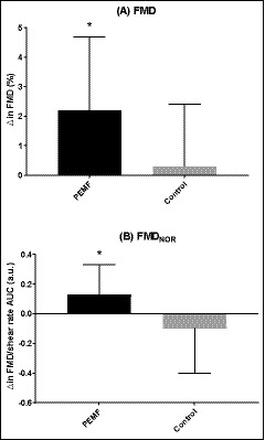
At pre‐assessment, the two groups demonstrated no differences in FMD, FMDNOR, SBP, DBP, and MAP (P > .05, Table 1). After PEMF therapy, the change in FMD from pre‐ to post‐assessment trended toward being significantly different between groups (P = .05). The PEMF group increased FMD (P < .01, Table 2), but the control group did not change (P > .05, Table 2). Figure 1A illustrates the changes in FMD in the PEMF and the control groups. This significant difference between groups remained after normalizing FMD for hyperemia (Figure 1B). There was an increase in FMDNOR in the PEMF group (P < .05, table 2) after treatment; however, FMDNOR in the control group remained the same (P > .05, table 2).
TABLE 2.
Brachial artery measures pre‐ and post‐assessment
| PEMF group (n = 15) | Control group (n = 11) | |
|---|---|---|
| Pre‐assessment | ||
| Baseline diameter (mm) | 4.17 ± 0.74 | 4.51 ± 0.71 |
| Peak diameter (mm) | 4.37 ± 0.73 | 4.73 ± 0.70 |
| FMD (%) | 4.88 ± 3.07 | 4.93 ± 2.17 |
| SRAUC | 27 048 ± 17 148 | 21 974 ± 25 294 |
| FMDNOR (FMD/SRAUC, 10−3) | 0.31 ± 0.3 | 0.45 ± 0.36 |
| Post‐assessment | ||
| Baseline diameter (mm) | 4.21 ± 0.74 | 4.72 ± 0.66 |
| Peak diameter (mm) | 4.51 ± 0.77 | 4.97 ± 0.68 |
| FMD (%)* | 7.09 ± 3.05 # | 5.27 ± 1.84 |
| SRAUC | 28 272 ± 16 576 | 20 071 ± 13 729 |
| FMDNOR (FMD/SRAUC, 10‐3)* | 0.44 ± 0.42 # | 0.38 ± 0.25 |
The data are presented by mean ± standard deviation (SD).
Abbreviations: FMD, Flow‐mediated dilation; PEMF, Pulsed electromagnetic field; SR, Shear rate.
Significant effect of group‐time interaction (ANOVA).
Significant difference between pre‐ and post‐assessment (post hoc comparison).
FIGURE 1.

The changes from pre‐ to post‐assessment. (A) depicts an absolute change (∆) in flow‐mediated dilatation (FMD), and (B) depicts an absolute ∆ in normalized FMD for hyperemia (FMDNOR). * denotes significant changes from pre‐ to post‐assessments (P < .05). The pulsed electromagnetic field group (PEMF, black bar) and the control group (Control, gray bar)
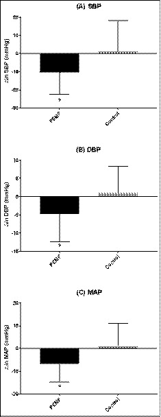
The PEMF group reduced SBP significantly (pre vs post: 144 ± 15 vs 133 ± 10 mm Hg, P < .01) after treatment; however, the control group did not change (pre vs post: 143 ± 11 vs 145 ± 17 mm Hg, P > .05). In addition, while MAP was reduced in the PEMF group (pre vs post: 104 ± 8 vs 98 ± 6 mm Hg, P < .01), it was not altered significantly in the control group (pre vs post: 103 ± 7 vs 105 ± 11 mm Hg, P > .05). Finally, the PEMF group reduced DBP (pre vs post: 85 ± 7 vs 80 ± 6 mm Hg, P < .05); however, the control did not change (pre vs post: 83 ± 7 vs 85 ± 9 mm Hg, P > .05). Figure 2 illustrates the alterations in SBP, DBP, and MAP from pre‐ to post‐assessments. However, there were no direct relationships between the change in FMDNOR and SBP (r = −.289, P > .05) and the change in FMDNOR and MAP (r = −.279, P > .05).
FIGURE 2.

The changes from pre‐ to post‐assessments. (A) depicts an absolute change (Δ) in systolic blood pressure (SBP), (B) depicts an absolute ∆ in diastolic blood pressure (DBP), and (C) depicts an absolute ∆ in mean arterial pressure (MAP). * denotes significant changes from pre‐ to post‐assessments (P < .05). The pulsed electromagnetic field group (PEMF, black bar) and the control group (Control, gray bar)
3.2. Circulating nitric oxide
Nitric oxide level at pre‐assessment was not different between the PEMF group and the control group (P > .05). An analysis with all participants from both groups (n = 26) revealed trends toward moderate inverse relationships between alterations in NO and BP from pre‐ and post‐assessment; NO and SBP (r = −.372, P = .07), NO and DBP (r = −.364, P = .08), and NO and MAP (r = −0.374, P = .07). The PEMF group demonstrated a greater change in NO (pre vs post: 16.7 ± 5.5 vs 22.2 ± 13.1 µmol) than the control group (pre vs post: 18.3 ± 11.1 vs 18.6 ± 13.1 µmol); however, no significant difference was observed (P > .05).
3.3. Heart rate and oxygen saturation
There was a drop in resting heart rate (HR) for those in the PEMF group (pre vs post: 75 ± 14 vs 67 ± 7 bpm) compared to no real change for individuals in the control group (pre vs post: 64 ± 11 vs 62 ± 10 bpm); however, this was not significant (P > .05). No significant alteration in SpO2 was observed in the PEMF group (pre vs post: 99.1 ± 1.0 vs 99.4 ± 0.7%, P > .05) and the control group (pre vs post: 98.7 ± 0.9 vs 99.3 ± 0.8%, P > .05).
4. DISCUSSION
The present study investigated the impact of PEMF therapy on endothelial vascular function and BP in hypertensive humans. In this study, FMD was improved following PEMF therapy and this improvement remained after normalizing FMD for the hyperemic stimulus. In addition, PEMF therapy improved SBP, DBP, and MAP. However, the improvements in FMD and BP were not significantly related. This study supports the hypothesis that 12 weeks of PEMF therapy can improve endothelial vascular function and blood pressure in hypertensive individuals.
Endothelial vascular dysfunction is often impaired in individuals with hypertension, 11 and moreover, an impaired FMD can be a prognostic marker in cardiovascular diseases. 25 Likewise, in the present study both PEMF and the control groups demonstrated an impaired FMD at baseline (pre‐assessment). According to a large community‐based study by Maruhashi et al 7 that investigated the relationship between FMD and cardiovascular (CV) risk factors, FMD decreased as Framingham CV risk factor score increased (FMD as Framingham score increased: ~7.3%, ~6.3%, ~5.8%, and 4.9%, respectively). In addition, a previous study by Dalli et al 26 that investigated the difference in FMD response in healthy individuals, individuals with cardiovascular (CV) risk factor(s), and patients with acute myocardial infarction (AMI), FMD was ~7.8% in healthy individuals, ~5.0% in individuals with CV risk factors, and ~3.3% in patients with AMI. As referred to those studies, an FMD of ~4.88% in the PEMF group and ~4.93% in the control group suggested that both groups demonstrated impairment in endothelial vascular function. Although a direct comparison is somewhat difficult due to inter‐study variations, it can be hypothesized that the change in FMD from 4.88% to ~7.09% in the PEMF group (albeit with a larger degree of variability SD = 3.05%) may be a remarkable improvement and this suggests that PEMF can be a possible technique to improve endothelial vascular function. Plasma NO, which is a suggested mechanism of FMD, was measured in this study but it was not significantly altered following PEMF therapy. This unexpected outcome might be due to several reasons. However, we assume that a smaller sample size in this study seems to be one of the major causes. The measurement of FMD is often thought to be an indirect assessment for NO, and other studies have confirmed this phenomenon. 27 , 28 , 29 , 30 In addition, previous studies demonstrated that PEMF elicited an increase in NO bioavailability. 16 , 19 Other studies demonstrated that PEMF increased potent vasodilators including calcitonin gene‐related peptide (CGRP) 17 , 31 and adenosine A2A receptor, 17 , 32 which consequently lead to an upregulation of NO availability. Indeed, although it was not significant, NO (on average) was increased by ~33% in the PEMF group after the therapy while no change occurred in the control group. Furthermore, a moderate relationship between NO and BP was observed. Accordingly, it is speculated that the sample size of this study was not sufficiently powered to determine significant alterations in NO following PEMF therapy, despite prior observations of increased NO with PEMF therapy. 33 This may explain a confounding outcome in this study that vascular reactivity improved without significant change in NO. In this study, a diameter of the brachial artery was not significantly changed following therapy in both groups; however, FMD was improved only in the PEMF group. This indicates that the PEMF group demonstrated an improved vascular reactivity, which may be related to NO, without a change in diameter.
Alongside the improvements in endothelial vascular function, SBP, DBP, and MAP were also reduced after PEMF therapy. Endothelial vascular function is associated with cardiovascular risk factors in general population. 34 Furthermore, it appears to be impaired in hypertensive population 11 and is significantly associated with BP. 7 A study by Rossi et al 35 demonstrated that vascular dysfunction is more likely a consequence than a cause of hypertension. Nevertheless, a relationship between an improvement in FMD and a reduction in BP has still not been clearly determined. In the present study, although PEMF therapy improved measures of both FMD and BP, improvements in FMD did not correlate with reductions in BP. This may be due to two possible reasons: (a) According to the study by Liu et al, 36 FMD (measured at the brachial artery) is more associated with central aortic pressure than brachial BP, and thus, PEMF therapy improved both FMD and BP, but the magnitude of improvements could be different. (b) FMD was assessed in a supine position, and BP was measure in a sitting position. Brachial BP is often lower with supine position rather than sitting position. 37 Thus postural differences could have influenced a potential relationship between these variables.
This study has some limitations. During participating in this study, participants' medication was not controlled and this might influence the outcomes. However, it also can be assumed that PEMF may possibly provide a synergic effect with anti‐hypertensive drugs on treatment of BP. There were more participants on diabetic medication in the PEMF group than the control group. This might be a confounding factor. In addition, when considering the outcomes from the present study, a larger sample size would be effective to determine the putative mechanism(s) for improvements FMD and BP following PEMF therapy. Aforementioned, the postural difference during measurements of FMD and BP could have limited outcomes.
5. CONCLUSION
Twelve weeks of PEMF therapy improved endothelial vascular function and reduced BP in hypertensive participants. This result may indicate that PEMF therapy can be a potential non‐pharmacological and non‐invasive strategy to manage vascular function and BP in cohorts with peripheral vascular disease as well as hypertension. However, since the optimal strength, frequency, and duration of electromagnetic field emittance have not been determined, therapeutic benefits may vary based on those variables. Accordingly, further studies should attempt to manipulate these variables while examining the putative mechanism(s) of action. Finally, larger clinical studies are needed to assess the clinical applications of PEMF therapy as a targeted non‐pharmacological and non‐invasive option and the influence of confounding factors (eg, obesity and age) on the effects of PEMF therapy.
CONFLICT OF INTEREST
None of authors have financial interests in the research project.
ACKNOWLEDGMENT
The present study was supported by Biomobie Regenerative Medicine, Shanghai, China. The authors would like to thank the participants for their dedicated participation in the study and thank to Kelsey E. Joyce, Girish Pathangey and Jesse C. Schwartz for their help on study coordination and data collection.
Stewart GM, Wheatley‐Guy CM, Johnson BD, Shen WK, Kim C‐H. Impact of pulsed electromagnetic field therapy on vascular function and blood pressure in hypertensive individuals. J Clin Hypertens. 2020;22:1083–1089. 10.1111/jch.13877
Funding information
The present study was funded by Biomobie Regenerative Medicine (Shanghai, China). It is clarified that the sponsor did not involve in the conduction of the study, analysis of the data, interpretation of the data, and drafting, editing, and approving the manuscript.
REFERENCES
- 1. Whelton PK, Carey RM, Aronow WS, et al. 2017 ACC/AHA/AAPA/ABC/ACPM/AGS/APhA/ASH/ASPC/NMA/PCNA guideline for the prevention, detection, evaluation, and management of high blood pressure in adults: a report of the American College of Cardiology/American Heart Association Task Force on Clinical Practice Guidelines. J Am Coll Cardiol. 2018;71(19):e127‐e248. [DOI] [PubMed] [Google Scholar]
- 2. He J, Whelton PK. Elevated systolic blood pressure and risk of cardiovascular and renal disease: overview of evidence from observational epidemiologic studies and randomized controlled trials. Am Heart J. 1999;138(3):S211‐S219. [DOI] [PubMed] [Google Scholar]
- 3. Kasiske BL, Ma JZ, Kalil RSN, Louis TA. Effects of antihypertensive therapy on serum‐lipids. Ann Intern Med. 1995;122(2):133‐141. [DOI] [PubMed] [Google Scholar]
- 4. Oelze M, Knorr M, Kroller‐Schon S, et al. Chronic therapy with isosorbide‐5‐mononitrate causes endothelial dysfunction, oxidative stress, and a marked increase in vascular endothelin‐1 expression. Eur Heart J. 2013;34(41):3206‐3216. [DOI] [PubMed] [Google Scholar]
- 5. Munzel T, Daiber A, Mulsch A. Explaining the phenomenon of nitrate tolerance. Circ Res. 2005;97(7):618‐628. [DOI] [PubMed] [Google Scholar]
- 6. Heitzer T, Schlinzig T, Krohn K, Meinertz T, Münzel T. Endothelial dysfunction, oxidative stress, and risk of cardiovascular events in patients with coronary artery disease. Circulation. 2001;104(22):2673‐2678. [DOI] [PubMed] [Google Scholar]
- 7. Maruhashi T, Soga J, Fujimura N, et al. Relationship between flow‐mediated vasodilation and cardiovascular risk factors in a large community‐based study. Heart. 2013;99(24):1837‐1842. [DOI] [PMC free article] [PubMed] [Google Scholar]
- 8. Panza JA, Casino PR, Kilcoyne CM, Quyyumi AA. Role of endothelium‐derived nitric oxide in the abnormal endothelium‐dependent vascular relaxation of patients with essential hypertension. Circulation. 1993;87(5):1468‐1474. [DOI] [PubMed] [Google Scholar]
- 9. Panza JA, Quyyumi AA, Brush JE Jr, Epstein SE. Abnormal endothelium‐dependent vascular relaxation in patients with essential hypertension. New Engl J Med. 1990;323(1):22‐27. [DOI] [PubMed] [Google Scholar]
- 10. Taddei S, Virdis A, Mattei P, et al. Hypertension causes premature aging of endothelial function in humans. Hypertension. 1997;29(3):736‐743. [DOI] [PubMed] [Google Scholar]
- 11. Panza JA, Quyyumi AA, Brush JE, Epstein SE. Abnormal endothelium‐dependent vascular relaxation in patients with essential‐hypertension. New Engl J Med. 1990;323(1):22‐27. [DOI] [PubMed] [Google Scholar]
- 12. Bassett A. Biological Effects of Electrical and Magnetic Fields. San Diego, CA: Academic Press Inc.; 1994. [Google Scholar]
- 13. Selvam R, Ganesan K, Raju KN, Gangadharan AC, Manohar BM, Puvanakrishnan R. Low frequency and low intensity pulsed electromagnetic field exerts its antiinflammatory effect through restoration of plasma membrane calcium ATPase activity. Life Sci. 2007;80(26):2403‐2410. [DOI] [PubMed] [Google Scholar]
- 14. Iannitti T, Fistetto G, Esposito A, Rottigni V, Palmieri B. Pulsed electromagnetic field therapy for management of osteoarthritis‐related pain, stiffness and physical function: clinical experience in the elderly. Clin Interv Aging. 2013;8:1289‐1293. [DOI] [PMC free article] [PubMed] [Google Scholar]
- 15. Nelson FR, Zvirbulis R, Pilla AA. Non‐invasive electromagnetic field therapy produces rapid and substantial pain reduction in early knee osteoarthritis: a randomized double‐blind pilot study. Rheumatol Int. 2013;33(8):2169‐2173. [DOI] [PubMed] [Google Scholar]
- 16. Diniz P, Soejima K, Ito G. Nitric oxide mediates the effects of pulsed electromagnetic field stimulation on the osteoblast proliferation and differentiation. Nitric Oxide‐Biol Ch. 2002;7(1):18‐23. [DOI] [PubMed] [Google Scholar]
- 17. Graak V, Chaudhary S, Bal BS, Sandhu JS. Evaluation of the efficacy of pulsed electromagnetic field in the management of patients with diabetic polyneuropathy. Int J Diabetes Dev C. 2009;29(2):56‐61. [DOI] [PMC free article] [PubMed] [Google Scholar]
- 18. Heden P, Pilla AA. Effects of pulsed electromagnetic fields on postoperative pain: a double‐blind randomized pilot study in breast augmentation patients. Aesthet Plast Surg. 2008;32(4):660‐666. [DOI] [PubMed] [Google Scholar]
- 19. Bragin DE, Statom GL, Hagberg S, Nemoto EM. Increases in microvascular perfusion and tissue oxygenation via pulsed electromagnetic fields in the healthy rat brain. J Neurosurg. 2015;122(5):1239‐1247. [DOI] [PMC free article] [PubMed] [Google Scholar]
- 20. Kwan RL, Wong WC, Yip SL, Chan KL, Zheng YP, Cheing GL. Pulsed electromagnetic field therapy promotes healing and microcirculation of chronic diabetic foot ulcers: a pilot study. Adv Skin Wound Care. 2015;28(5):212‐219. [DOI] [PubMed] [Google Scholar]
- 21. Mayrovitz HN, Larsen PB. Effects of pulsed electromagnetic‐fields on skin microvascular blood perfusion. Wounds. 1992;4(5):197‐202. [Google Scholar]
- 22. Mckay JC, Prato FS, Thomas AW. A literature review: the effects of magnetic field exposure on blood flow and blood vessels in the microvasculature. Bioelectromagnetics. 2007;28(2):81‐98. [DOI] [PubMed] [Google Scholar]
- 23. Corretti MC, Anderson TJ, Benjamin EJ, et al. Guidelines for the ultrasound assessment of endothelial‐dependent flow‐mediated vasodilation of the brachial artery: a report of the International Brachial Artery Reactivity Task Force. J Am Coll Cardiol. 2002;39(2):257‐265. [DOI] [PubMed] [Google Scholar]
- 24. Thijssen DH, Black MA, Pyke KE, et al. Assessment of flow‐mediated dilation in humans: a methodological and physiological guideline. Am J Physiol‐Heart C. 2011;300(1):H2. [DOI] [PMC free article] [PubMed] [Google Scholar]
- 25. Genta FT, Eleuteri E, Temporelli PL, et al. Flow‐mediated dilation normalization predicts outcome in chronic heart failure patients. J Card Fail. 2013;19(4):260‐267. [DOI] [PubMed] [Google Scholar]
- 26. Dalli E, Segarra L, Ruvira J, et al. Brachial artery flow‐mediated dilation in healthy men, men with risk factors, and men with acute myocardial infarction. Importance of occlusion‐cuff position. Rev Esp Cardiol. 2002;55(9):928‐935. [DOI] [PubMed] [Google Scholar]
- 27. Black MA, Cable NT, Thijssen DHJ, Green DJ. Impact of age, sex, and exercise on brachial artery flow‐mediated dilatation. Am J Physiol‐Heart C. 2009;297(3):H1109‐H1116. [DOI] [PMC free article] [PubMed] [Google Scholar]
- 28. Green DJ, Dawson EA, Groenewoud HMM, Jones H, Thijssen DHJ. Is flow‐mediated dilation nitric oxide mediated? A meta‐analysis. Hypertension. 2014;63(2):376‐382. [DOI] [PubMed] [Google Scholar]
- 29. Joannides R, Haefeli WE, Linder L, et al. Nitric‐oxide is responsible for flow‐dependent dilatation of human peripheral conduit arteries in‐vivo. Circulation. 1995;91(5):1314‐1319. [DOI] [PubMed] [Google Scholar]
- 30. Kooijman M, Thijssen DHJ, de Groot PCE, et al. Flow‐mediated dilatation in the superficial femoral artery is nitric oxide mediated in humans. J Physiol‐London. 2008;586(4):1137‐1145. [DOI] [PMC free article] [PubMed] [Google Scholar]
- 31. Nilsson LAJ, Edvinsson L. Effect of pulsed electromagnetic fields versus placebo on distal microcirculation and release of CGRP in diabetic patients with critical ischemia: A double blind, randomised, cross‐over pilot study. 1997.
- 32. Varani K, Gessi S, Merighi S, et al. Effect of low frequency electromagnetic fields on A(2A) adenosine receptors in human neutrophils. Brit J Pharmacol. 2002;136(1):57‐66. [DOI] [PMC free article] [PubMed] [Google Scholar]
- 33. Kim CH, Wheatley‐Guy CM, Stewart GM, Yeo D, Shen WK, Johnson BD. The impact of pulsed electromagnetic field therapy on blood pressure and circulating nitric oxide levels: a double blind, randomized study in subjects with metabolic syndrome. Blood Press. 2019;1–8. [DOI] [PubMed] [Google Scholar]
- 34. Benjamin EJ, Larson MG, Keyes MJ, et al. Clinical correlates and heritability of flow‐mediated dilation in the community ‐ The Framingham Heart Study. Circulation. 2004;109(5):613‐619. [DOI] [PubMed] [Google Scholar]
- 35. Rossi R, Chiurlia E, Nuzzo A, Cioni E, Origliani G, Modena MG. Flow‐mediated vasodilation and the risk of developing hypertension in healthy postmenopausal women. J Am Coll Cardiol. 2004;44(8):1636‐1640. [DOI] [PubMed] [Google Scholar]
- 36. Liu K, Cui R, Eshak ES, et al. Associations of central aortic pressure and brachial blood pressure with flow mediated dilatation in apparently healthy Japanese men: The Circulatory Risk in Communities Study (CIRCS). Atherosclerosis. 2017;259:46‐50. [DOI] [PubMed] [Google Scholar]
- 37. Netea RT, Smits P, Lenders JWM, Thien T. Does it matter whether blood pressure measurements are taken with subjects sitting or supine? J Hypertens. 1998;16(3):263‐268. [DOI] [PubMed] [Google Scholar]


