PLOS ONE logoLink to PLOS ONE
. 2021 Apr 8;16(4):e0249521. doi: 10.1371/journal.pone.0249521

Effects of supplemental feeding on the fecal bacterial communities of Rocky Mountain elk in the Greater Yellowstone Ecosystem

Claire E Couch 1,*, Benjamin L Wise 2, Brandon M Scurlock 2, Jared D Rogerson 3, Rebecca K Fuda 4, Eric K Cole 5, Kimberly E Szcodronski 6, Adam J Sepulveda 6, Patrick R Hutchins 6, Paul C Cross 6
Editor: Suzanne L Ishaq7
PMCID: PMC8031386  PMID: 33831062

Abstract

Supplemental feeding of wildlife is a common practice often undertaken for recreational or management purposes, but it may have unintended consequences for animal health. Understanding cryptic effects of diet supplementation on the gut microbiomes of wild mammals is important to inform conservation and management strategies. Multiple laboratory studies have demonstrated the importance of the gut microbiome for extracting and synthesizing nutrients, modulating host immunity, and many other vital host functions, but these relationships can be disrupted by dietary perturbation. The well-described interplay between diet, the microbiome, and host health in laboratory and human systems highlights the need to understand the consequences of supplemental feeding on the microbiomes of free-ranging animal populations. This study describes changes to the gut microbiomes of wild elk under different supplemental feeding regimes. We demonstrated significant cross-sectional variation between elk at different feeding locations and identified several relatively low-abundance bacterial genera that differed between fed versus unfed groups. In addition, we followed four of these populations through mid-season changes in supplemental feeding regimes and demonstrated a significant shift in microbiome composition in a single population that changed from natural forage to supplementation with alfalfa pellets. Some of the taxonomic shifts in this population mirrored changes associated with ruminal acidosis in domestic livestock. We discerned no significant changes in the population that shifted from natural forage to hay supplementation, or in the populations that changed from one type of hay to another. Our results suggest that supplementation with alfalfa pellets alters the native gut microbiome of elk, with potential implications for population health.

Introduction

Supplemental feeding of wildlife is a widespread but controversial practice that occurs at human-wildlife interfaces across the globe [1]. Feeding may be undertaken for recreational purposes such as wildlife viewing [2] or hunting [3], or for management purposes such as increasing population density [4] or diverting wildlife movement and feeding patterns to reduce conflict [5]. However, feeding can have unintended consequences such as increased disease transmission [5] or altered species interactions [6]. Understanding how supplemental feeding impacts cryptic aspects of host health is key to optimizing conservation and management decisions as the human-wildlife interface continues to expand and change.

In the past two decades, multiple studies in humans and domestic animals have shown that diet is a key driver of variation in the gut microbiome [710], and aspects of this variation have in turn been shown to associate with host health and disease [11,12]. The gut microbiome plays crucial roles in multiple host functions including nutrient extraction [13], immunity [14], and hormone regulation [15]. An emerging body of research is beginning to suggest intriguing patterns of microbiome variation in wild mammalian populations that may be relevant to conservation [16,17]. There is potential for microbiomes to serve as a tool for conservation efforts such as surveying population health and immunity [18], understanding connectivity between individuals [19] and populations [20], and improving survival prospects of reintroduced or translocated individuals [2123]. However, there is little research directly connecting current management actions, microbiome dynamics, and consequences for host health. The impacts of supplemental feeding programs on the microbiomes of wildlife populations have been directly addressed in only a few studies [24,25], and their findings underscore the importance of clarifying the links between anthropogenic diet inputs, gut microbiome shifts, and downstream impacts on wildlife health.

Covariation between diet and gut microbiome in wildlife depend largely on host phylogeny and environment [26,27]. Temporal variation in gut microbiome communities has been shown to correlate with seasonal fluctuations in diet composition within wild mammalian populations [24,28,29]. However, because diet, social structure, and environmental conditions such as temperature and precipitation often covary alongside seasonal dietary changes, it can be challenging to disentangle their relative effects on microbiome communities. A number of studies have identified gut microbiome discrepancies between captive versus wild populations of conspecific mammals [3032], presumably related to differences in diet. Again though, it is difficult to determine whether diet or one of the other manifold environmental or social differences between captive versus wild populations drives these differences. Findings from the few studies that have addressed the impacts of supplemental feeding in wild populations support the hypothesis that feeding can significantly alter gut community structure in wild hosts [24,25,33]. Because supplemental feeding is a widely used management strategy that is often intended to increase population numbers or reduce human-wildlife conflict, understanding the consequences of supplemental feeding on gut microbiome communities in wildlife, and the consequences for host health, would be of great value to wildlife managers.

Each winter, elk in the Greater Yellowstone Ecosystem are provided with supplemental feed at more than 20 locations (feedgrounds) throughout western Wyoming. Most state-operated feedgrounds provide loose grass, alfalfa, or mixed alfalfa/grass hay beginning in December or January, depending on snowfall conditions, and continues until elk disperse to seek springtime forage in March or April [34]. On the U.S. Fish and Wildlife Service’s National Elk Refuge (NER) near Jackson, WY, USA, elk are provided with compressed alfalfa pellets which provide more concentrated nutritional value than loose hay. Supplemental feeding of elk is highly controversial. Although feeding can mitigate human-wildlife conflict by reducing comingling with livestock and can support large populations in lieu of native habitat, there is concern that feedgrounds act as hotspots for disease transmission [35,36]. Research on the impacts of feedgrounds on disease dynamics in this system is ongoing [37], but other cryptic impacts of feeding, including potential impacts on gut microbiota, have not been explored.

In this study, we assess the impacts of supplemental winter feeding on gut microbiome dynamics among Rocky Mountain elk (Cervus canadensis nelsoni) attending feedgrounds in western Wyoming by describing commensal gut microbiome variation related to supplemental feeding regimes and exploring potential implications for elk population health and disease. We compared cross-sectional samples from active feedgrounds and unfed control groups and assessed longitudinal changes in four of these populations that experienced mid-season changes to feeding regime. We hypothesized that microbiome composition would differ based on feed type in the cross-sectional comparison, and that compositional shifts would correlate with feed regime changes in the longitudinal study. Additionally, we explored possible correlations between diet-driven microbiome changes and elk population health and disease. As part of this exploratory work, we developed an elk-specific assay to assess prevalence and abundance of Fusobacterium necrophorum, a ubiquitous resident of ruminant gastrointestinal (GI) tract microbiomes that has been linked with hoof rot and necrotizing stomatitis in ruminants [38,39] and is a pathogen of concern for elk on Wyoming feedgrounds [40]. Overall, we sought to describe diet-driven alterations to the gut microbiome and identify priorities for future research linking the gut microbiome with elk population health.

Materials & methods

Sample collection & storage

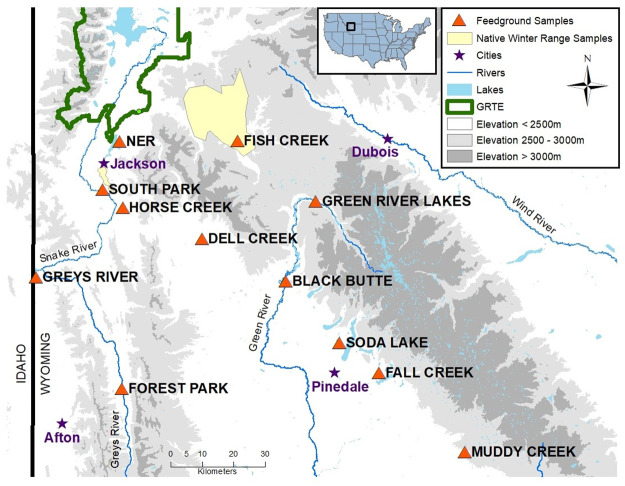
Fresh fecal pellets were collected from elk at twelve feedgrounds and two native winter range sites (Fig 1, Table 1). GPS collar data demonstrates that the vast majority of elk remain at a single feeding location for the duration of winter, rarely dispersing more than 5 km [41], For elk on feedgrounds, sampling was conducted noninvasively from the ground or by habituating elk to feeding in corrals for capture and then directly collecting feces from the rectum. Sub-freezing temperatures typical of western Wyoming during the feeding season enabled us to assess freshness of noninvasively collected feces. We collected samples that were still warm and moist from snow-covered ground and assumed that, under sub-freezing conditions, these samples were likely less than one hour old. Elk are estimated to defecate approximately once every 2–2.5 hours while grazing [42], therefore we assumed that samples at each time point came from different individuals. Fecal samples were collected using sterile gloves and placed in individual whirl-pack sample bags or 50 ml conical tubes. For elk on native winter range, samples were opportunistically collected directly from the rectum when animals were captured via net guns for collaring. On the NER, samples from the first time points (including the cross-sectional time point) were collected noninvasively prior to the initiation of feeding operations, and the following three time points were collected at two, four, and six weeks after feeding commenced. Cross-sectional samples were collected between January 20–27, 2019, and longitudinal samples were collected opportunistically from November 2018-April 2019. Between 8–23 samples were collected per location per time point and stored at -20 °C until processing (Table 1). Hay samples were collected concurrently with cross-sectional fecal samples from feedgrounds, and alfalfa pellet samples were obtained from the NER after feeding commenced. Hay and alfalfa pellet samples were outsourced for nutrient content analysis (A&L Western Laboratories, Modesto, CA). Samples from state-run feedgrounds and native range elk were collected under the supervision of Wyoming Game and Fish Department during routine monitoring and captures, and samples from the NER were collected by USFW personnel during routine monitoring, therefore no project-specific permits were required.

Fig 1. Geographic locations of elk microbiome sample collection sites.

Fig 1

This study included elk from twelve feedgrounds (orange triangles), in addition to unfed elk on native winter range (filled yellow polygons). Longitudinal samples were collected from South Park, Horse Creek, Fish Creek, and the National Elk Refuge (map courtesy of the U.S. Geological Survey).

Table 1. Distribution of elk fecal samples across time, space, and sampling methodologies.

Location Date Collection method Number of samples Feed type
Fish Creek 12/20/2018 Corral 8 Alfalfa/grass hay mix
1/7/2019 Corral 23 Alfalfa/grass hay mix
1/20/2019 Noninvasive 10 Alfalfa/grass mix
Horse Creek 1/14/2019 Noninvasive 10 Grass hay
1/22/2019 Noninvasive 19 Grass hay
4/4/2019 Noninvasive 9 Alfalfa hay
South Park 1/14/2019 Noninvasive 10 Grass hay
1/22/2019 Noninvasive 10 Grass hay
3/11/2019 Noninvasive 10 Alfalfa hay
4/4/2019 Noninvasive 10 Alfalfa hay
National Elk Refuge (NER) 1/21/2019 Noninvasive 10 Natural
2/4/2019 Noninvasive 10 Natural
2/24/2019 Noninvasive 10 Alfalfa pellets
3/8/2019 Noninvasive 10 Alfalfa pellets
3/24/2019 Noninvasive 10 Alfalfa pellets
Black Butte 1/22/2019 Noninvasive 10 Grass hay
Green River Lakes 1/23/2019 Noninvasive 10 Grass hay
Soda Lake 1/25/2019 Noninvasive 10 Grass hay
Grey’s River 1/19/2019 Noninvasive 9 Alfalfa/grass mix
Forest Park 1/19/2019 Noninvasive 10 Alfalfa/grass mix
Alpine 1/19/2019 Noninvasive 9 Alfalfa/grass mix
Muddy Creek 1/22/2019 Noninvasive 10 Alfalfa hay
Dell Creek 1/24/2019 Noninvasive 10 Alfalfa hay
Fall Creek Feedground 1/24/2019 Noninvasive 10 Alfalfa hay
South Jackson Native Winter Range (near South Park) 1/27/2019 Net 9 Natural
Gros Ventre Native Winter Range (near Fish Creek) 11/5/2018 Net 16 Natural

Samples from elk on native winter range were obtained directly from the animals following net-gun capture (Net). Samples from elk on feedgrounds were collected either from the ground (Noninvasive), or by habituating elk to feeding in an enclosure for capture and direct sampling (Corral).

Sample processing & sequencing

DNA extraction, PCR amplification, and 16S sequencing were performed by the Center for Genome Research & Biocomputing at Oregon State University. For each sample, a single fecal pellet was homogenized, and then a 200 mg aliquot was used for DNA extraction according to the Earth Microbiome Protocol [43]. PCR and sequencing of the 16S V4 region were performed according to the Earth Microbiome protocol using amplification primers 515F and 806R [44,45]. Samples were split equally between two MiSeq runs that included a total of 315 elk fecal samples, including the 282 samples used in this study. Details of the F. necrophorum qPCR assay development and validation are provided in S1 Appendix and S2 Table. In addition to nonspecific 16S sequencing, we ran a targeted qPCR assay to detect two subspecies of F. necrophorum (ssp. necrophorum and ssp. funduliforme) [46] while normalizing based on host DNA content [47] (see S1 Appendix for methods).

Statistical analysis

DADA2 (version 1.12.1) was used to identify amplicon sequence variants (ASVs), trim adapter sequences, and remove chimeras [48]. Raw sequence data were processed through the DADA2 pipeline using the following trimming parameters: truncLen = c(240, 200), maxN = 0, maxEE = c(2,2), truncQ = 2, rm.phix = TRUE. Default parameters were used for estimating error parameters using learnErrors(), and chimeras were removed using removeBimeraDenova (method = “consensus”). A total of 22,620,453 reads were obtained from 315 samples following initial preprocessing steps. Prior to statistical analyses, samples with less than 20,000 reads were removed, and the remaining 282 samples were rarified to the minimum sequencing depth of 29,710 reads per sample. All statistical analyses were performed in R version 3.6.3 unless otherwise specified [49].

Microbiome richness was calculated as number of unique ASVs in each sample. Richness and relative taxonomic abundance from phylum-genus ranks were calculated and visualized in the phyloseq package (version 1.30.0) [50]. Inverse Simpson and Shannon diversity indices, both of which incorporate taxonomic evenness in addition to richness, were also calculated in phyloseq [50] to assess whether alpha diversity results were especially sensitive to changes in rare taxa (Shannon) or common taxa (Inverse Simpson). Cross-sectional variation in microbiome richness across feed regimes was assessed using generalized linear mixed models (GLMMs) with feed as a categorical fixed effect and location as a random effect. This model was compared to a null model containing only location as a random effect using a chi-squared test. Both models were generated using the lmer function in the lme4 package based on a Poisson distribution. Results from the richness model were verified using Kruskal-Wallis tests and pairwise Wilcoxon rank-sum tests. For the Inverse Simpson and Shannon diversity metrics, GLMMs with a random effect for location resulted in singularities due to insufficient variance among locations, therefore we relied on Kruskal-Wallis tests and pairwise Wilcoxon rank-sum tests to assess diet-associated variation in these indices. For all pairwise Wilcoxon rank-sum tests, we applied false discovery rate (FDR) correction to resulting p-values.

For microbiome compositional analysis, ASVs were merged by genus for ease of interpretation and to reduce computational intensity. In order to assess inter-group variation among feed types and sampling locations, the nested.npermanova function in the BiodiversityR package (version 2.11–3) [51] was used to perform nested PERMANOVA tests with sample location nested within feed type. To visualize compositional differences between feed types, principal coordinate analysis (PCoA) was run on Bray-Curtis distances between all cross-sectional samples using the ordinate function in phyloseq, and the first three axes were plotted using plot_ordination. To identify taxa that differed significantly between fed versus unfed elk, we used the linear discriminate analysis effect size (LEfSe) approach [52]. Briefly, this method performs non-parametric tests between classes (i.e. feed status) that are consistent among subclasses (i.e. location) to identify significantly different taxa, and then uses linear discriminate analysis to estimate the effect size of each differentially abundant taxon. Taxa that showed significant differences between classes, and were consistent among subclasses, were reported if the effect size was greater than log 2-fold between the two classes. To assess longitudinal changes in microbiome communities associated with changes in diet regime within the four longitudinally sampled populations, we performed PCoA on sample-wise Bray-Curtis distances for each population for visualization and performed nested PERMANOVA tests with collection date nested within feed type. In the population where diet change significantly associated with microbiome shifts, we used LEfSe to identify the significantly different taxa from the phylum through genus levels as described above but using diet as class and collection date as subclass.

Results

Cross-sectional variation in alpha diversity and composition

Among the cross-sectional samples, microbiome richness ranged from 434–1299 unique amplicon sequence variants (ASVs) per sample. The GLMM that included feed type as a fixed effect was significantly better than the location-only null model (chi-square p = 0.0028), indicating that feed regime is a significant driver of variation in richness between locations. A pairwise Wilcoxon rank-sum test supported these results, indicating that richness was significantly lower among unfed compared with supplementally fed elk (p < 0.005 for each pairwise comparison), but that among fed elk, different feed types did not significantly impact richness (Fig 2). Pairwise Wilcoxon rank-sum tests also demonstrated significantly lower alpha diversity index values in unfed versus fed elk (Inverse Simpson FDR-corrected p <0.05 and Shannon FDR-corrected p <0.001 for each pairwise comparison). The top twenty most abundant genera across all populations belonged to seven families comprised in three unique orders representing phyla Firmicutes, Bacteroidetes, and Verrucumicrobia (Fig 2). F. necrophorum funduliforme was identified in only a single fecal sample, and F. necrophorum necrophorum was not detected in any samples, suggesting that these species are rarely or never shed in elk feces.

Fig 2. Relative abundance for the top 20 most abundant genera are shown for each population in the cross-sectional study.
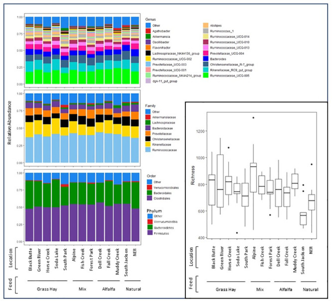
Fig 2

Fill color indicates genus (top left), family (middle left) or order/phylum (bottom left). Amplicon sequence variant-level richness and alpha diversity for each population is shown in the inset (bottom right) where the midline indicates the median values, hinges indicate the first and third quartiles, whiskers extend up to 1.5 the interquartile range, and outliers beyond this range are represented as individual points. Populations are grouped by diet at the time of sample collection. Note that cross-sectional samples from the National Elk Refuge (NER) were collected prior to commencement of feeding at that location.

Diet-related microbiome differences among populations

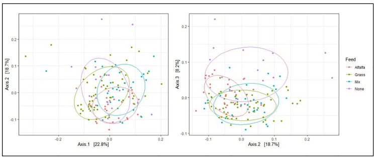
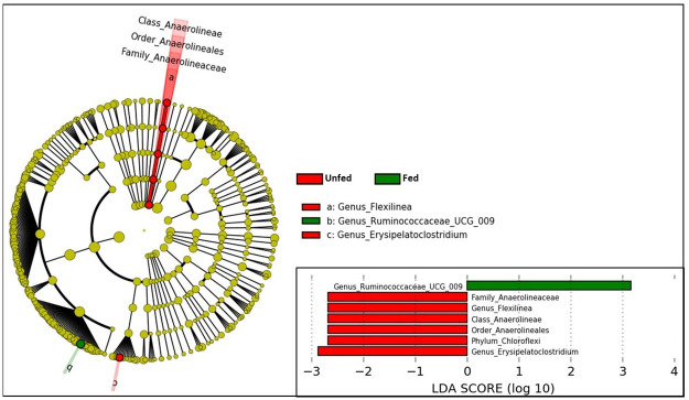
Significant compositional differences between location were observed (p = 0.0001) based on nested PERMANOVA tests, but differences between feed type were marginal (p = 0.07). Based on visualization of PCoA axes 1–2, which collectively accounted for 41.5% of the variance among samples, differences among feed types were not visually apparent (Fig 3). This pattern aligns with Fig 2, which shows no obvious compositional differences related to feed type in the most abundant genera. However, separation along PCoA axis 3, which accounted for 8.2% of the variance among samples, suggested that unfed elk differed from other feed regimes along that axis. In support of this finding, LEfSe revealed that several low-abundance taxa significantly differed among fed vs unfed elk after accounting for location (Fig 4). Genus Ruminococcaceae UCG-009 was enriched in fed elk, whereas genera Erysipelatoclostridium and Flexilinea (and parent clades through the phylum level) were enriched in unfed elk (Fig 4).

Fig 3. Principal coordinate analysis of cross-sectional elk gut microbiomes sampled from 13 locations, including 11 feedgrounds stratified among three different feed types and two unfed control groups.

Fig 3

The left panel shows the first two principal coordinate axes, and the right panel shows the second and third axes. Collectively, the first three axes explained 49.7% of the variance among samples. Ellipses are drawn around 70% of the data points in each feed group.

Fig 4. Differentially abundant microbial taxa between fed and unfed elk.

Fig 4

Linear discriminate analysis was used to identify taxa (phylum-genus levels) that exhibited log-two-fold abundance changes between the two groups.

Longitudinal microbiome shifts related to feed change

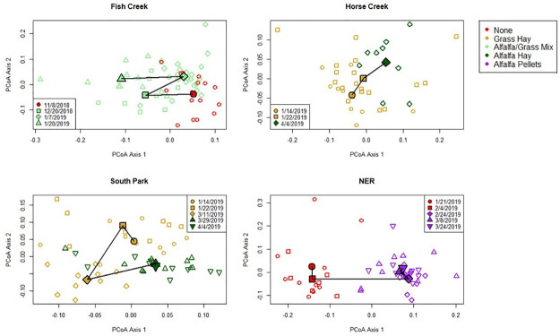
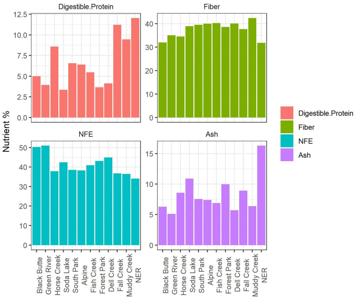
We longitudinally sampled elk at 4 different locations before and after feed regime transitions to determine whether the change in feed type resulted in a change to microbial communities. In the longitudinal series, CCA and nested PERMANOVA tests revealed significant differences between pre- and post-feed samples only within the NER population, which transitioned from no supplemental feed (“natural diet”) to pelleted alfalfa (Fig 5). No significant taxonomic changes were identified in the population that transitioned from natural diet to alfalfa/grass mix (Fish Creek) or in either of the populations that transitioned from grass hay to alfalfa hay (South Park & Horse Creek). Nutrient analysis demonstrated that that pelleted alfalfa had lower fiber content and somewhat lower NFE (soluble carbohydrates), but higher protein and ash (mineral) content than any of the supplemental hay for which nutrient analysis was conducted (Fig 6, see S1 Table for full nutrient analysis results). The compositional shift in the NER population was characterized by phylum-level increases in Firmicutes, Spirochaetes, and Tenericutes, and decreases in Bacteroidetes, Chloroflexi, Plantomycetes, Proteobacteria, and Verrucromicrobia (Fig 7). Additional shifts at lower taxonomic levels are shown in S1 Fig.

Fig 5. Principal coordinate analysis plots of gut microbiome samples from populations that underwent mid-season shifts in feeding regime over the course of the study.

Fig 5

Principal coordinate analysis was performed on Bray-Curtis distances separately for each population, therefore axes do not represent the same dimensions for each plot. Individual samples are represented by unfilled symbols, and population centroids for each time point are represented by filled symbols. Color represents feed regime, and shape indicates sample date. Centroids for each sampling date are connected in temporal order with solid lines to highlight compositional shifts over time.

Fig 6. Comparison of macronutrient levels in the hay and pellets fed to elk at the Wyoming feedgrounds in this study.

Fig 6

We measured percentages of digestible protein, fiber, nitrogen-free extract (soluble, non-fiber carbohydrates), and ash (total mineral content). Feed samples from Horse Creek and South Park were collected before the transition from grass hay to alfalfa hay, and feed samples from the National Elk Refuge were collected following the commencement of feeding operations in the longitudinal study.

Fig 7. Linear discriminate analysis effect size (LEfSe) results showing bacterial lineages that differed significantly in a population before and after supplemental feeding with concentrated alfalfa pellets.
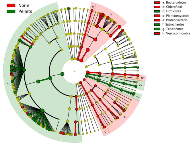
Fig 7

Each node in the tree represents a taxon from phylum level through genus level (tips). Nodes are colored red if they were enriched under the natural diet regime, and green if they were enriched under the pelleted feed regime. Yellow nodes did not differ significantly across regimes. Branches are highlighted red if they belong to phyla that were enriched under the natural diet regime, and green if they belong to phyla that were enriched in pellet-fed elk.

Discussion

We assessed cross-sectional and longitudinal variation in microbiome diversity and composition among wild elk under supplemental feeding regimes compared with those under natural foraging conditions. Our results suggest that feeding supplemental loose hay (grass, alfalfa, or mix) associates with changes to only a few low-abundance taxa, and that location is more predictive of gut microbiome than feeding regime for hay-based supplemented diets. In contrast, feeding concentrated alfalfa pellets appears to generate significant shifts in gut microbiome composition compared to natural foraging conditions on the National Elk Refuge, such that these microbiome shifts associated with concentrated feed could have implications for elk population health.

Microbiome communities across locations in the cross-sectional study demonstrated compositional patterns consistent with those reported in studies of other wild ungulates. Across populations, the top twenty bacterial genera were predominantly from phylum Firmicutes, followed by Bacteroidetes, with a small proportion from phylum Veruccumicrobia. This result aligns with microbiome composition from fecal samples previously described in elk [53]. Richness and alpha diversity were lower in unfed elk relative to fed elk, but beta diversity was not significantly associated with diet after controlling for location. The number of population replicates in the cross-sectional study was limited, and future studies should include additional replicates from each feed group, particularly with more samples from unfed control animals, in order to robustly assess the impacts of hay supplementation on microbiome alpha diversity and composition. Although overall beta diversity estimates did not depend on diet, a few genera differed between fed and unfed populations. Genus Ruminococcus UCG-009 was enriched in fed elk, whereas Flexilinea and Erysipelatoclostridium were enriched in unfed elk. Ruminococcus UCG-009 has also been shown to be enriched in captive versus wild Pere David’s deer [31], and Flexilinea and Erysipelatoclostridium vary temporally in other wild herbivores [54,55], presumably due to fluctuating forage availability. Overall, these findings suggest that elk gut microbiome composition is relatively robust to dietary changes associated with hay supplementation, but changes to a few key taxa are consistent with patterns identified in studies of other wild herbivore species.

Significant longitudinal shifts in microbiome composition occurred in the NER population after transitioning from a natural diet to supplementation with alfalfa pellets, possibly due to reduced fiber or increased protein or mineral content relative to other supplemental feed types (Fig 6). LEfSE analysis demonstrated significant increases in 38 taxa and decreases in 49 taxa following the transition to supplemental pellets. At the phylum level, taxonomic shifts included a reduction of Bacteroidetes, Chloroflexi, Plantomycetes, Verrucumicrobia, and Proteobacteria, and an increase in Firmicutes, Spirochaetes, and Tenericutes following the transition from natural diet to supplementation with concentrated alfalfa pellets. Interestingly, a subset of these taxa is associated with host immunity in laboratory systems. For example, members of phylum Bacteroidetes contribute to the development of gut-associated lymphoid tissues [56], activating the T-cell-dependent immune response [57], and other host immune functions [58]. Recent research also suggests that members of Verrucumicrobia have the potential to induce regulatory immunity in horses [59]. Laboratory studies demonstrate that increasing the Firmicutes:Bacteroidetes ratio reduces short-chain fatty acid production, and it is speculated that these shifts could reduce microglial activity and promote prion diseases; however, this association has not been confirmed [60]. It is therefore possible that some of the diet-driven changes in bacterial relative abundance observed in elk on the NER may associate with changes in immune function, but further research is necessary to directly relate immunity with microbiome community structure in elk.

In addition to potential associations with immunity, the gut microbiome dynamics of elk on the NER may offer other insights into the physiological impacts of supplemental feeding. Elk are intermediate (mixed) ruminants with relatively flexible feeding strategies [61] and significant reliance on microbes in the reticulorumen and hindgut for nutrient extraction. In domestic mixed ruminants, the gastrointestinal microbiome adapts rapidly to diet change [62], and some microbial changes induced by dramatic feed alteration have been linked to rumen acidosis [63] The reduction we observed in Proteobacteria and Verrucomicrobia in elk fed alfalfa pellets reflect some of the changes associated with rumen acidosis in the fecal microbiomes of domestic cattle [64], and the increase in Firmicutes mirrors the change in abundance of this phylum in the rumen of cattle with this condition [64]. Due to physiological differences between elk and cattle, we cannot assume analogous rumen acidosis microbiome phenotypes, but this finding warrants further exploration. Previous work has shown that rumen acidosis is a leading cause of death among captive elk [65], and gastritis of unknown etiology was observed in necropsies of approximately 20% of apparently ill, pellet-fed elk during winter feeding operations at the NER from 2009–2013 (L. Jones, prs. comm.). Understanding the impact of supplemental feeding on this syndrome is crucial to informing management practices. More research is needed to characterize the fecal microbiome shifts associated with rumen acidosis in elk, a question which could be addressed by collecting and comparing rumen and microbiome samples in intensively studied wild or captive elk. Future studies should also account for host demographics, including age and sex, which were not included in this study. This information could then be used to assess the impacts of feed on rumen acidosis via noninvasive fecal microbiome sampling.

The elk microbiome is known to vary significantly along the gastrointestinal tract, thus the relative robustness of the fecal microbiome to hay supplementation does not necessarily reflect robustness along the entire GI tract [53]. In domestic ruminants, the foregut microbiome has higher richness and may be more responsive to feed changes than the fecal microbiome [66] (Lourenco et al. 2020). Therefore, while the fecal microbiomes of ruminants are easily sampled noninvasively, they represent only a subset of the complex and variable gastrointestinal tract microbiome and must be interpreted with care. Notably, some commensal GI bacteria are ubiquitous among their hosts but are rarely shed in the feces, and therefore any potential effects of supplemental feeding on these bacteria remain cryptic. Based on a recent study in domestic sheep, this is likely the case with F. necrophorum [67], which would account for the non-detection of this widespread ruminant commensal in elk feces. Future studies should assess changes to the microbiome of the rumen and other sites along the GI tract that occur as a result of hay supplementation, including changes in F. necrophorum abundance and distribution.

Our work suggests that supplementation with hay (grass, alfalfa, or mix) has a much smaller impact on fecal microbiome composition than concentrated alfalfa pellets. Shifts in microbiome composition observed in an elk population that transitioned from natural feed to supplemental concentrate may be related to immune functioning or to subacute rumen acidosis in elk and therefore warrant further investigation. More broadly, this study underscores the potential of gut microbiome studies as a tool for noninvasive monitoring of population health in wildlife conservation efforts.

Supporting information

S1 Fig. LEfSe results showing bacterial taxa that differed significantly in elk gut microbiomes before and after supplementary feeding with concentrated alfalfa pellets.

(TIF)

S1 Table. Full nutrient analysis results.

(DOCX)

S2 Table. Oligonucleotides used in multiplex qPCR assays for F. necrophorum.

(DOCX)

S1 Appendix. Design and optimization of Fusobacterium necrophorum qPCR assay.

(DOCX)

Acknowledgments

We thank Anna Jolles, Thomas Sharpton, Hank Edwards, and Lee Jones for their guidance with manuscript preparation. Any use of trade, firm, or product names is for descriptive purposes only and does not imply endorsement by the U.S. Government.

Data Availability

Sequencing data are available in the NCBI Sequence Read Archive and publicly accessible under BioProject ID PRJNA629905.

Funding Statement

Funding for this project was provided by the NSF Graduate Research Internship Program (https://www.nsf.gov/funding/pgm_summ.jsp?pims_id=505127, awarded to CEC) and the USGS Ecosystems and Environmental Health Mission Areas (https://www.usgs.gov/mission-areas/environmental-health, awarded to PCC). The funders had no role in study design, data collection and analysis, decision to publish, or preparation of the manuscript.

References

  • 1.Steyaert SM, Kindberg J, Jerina K, Krofel M, Stergar M, Swenson JE, et al. Behavioral correlates of supplementary feeding of wildlife: can general conclusions be drawn? Basic Appl Ecol. 2014; 15(8):669–76. [Google Scholar]
  • 2.Galbraith JA, Jones DN, Beggs JR, Parry K, Stanley MC. Urban bird feeders dominated by a few species and individuals. Front Ecol Evol. 2017;5:81. [Google Scholar]
  • 3.Draycott RAH, Woodburn MIA, Carroll JP, Sage RB. Effects of spring supplementary feeding on population density and breeding success of released pheasants Phasianus colchicus in Britain. Wildlife Biol. 2005;11(3):177–82. [Google Scholar]
  • 4.Elliott GP, Merton DV, Jansen PW. Intensive management of a critically endangered species: the kakapo. Biol Conserv. 2001;99(1):121–33. [Google Scholar]
  • 5.Putman RJ, Staines BW. Supplementary winter feeding of wild red deer Cervus elaphus in Europe and North America: justifications, feeding practice and effectiveness. Mamm Rev. 2004;34(4):285–306. [Google Scholar]
  • 6.Cortés-Avizanda A, Carrete M, Serrano D, Donázar JA. Carcasses increase the probability of predation of ground-nesting birds: a caveat regarding the conservation value of vulture restaurants. Anim Conserv. 2009;12:85–8. [Google Scholar]
  • 7.David LA, Maurice CF, Carmody RN, Gootenberg DB, Button JE, Wolfe BE, et al. Diet rapidly and reproducibly alters the human gut microbiome. Nature. 2014;505(7484):559–63. 10.1038/nature12820 [DOI] [PMC free article] [PubMed] [Google Scholar]
  • 8.Wu GD, Chen J, Hoffmann C, Bittinger K, Chen Y-Y, Keilbaugh SA, et al. Linking long-term dietary patterns with gut microbial enterotypes. Science. 2011;334(6052):105–8. 10.1126/science.1208344 [DOI] [PMC free article] [PubMed] [Google Scholar]
  • 9.Campbell SC, Wisniewski PJ, Noji M, McGuinness LR, Häggblom MM, Lightfoot SA, et al. The effect of diet and exercise on intestinal integrity and microbial diversity in mice. PLoS One. 2016;11(3):e0150502. 10.1371/journal.pone.0150502 [DOI] [PMC free article] [PubMed] [Google Scholar]
  • 10.Liu J, Xue C, Sun D, Zhu W, Mao S. Impact of high-grain diet feeding on mucosa-associated bacterial community and gene expression of tight junction proteins in the small intestine of goats. MicrobiologyOpen. 2019; 8:e00745. 10.1002/mbo3.745 [DOI] [PMC free article] [PubMed] [Google Scholar]
  • 11.Honda K, Littman DR. The microbiome in infectious disease and inflammation. Annu Rev Immunol. 2012;30:759–95. 10.1146/annurev-immunol-020711-074937 [DOI] [PMC free article] [PubMed] [Google Scholar]
  • 12.Gentile CL, Weir TL. The gut microbiota at the intersection of diet and human health. Science. 2018;362(6416):776–80. 10.1126/science.aau5812 [DOI] [PubMed] [Google Scholar]
  • 13.Krajmalnik-Brown R, Ilhan Z-E, Kang D-W, DiBaise JK. Effects of gut microbes on nutrient absorption and energy regulation. Nutr Clin Pract. 2012;27(2):201–14. 10.1177/0884533611436116 [DOI] [PMC free article] [PubMed] [Google Scholar]
  • 14.Kau AL, Ahern PP, Griffin NW, Goodman AL, Gordon JI. Human nutrition, the gut microbiome and the immune system. Nature. 2011;474(7351):327–36. 10.1038/nature10213 [DOI] [PMC free article] [PubMed] [Google Scholar]
  • 15.Markle JGM, Frank DN, Mortin-Toth S, Robertson CE, Feazel LM, Rolle-Kampczyk U, et al. Sex differences in the gut microbiome drive hormone-dependent regulation of autoimmunity. Science. 2013;339(6123):1084–8. 10.1126/science.1233521 [DOI] [PubMed] [Google Scholar]
  • 16.Bahrndorff S, Alemu T, Alemneh T, Lund Nielsen J. The microbiome of animals: implications for conservation biology. Int J Genomics Proteomics. 2016;2016:5304028. 10.1155/2016/5304028 [DOI] [PMC free article] [PubMed] [Google Scholar]
  • 17.Trevelline BK, Fontaine SS, Hartup BK, Kohl KD. Conservation biology needs a microbial renaissance: a call for the consideration of host-associated microbiota in wildlife management practices. Proc Biol Sci. 2019;286(1895):20182448. 10.1098/rspb.2018.2448 [DOI] [PMC free article] [PubMed] [Google Scholar]
  • 18.Apprill A, Robbins J, Eren AM, Pack AA, Reveillaud J, Mattila D, et al. Humpback whale populations share a core skin bacterial community: towards a health index for marine mammals? PLoS One. 2014;9(3):e90785. 10.1371/journal.pone.0090785 [DOI] [PMC free article] [PubMed] [Google Scholar]
  • 19.Tung J, Barreiro LB, Burns MB, Grenier J-C, Lynch J, Grieneisen LE, et al. Social networks predict gut microbiome composition in wild baboons. Elife. 2015;; 4:e05224. 10.7554/eLife.05224 [DOI] [PMC free article] [PubMed] [Google Scholar]
  • 20.Couch CE, Arnold HK, Crowhurst RS, Jolles AE, Sharpton TJ, Witczak MF, et al. Bighorn sheep gut microbiomes associate with genetic and spatial structure across a metapopulation. Sci Rep. 2020;10(1):6582. 10.1038/s41598-020-63401-0 [DOI] [PMC free article] [PubMed] [Google Scholar]
  • 21.Xie Y, Xia P, Wang H, Yu H, Giesy JP, Zhang Y, et al. Effects of captivity and artificial breeding on microbiota in feces of the red-crowned crane (Grus japonensis). Sci Rep. 2016;6:33350. 10.1038/srep33350 [DOI] [PMC free article] [PubMed] [Google Scholar]
  • 22.Yao R, Xu L, Hu T, Chen H, Qi D, Gu X, et al. The “wildness” of the giant panda gut microbiome and its relevance to effective translocation. Glob Ecol Conserv. 2019;18:e00644. [Google Scholar]
  • 23.Chong R, Grueber CE, Fox S, Wise P, Barrs VR, Hogg CJ, et al. Looking like the locals—gut microbiome changes post-release in an endangered species. Anim Microbiome. 2019;1(1):8. 10.1186/s42523-019-0012-4 [DOI] [PMC free article] [PubMed] [Google Scholar]
  • 24.Ren T, Boutin S, Humphries MM, Dantzer B, Gorrell JC, Coltman DW, et al. Seasonal, spatial, and maternal effects on gut microbiome in wild red squirrels. Microbiome. 2017;5(1):163. 10.1186/s40168-017-0382-3 [DOI] [PMC free article] [PubMed] [Google Scholar]
  • 25.Ricci S, Sandfort R, Pinior B, Mann E, Wetzels SU, Stalder G. Impact of supplemental winter feeding on ruminal microbiota of roe deer Capreolus capreolus. Wildlife Biol. 2019;1:1–11. [Google Scholar]
  • 26.Youngblut ND, Reischer GH, Walters W, Schuster N, Walzer C, Stalder G, et al. Host diet and evolutionary history explain different aspects of gut microbiome diversity among vertebrate clades. Nat Commun. 2019;10(1):2200. 10.1038/s41467-019-10191-3 [DOI] [PMC free article] [PubMed] [Google Scholar]
  • 27.Kartzinel TR, Hsing JC, Musili PM, Brown BRP, Pringle RM. Covariation of diet and gut microbiome in African megafauna. Proc Natl Acad Sci U S A. 2019;116(47):23588–93. 10.1073/pnas.1905666116 [DOI] [PMC free article] [PubMed] [Google Scholar]
  • 28.Hicks AL, Lee KJ, Couto-Rodriguez M, Patel J, Sinha R, Guo C, et al. Gut microbiomes of wild great apes fluctuate seasonally in response to diet. Nat Commun. 2018;9(1):1786. 10.1038/s41467-018-04204-w [DOI] [PMC free article] [PubMed] [Google Scholar]
  • 29.Bergmann GT, Craine JM, Robeson MS 2nd, Fierer N. Seasonal shifts in diet and gut microbiota of the American bison (Bison bison). PLoS One. 2015;10(11):e0142409. 10.1371/journal.pone.0142409 [DOI] [PMC free article] [PubMed] [Google Scholar]
  • 30.McKenzie VJ, Song SJ, Delsuc F, Prest TL, Oliverio AM, Korpita TM, et al. The effects of captivity on the mammalian gut microbiome. Integr Comp Biol. 2017;57(4):690–704. 10.1093/icb/icx090 [DOI] [PMC free article] [PubMed] [Google Scholar]
  • 31.Sun C-H, Liu H-Y, Liu B, Yuan B-D, Lu C-H. Analysis of the gut microbiome of wild and captive Père David’s deer. Front Microbiol. 2019;10:2331. 10.3389/fmicb.2019.02331 [DOI] [PMC free article] [PubMed] [Google Scholar]
  • 32.Guan Y, Yang H, Han S, Feng L, Wang T, Ge J. Comparison of the gut microbiota composition between wild and captive sika deer (Cervus nippon hortulorum) from feces by high-throughput sequencing. AMB Express. 2017;7(1):212. 10.1186/s13568-017-0517-8 [DOI] [PMC free article] [PubMed] [Google Scholar]
  • 33.Menke S, Heurich M, Henrich M, Wilhelm K, Sommer S. Impact of winter enclosures on the gut bacterial microbiota of red deer in the Bavarian Forest National Park. Wildlife Biol. 2019;1:1–10. [Google Scholar]
  • 34.Cross PC, Maichak EJ, Rogerson JD, Irvine KM, Jones JD, Heisey DM, et al. Estimating the phenology of elk brucellosis transmission with hierarchical models of cause-specific and baseline hazards. J Wildl Manage. 2015;79(5):739–48. [Google Scholar]
  • 35.Cross PC, Edwards WH, Scurlock BM, Maichak EJ, Rogerson JD. Effects of management and climate on elk brucellosis in the Greater Yellowstone Ecosystem. Ecol Appl. 2007;17(4):957–64. 10.1890/06-1603 [DOI] [PubMed] [Google Scholar]
  • 36.Hines AM, Ezenwa VO, Cross P, Rogerson JD. Effects of supplemental feeding on gastrointestinal parasite infection in elk (Cervus elaphus): preliminary observations. Vet Parasitol. 2007;148(3–4):350–5. 10.1016/j.vetpar.2007.07.006 [DOI] [PubMed] [Google Scholar]
  • 37.Cotterill GG, Cross PC, Cole EK, Fuda RK, Rogerson JD, Scurlock BM, et al. Winter feeding of elk in the Greater Yellowstone Ecosystem and its effects on disease dynamics. Philos Trans R Soc Lond B Biol Sci. 2018;373(1745). 10.1098/rstb.2017.0093 [DOI] [PMC free article] [PubMed] [Google Scholar]
  • 38.Bennett G, Hickford J, Sedcole R, Zhou H. Dichelobacter nodosus, Fusobacterium necrophorum and the epidemiology of footrot. Anaerobe. 2009;15(4):173–6. 10.1016/j.anaerobe.2009.02.002 [DOI] [PubMed] [Google Scholar]
  • 39.Yeruham I, Elad D. Necrotizing stomatitis associated with Fusobacterium necrophorum in two goats. J Vet Med B Infect Dis Vet Public Health. 2004;51(1):46–7. 10.1046/j.1439-0450.2003.00717.x [DOI] [PubMed] [Google Scholar]
  • 40.Wyoming Game & Fish Department 2015 Annual Report. Cheyanne, WY: Wyoming Game & Fish Department, 2015.
  • 41.Merkle JA, Cross PC, Scurlock BM, Cole EK, Courtemanch AB, Dewey SR, et al. Linking spring phenology with mechanistic models of host movement to predict disease transmission risk. J Appl Ecol. 2018;55: 810–819. [Google Scholar]
  • 42.Collins WB, Urness P. Elk pellet group distributions and rates of deposition in aspen and lodgepole pine habitats. 1979. PhD Dissertation. https://digitalcommons.usu.edu/cgi/viewcontent.cgi?article=5691&context=aspen_bib.
  • 43.Marotz C, Amir A, Humphrey G, Gaffney J, Gogul G, Knight R. DNA extraction for streamlined metagenomics of diverse environmental samples. Biotechniques. 2017;62(6):290–3. 10.2144/000114559 [DOI] [PubMed] [Google Scholar]
  • 44.Caporaso JG, Lauber CL, Walters WA, Berg-Lyons D, Lozupone CA, Turnbaugh PJ, et al. Global patterns of 16S rRNA diversity at a depth of millions of sequences per sample. Proc Natl Acad Sci U S A. 2011;108 Suppl 1:4516–22. [DOI] [PMC free article] [PubMed] [Google Scholar]
  • 45.Caporaso JG, Lauber CL, Walters WA, Berg-Lyons D, Huntley J, Fierer N, et al. Ultra-high-throughput microbial community analysis on the Illumina HiSeq and MiSeq platforms. ISME J. 2012;6(8):1621–4. 10.1038/ismej.2012.8 [DOI] [PMC free article] [PubMed] [Google Scholar]
  • 46.Jensen A, Hagelskjaer Kristensen L, Prag J. Detection of Fusobacterium necrophorum subsp. funduliforme in tonsillitis in young adults by real-time PCR. Clin Microbiol Infect. 2007;13(7):695–701. 10.1111/j.1469-0691.2007.01719.x [DOI] [PubMed] [Google Scholar]
  • 47.Kaltenbrunner M, Hochegger R, Cichna-Markl M. Red deer (Cervus elaphus)-specific real-time PCR assay for the detection of food adulteration. Food Control. 2018;89:157–66. [DOI] [PubMed] [Google Scholar]
  • 48.Callahan BJ, McMurdie PJ, Rosen MJ, Han AW, Johnson AJA, Holmes SP. DADA2: High-resolution sample inference from Illumina amplicon data. Nat Methods. 2016;13(7):581–3. 10.1038/nmeth.3869 [DOI] [PMC free article] [PubMed] [Google Scholar]
  • 49.R Core Team. R: A language and environment for statistical computing. 2020. https://www.R-project.org/.
  • 50.Callahan BJ, Sankaran K, Fukuyama JA, McMurdie PJ, Holmes SP. Bioconductor workflow for microbiome data analysis: from raw reads to community analyses. F1000Res. 2016;5:1492. 10.12688/f1000research.8986.2 [DOI] [PMC free article] [PubMed] [Google Scholar]
  • 51.Kindt R, Coe R. Tree diversity analysis: a manual and software for common statistical methods for ecological and biodiversity studies. World Agroforestry Centre. 2005; 196. [Google Scholar]
  • 52.Segata N, Izard J, Waldron L, Gevers D, Miropolsky L, Garrett WS, et al. Metagenomic biomarker discovery and explanation. Genome Biol. 2011;12(6):R60. 10.1186/gb-2011-12-6-r60 [DOI] [PMC free article] [PubMed] [Google Scholar]
  • 53.Kim J-H, Hong SW, Park B-Y, Yoo JG, Oh M-H. Characterisation of the bacterial community in the gastrointestinal tracts of elk (Cervus canadensis). Antonie Van Leeuwenhoek. 2019;112(2):225–35. 10.1007/s10482-018-1150-5 [DOI] [PubMed] [Google Scholar]
  • 54.Gao H, Chi X, Li G, Qin W, Song P, Jiang F, et al. Gut microbial diversity and stabilizing functions enhance the plateau adaptability of Tibetan wild ass (Equus kiang). MicrobiologyOpen. 2020;e1025. [DOI] [PMC free article] [PubMed] [Google Scholar]
  • 55.Haworth SE, White KS, Côté SD, Shafer ABA. Space, time and captivity: quantifying the factors influencing the fecal microbiome of an alpine ungulate. FEMS Microbiol Ecol. 2019;95(7). 10.1093/femsec/fiz095 [DOI] [PubMed] [Google Scholar]
  • 56.Rhee K-J, Sethupathi P, Driks A, Lanning DK, Knight KL. Role of commensal bacteria in development of gut-associated lymphoid tissues and preimmune antibody repertoire. J Immunol; 172:1118–24. 10.4049/jimmunol.172.2.1118 [DOI] [PubMed] [Google Scholar]
  • 57.Mazmanian SK, Liu CH, Tzianabos AO, Kasper DL. An immunomodulatory molecule of symbiotic bacteria directs maturation of the host immune system. Cell. 2005;122(1):107–18. 10.1016/j.cell.2005.05.007 [DOI] [PubMed] [Google Scholar]
  • 58.Wexler HM. Bacteroides: the good, the bad, and the nitty-gritty. Clin Microbiol Rev. 2007;20(4):593–621. 10.1128/CMR.00008-07 [DOI] [PMC free article] [PubMed] [Google Scholar]
  • 59.Lindenberg F, Krych L, Fielden J, Kot W, Frøkiær H, van Galen G, et al. Expression of immune regulatory genes correlate with the abundance of specific Clostridiales and Verrucomicrobia species in the equine ileum and cecum. Sci Rep. 2019;9(1):12674. 10.1038/s41598-019-49081-5 [DOI] [PMC free article] [PubMed] [Google Scholar]
  • 60.D’Argenio V, Sarnataro D. Microbiome Influence in the Pathogenesis of Prion and Alzheimer’s Diseases. Int J Mol Sci. 2019;20(19). 10.3390/ijms20194704 [DOI] [PMC free article] [PubMed] [Google Scholar]
  • 61.Hofmann RR, Steward DRM. Grazer or browser: a classification based on the stomach-structure and feeding habits of east African ruminants. Mamalia. 1972; 36:226–240. [Google Scholar]
  • 62.Saro C, Ranilla MJ, Tejido ML, Carro MD. Influence of forage type in the diet of sheep on rumen microbiota and fermentation characteristics. Livest Sci. 2014; 160:52–59. [Google Scholar]
  • 63.Khafipour E, Shucong L, Plaizier JC, Krause DO. Rumen microbiome composition determined using two nutritional models of subacute ruminal acidosis. Appl Environ Microbiol. 2009; 75(22):7115–24. 10.1128/AEM.00739-09 [DOI] [PMC free article] [PubMed] [Google Scholar]
  • 64.Plaizier JC, Li S, Danscher AM, Derakshani H, Andersen PH, Khafipour E. Changes in microbiota in rumen digesta and feces due to a grain-based subacute ruminal acidosis (SARA) challenge. Microb Ecol. 2017;74(2):485–95. 10.1007/s00248-017-0940-z [DOI] [PubMed] [Google Scholar]
  • 65.Hattel AL, Shaw DP, Fisher JS, Brooks JW, Love BC, Drake TR, et al. Mortality in Pennsylvania captive elk (Cervus elaphus): 1998–2006. J Vet Diagn Invest. 2007;19(3):334–7. 10.1177/104063870701900322 [DOI] [PubMed] [Google Scholar]
  • 66.Lourenco JM, Kieran TJ, Seidel DS, et al. Comparison of the ruminal and fecal microbiotas in beef calves supplemented or not with concentrate. PLoS One. 2020; 15(4): e0231533. 10.1371/journal.pone.0231533 [DOI] [PMC free article] [PubMed] [Google Scholar]
  • 67.Clifton R, Giebel K, Liu NLBH, Purdy KJ, Green LE. Sites of persistence of Fusobacterium necrophorum and Dichelobacter nodosus: a paradigm shift in understanding the epidemiology of footrot in sheep. Sci Rep. 2019;9(1):14429. 10.1038/s41598-019-50822-9 [DOI] [PMC free article] [PubMed] [Google Scholar]

Decision Letter 0

Juan J Loor

30 Jun 2020

PONE-D-20-13745

Effects of supplemental feeding on the gut microbiomes of Rocky Mountain elk in the Greater Yellowstone Ecosystem

PLOS ONE

Dear Dr. Couch,

Thank you for submitting your manuscript to PLOS ONE. After careful consideration, we feel that it has merit but does not fully meet PLOS ONE’s publication criteria as it currently stands. Therefore, we invite you to submit a revised version of the manuscript that addresses the points raised during the review process.

Please submit your revised manuscript by Aug 14 2020 11:59PM. If you will need more time than this to complete your revisions, please reply to this message or contact the journal office at plosone@plos.org. When you're ready to submit your revision, log on to https://www.editorialmanager.com/pone/ and select the 'Submissions Needing Revision' folder to locate your manuscript file.

Please include the following items when submitting your revised manuscript:

  • A rebuttal letter that responds to each point raised by the academic editor and reviewer(s). You should upload this letter as a separate file labeled 'Response to Reviewers'.

  • A marked-up copy of your manuscript that highlights changes made to the original version. You should upload this as a separate file labeled 'Revised Manuscript with Track Changes'.

  • An unmarked version of your revised paper without tracked changes. You should upload this as a separate file labeled 'Manuscript'.

If you would like to make changes to your financial disclosure, please include your updated statement in your cover letter. Guidelines for resubmitting your figure files are available below the reviewer comments at the end of this letter.

If applicable, we recommend that you deposit your laboratory protocols in protocols.io to enhance the reproducibility of your results. Protocols.io assigns your protocol its own identifier (DOI) so that it can be cited independently in the future. For instructions see: http://journals.plos.org/plosone/s/submission-guidelines#loc-laboratory-protocols

We look forward to receiving your revised manuscript.

Kind regards,

Juan J Loor

Academic Editor

PLOS ONE

Journal Requirements:

When submitting your revision, we need you to address these additional requirements.

1. Please ensure that your manuscript meets PLOS ONE's style requirements, including those for file naming. The PLOS ONE style templates can be found at

https://journals.plos.org/plosone/s/file?id=wjVg/PLOSOne_formatting_sample_main_body.pdf and

https://journals.plos.org/plosone/s/file?id=ba62/PLOSOne_formatting_sample_title_authors_affiliations.pdf

2. In your Methods section, please provide additional information regarding the permits you obtained for the work. Please ensure you have included the full name of the authority that approved the field site access and, if no permits were required, a brief statement explaining why.

3. We note that you have stated that you will provide repository information for your data at acceptance. Should your manuscript be accepted for publication, we will hold it until you provide the relevant accession numbers or DOIs necessary to access your data. If you wish to make changes to your Data Availability statement, please describe these changes in your cover letter and we will update your Data Availability statement to reflect the information you provide.

4. We note that Figure1 in your submission contain map images which may be copyrighted. All PLOS content is published under the Creative Commons Attribution License (CC BY 4.0), which means that the manuscript, images, and Supporting Information files will be freely available online, and any third party is permitted to access, download, copy, distribute, and use these materials in any way, even commercially, with proper attribution. For these reasons, we cannot publish previously copyrighted maps or satellite images created using proprietary data, such as Google software (Google Maps, Street View, and Earth). For more information, see our copyright guidelines: http://journals.plos.org/plosone/s/licenses-and-copyright.

We require you to either (1) present written permission from the copyright holder to publish these figures specifically under the CC BY 4.0 license, or (2) remove the figures from your submission:

4.1.    You may seek permission from the original copyright holder of Figure 1 to publish the content specifically under the CC BY 4.0 license.

We recommend that you contact the original copyright holder with the Content Permission Form (http://journals.plos.org/plosone/s/file?id=7c09/content-permission-form.pdf) and the following text:

“I request permission for the open-access journal PLOS ONE to publish XXX under the Creative Commons Attribution License (CCAL) CC BY 4.0 (http://creativecommons.org/licenses/by/4.0/). Please be aware that this license allows unrestricted use and distribution, even commercially, by third parties. Please reply and provide explicit written permission to publish XXX under a CC BY license and complete the attached form.”

Please upload the completed Content Permission Form or other proof of granted permissions as an "Other" file with your submission.

In the figure caption of the copyrighted figure, please include the following text: “Reprinted from [ref] under a CC BY license, with permission from [name of publisher], original copyright [original copyright year].”

4.2.    If you are unable to obtain permission from the original copyright holder to publish these figures under the CC BY 4.0 license or if the copyright holder’s requirements are incompatible with the CC BY 4.0 license, please either i) remove the figure or ii) supply a replacement figure that complies with the CC BY 4.0 license. Please check copyright information on all replacement figures and update the figure caption with source information. If applicable, please specify in the figure caption text when a figure is similar but not identical to the original image and is therefore for illustrative purposes only.

The following resources for replacing copyrighted map figures may be helpful:

USGS National Map Viewer (public domain): http://viewer.nationalmap.gov/viewer/

The Gateway to Astronaut Photography of Earth (public domain): http://eol.jsc.nasa.gov/sseop/clickmap/

Maps at the CIA (public domain): https://www.cia.gov/library/publications/the-world-factbook/index.html and https://www.cia.gov/library/publications/cia-maps-publications/index.html

NASA Earth Observatory (public domain): http://earthobservatory.nasa.gov/

Landsat: http://landsat.visibleearth.nasa.gov/

USGS EROS (Earth Resources Observatory and Science (EROS) Center) (public domain): http://eros.usgs.gov/#

Natural Earth (public domain): http://www.naturalearthdata.com/

5. Please include captions for your Supporting Information files at the end of your manuscript, and update any in-text citations to match accordingly. Please see our Supporting Information guidelines for more information: http://journals.plos.org/plosone/s/supporting-information.

[Note: HTML markup is below. Please do not edit.]

Reviewers' comments:

Reviewer's Responses to Questions

Comments to the Author

1. Is the manuscript technically sound, and do the data support the conclusions?

The manuscript must describe a technically sound piece of scientific research with data that supports the conclusions. Experiments must have been conducted rigorously, with appropriate controls, replication, and sample sizes. The conclusions must be drawn appropriately based on the data presented.

Reviewer #1: No

**********

2. Has the statistical analysis been performed appropriately and rigorously?

Reviewer #1: Yes

**********

3. Have the authors made all data underlying the findings in their manuscript fully available?

The PLOS Data policy requires authors to make all data underlying the findings described in their manuscript fully available without restriction, with rare exception (please refer to the Data Availability Statement in the manuscript PDF file). The data should be provided as part of the manuscript or its supporting information, or deposited to a public repository. For example, in addition to summary statistics, the data points behind means, medians and variance measures should be available. If there are restrictions on publicly sharing data—e.g. participant privacy or use of data from a third party—those must be specified.

Reviewer #1: No

**********

4. Is the manuscript presented in an intelligible fashion and written in standard English?

PLOS ONE does not copyedit accepted manuscripts, so the language in submitted articles must be clear, correct, and unambiguous. Any typographical or grammatical errors should be corrected at revision, so please note any specific errors here.

Reviewer #1: Yes

**********

5. Review Comments to the Author

Please use the space provided to explain your answers to the questions above. You may also include additional comments for the author, including concerns about dual publication, research ethics, or publication ethics. (Please upload your review as an attachment if it exceeds 20,000 characters)

Reviewer #1: The manuscript entitled Effects of supplemental feeding on the gut microbiomes of Rocky Mountain elk in the Greater Yellowstone Ecosystem by Couch and colleagues attempts to describe the influence of supplemental feeding on the gastrointestinal microbiota of Rocky mountain Elk in the greater Yellowstone ecosystem, specifically at several feeding grounds in Wyoming. The paper is well written, samples appear to have been appropriately processed and analyses are mostly appropriate but are let down by several frail assumptions that negate the conclusions of the paper.

First and foremost is that samples are ground-collected fecal pellets. While I am keenly aware of the challenges of sampling wild animals, including Elk, it should be noted that microbiota detected in fecal pellets are significantly different from those detected in the rumen (e.g. Perea et al. DOI: 10.2527/jas.2016.1222), the paper I have provided as an example here may be important to reconciling this first issue as it not only shows these differences explicitly but also points to the importance of the distal gut (incl. fecal) microbiota to nutrition, measured therein as Feed efficiency. That being said, the discussion, including in the abstract on ruminal acidosis is not only speculative it is irrelevant to the study design.

Next, the collection of pellets from the ground will certainly carryover soil and other environmental microbes, thus conclusions around 'low-abundance bacterial genera' can not conclusively be attributed to differences in gut microbial populations. Finally, there is no knowledge of how many elk are represented by the pellets collected - they could theoretically represent a single animal at each sampling site, in which case the differences could simply reflect inter-individual variation.

Secondly, the sample sites are all within 8 - 12 miles of one another; given Elk can move this distance in a day, and that diet-induced transitions of the gut microbiota can take 3-4 weeks, how can the authors be certain the samples are reflective of elk strictly consuming the supplemental feeds at each feed station and not elk that have recently moved to one or other feed station?

Other concerns:

Sampling depth appears excellent, but coverage is not reported

The authors report differences in richness, but oddly do not look at alpha diversity, which is one of the most reliable measures of dysbiosis.

The F. necrophorum qPCR data is oddly tacked on, shows nothing of consequence and should be removed.

**********

6. PLOS authors have the option to publish the peer review history of their article (what does this mean?). If published, this will include your full peer review and any attached files.

If you choose “no”, your identity will remain anonymous but your review may still be made public.

Do you want your identity to be public for this peer review? For information about this choice, including consent withdrawal, please see our Privacy Policy.

Reviewer #1: No

[NOTE: If reviewer comments were submitted as an attachment file, they will be attached to this email and accessible via the submission site. Please log into your account, locate the manuscript record, and check for the action link "View Attachments". If this link does not appear, there are no attachment files.]

While revising your submission, please upload your figure files to the Preflight Analysis and Conversion Engine (PACE) digital diagnostic tool, https://pacev2.apexcovantage.com/. PACE helps ensure that figures meet PLOS requirements. To use PACE, you must first register as a user. Registration is free. Then, login and navigate to the UPLOAD tab, where you will find detailed instructions on how to use the tool. If you encounter any issues or have any questions when using PACE, please email PLOS at figures@plos.org. Please note that Supporting Information files do not need this step.

PLoS One. 2021 Apr 8;16(4):e0249521. doi: 10.1371/journal.pone.0249521.r002

Author response to Decision Letter 0


20 Aug 2020

We are grateful to the editor and reviewers for their thorough and insightful comments and have addressed their concerns to the best of our abilities. Please note that, in addition to the changes suggested by the editor and reviewer, we have also implemented several minor changes suggested during an internal USGS review process.

Editor:

1. Please ensure that your manuscript meets PLOS ONE's style requirements, including those for file naming.

2. In your Methods section, please provide additional information regarding the permits you obtained for the work. Please ensure you have included the full name of the authority that approved the field site access and, if no permits were required, a brief statement explaining why.

Response: We have added an explanation to the methods section at lines 146-149 explaining why project-specific permits were not required.

3. We note that you have stated that you will provide repository information for your data at acceptance. Should your manuscript be accepted for publication, we will hold it until you provide the relevant accession numbers or DOIs necessary to access your data. If you wish to make changes to your Data Availability statement, please describe these changes in your cover letter and we will update your Data Availability statement to reflect the information you provide.

Response: We still intend to provide repository information prior to publication.

4. We note that Figure1 in your submission contain map images which may be copyrighted. All PLOS content is published under the Creative Commons Attribution License (CC BY 4.0), which means that the manuscript, images, and Supporting Information files will be freely available online, and any third party is permitted to access, download, copy, distribute, and use these materials in any way, even commercially, with proper attribution. For these reasons, we cannot publish previously copyrighted maps or satellite images created using proprietary data, such as Google software (Google Maps, Street View, and Earth). For more information, see our copyright guidelines: http://journals.plos.org/plosone/s/licenses-and-copyright.

Response: The DEM and river/hydrology layers are USGS "owned" and the feedground locations are public information, so to the best of our knowledge, the whole figure is not copywrite-able and is in the public domain. The creator of the figure, Kimberly Szcodronski, is an author on the paper. We have added the text “map courtesy of the U.S. Geological Survey” to the end of the figure caption for clarity.

5. Please include captions for your Supporting Information files at the end of your manuscript, and update any in-text citations to match accordingly.

Response: We have added the required captions and updated in-text citations to match.

Reviewer #1:

The manuscript entitled Effects of supplemental feeding on the gut microbiomes of Rocky Mountain elk in the Greater Yellowstone Ecosystem by Couch and colleagues attempts to describe the influence of supplemental feeding on the gastrointestinal microbiota of Rocky mountain Elk in the greater Yellowstone ecosystem, specifically at several feeding grounds in Wyoming. The paper is well written, samples appear to have been appropriately processed and analyses are mostly appropriate but are let down by several frail assumptions that negate the conclusions of the paper.

First and foremost is that samples are ground-collected fecal pellets. While I am keenly aware of the challenges of sampling wild animals, including Elk, it should be noted that microbiota detected in fecal pellets are significantly different from those detected in the rumen (e.g. Perea et al. DOI: 10.2527/jas.2016.1222), the paper I have provided as an example here may be important to reconciling this first issue as it not only shows these differences explicitly but also points to the importance of the distal gut (incl. fecal) microbiota to nutrition, measured therein as Feed efficiency. That being said, the discussion, including in the abstract on ruminal acidosis is not only speculative it is irrelevant to the study design.

Response: We understand the reviewer’s concerns regarding the biospatial delineations among GI tract microbiota, and we address this issue specifically regarding elk on line 347-350. However, the study we reference for ruminal acidosis-associated changes (line 339-340, Plaizier et al. 2017) reveals changes to the fecal microbiome in addition to the rumen microbiome, several of which are also observed in our study. Therefore, we regard the potential connection between the fecal microbiome and rumen acidosis in elk to be worthy of discussion and relevant to future research.

Moreover, although Perea et al. identifies biospatial differences in the GI tract microbiota of lambs, the study also identifies feed efficiency-associated shifts that are consistent between the rumen and fecal microbiomes (e.g. OTU 3 was found to be more abundant in both the rumen and the feces of efficient vs inefficient lambs). Therefore, it is conceivable while sites along the GI tract host distinct microbial communities, individual taxa may exhibit trends that are consistent between GI tract regions.

Next, the collection of pellets from the ground will certainly carryover soil and other environmental microbes, thus conclusions around 'low-abundance bacterial genera' can not conclusively be attributed to differences in gut microbial populations.

Response: Pellets were collected from snow-covered ground, which, while not sterile, likely precluded most soil microbes. We have added this information at line 130. Previous studies of snow surface microbiomes indicate extremely low biomass (<1,000 reads/ml snowmelt) and unique dominant taxa that were not present among the low-abundance taxa we discuss here (e.g. Michaud et al. 2014, DOI: 10.1371/journal.pone.0104505).

Finally, there is no knowledge of how many elk are represented by the pellets collected - they could theoretically represent a single animal at each sampling site, in which case the differences could simply reflect inter-individual variation.

This is an excellent point, and we recognize that we did not give sufficient information to address this concern in the original manuscript. Although possible, it is extremely unlikely that duplicate samples were collected from the same individual at any given time point, because we collected very fresh samples that were still warm and not frozen, and outside temperatures generally far below freezing at time of collection. Given that the defecation rate of elk is approximately once per hour when elk are awake and active (Clinton Epps, personal communication), we believe we may safely assume that samples were not repeated within individuals at each time point. We have added a more thorough explanation of our methods and assumptions at lines 130-134.

Secondly, the sample sites are all within 8 - 12 miles of one another; given Elk can move this distance in a day, and that diet-induced transitions of the gut microbiota can take 3-4 weeks, how can the authors be certain the samples are reflective of elk strictly consuming the supplemental feeds at each feed station and not elk that have recently moved to one or other feed station?

Response: This is another valid concern that was not addressed sufficiently in the original manuscript. GPS collar data demonstrates that the vast majority of elk remain at a single location all winter, rarely moving more than 5 km away (see Appendix 2 of Merkle et al. 2017). We are therefore confident that samples reflect the diets being fed at each feedground. We have improved our explanation of movement at lines 113-115.

Other concerns:

Sampling depth appears excellent, but coverage is not reported

Based on the context, it is unclear whether the reviewer is referring to “coverage” in terms of redundancy per base (averaged across bacterial sequence variants), or total number of usable reads from the sequencing machine. We have added the latter value to the manuscript at line 173, as it is likely to be of more general interest and can be used to estimate the former value if desired.

The authors report differences in richness, but oddly do not look at alpha diversity, which is one of the most reliable measures of dysbiosis.

Response: Species richness is itself a measure of alpha diversity, and arguably more transparent and easier to interpret than indices that incorporate evenness. Preliminary analyses demonstrated that Shannon diversity exhibited very similar patterns to observed richness. However, we opted to include only richness as we were primarily interested in beta diversity and composition, and including additional metrics of alpha diversity did not assist in addressing our hypotheses.

The F. necrophorum qPCR data is oddly tacked on, shows nothing of consequence and should be removed.

Response: We agree that the qPCR results appear somewhat tangential to the overall findings of the paper, however, the original intent of the study was to identify correlations between F. necrophorum abundance and microbiome community variation, and it was with this intent that we developed and validated this qPCR assay. The protocol we developed is unlikely to be published if not included in this paper, and we believe its publication will be valuable to other researchers studying F. necrophorum in elk. Given that PLOS One evaluates research on the basis of scientific validity rather than impact, we hope that we will be permitted to include the F. necrophorum protocol and results in this publication.

Attachment

Submitted filename: Reviewer_response.docx

Decision Letter 1

Juan J Loor

15 Oct 2020

PONE-D-20-13745R1

Effects of supplemental feeding on the gut microbiomes of Rocky Mountain elk in the Greater Yellowstone Ecosystem

PLOS ONE

Dear Dr. Couch,

Thank you for submitting your manuscript to PLOS ONE. After careful consideration, we have decided that your manuscript does not meet our criteria for publication and must therefore be rejected.

Specifically:

MAJOR ISSUES RAISED IN THE ORIGINAL REVIEW REMAIN AS IT SEEMS THAT AUTHORS DID NOT SERIOUSLY CONSIDER THEM.

I am sorry that we cannot be more positive on this occasion, but hope that you appreciate the reasons for this decision.

Yours sincerely,

Juan J Loor

Academic Editor

PLOS ONE

[Note: HTML markup is below. Please do not edit.]

Reviewers' comments:

Reviewer's Responses to Questions

Comments to the Author

1. If the authors have adequately addressed your comments raised in a previous round of review and you feel that this manuscript is now acceptable for publication, you may indicate that here to bypass the “Comments to the Author” section, enter your conflict of interest statement in the “Confidential to Editor” section, and submit your "Accept" recommendation.

Reviewer #1: (No Response)

**********

2. Is the manuscript technically sound, and do the data support the conclusions?

The manuscript must describe a technically sound piece of scientific research with data that supports the conclusions. Experiments must have been conducted rigorously, with appropriate controls, replication, and sample sizes. The conclusions must be drawn appropriately based on the data presented.

Reviewer #1: No

**********

3. Has the statistical analysis been performed appropriately and rigorously?

Reviewer #1: No

**********

4. Have the authors made all data underlying the findings in their manuscript fully available?

The PLOS Data policy requires authors to make all data underlying the findings described in their manuscript fully available without restriction, with rare exception (please refer to the Data Availability Statement in the manuscript PDF file). The data should be provided as part of the manuscript or its supporting information, or deposited to a public repository. For example, in addition to summary statistics, the data points behind means, medians and variance measures should be available. If there are restrictions on publicly sharing data—e.g. participant privacy or use of data from a third party—those must be specified.

Reviewer #1: No

**********

5. Is the manuscript presented in an intelligible fashion and written in standard English?

PLOS ONE does not copyedit accepted manuscripts, so the language in submitted articles must be clear, correct, and unambiguous. Any typographical or grammatical errors should be corrected at revision, so please note any specific errors here.

Reviewer #1: Yes

**********

6. Review Comments to the Author

Please use the space provided to explain your answers to the questions above. You may also include additional comments for the author, including concerns about dual publication, research ethics, or publication ethics. (Please upload your review as an attachment if it exceeds 20,000 characters)

Reviewer #1: Rather than addressing several significant concerns, the authors have elected to be dismissive leaving a manuscript that lacks rigor with conclusions built on multiple unprovable assumptions given the data provided and study design. Collectively it is hard to see what value it adds to the literature. This is perhaps best exemplified by the persistent inclusion of F. necrophorum qPCR data that, by the authors own admission are irrelevant because it is unlikely to be published elsewhere. For future it should also be noted that diversity by definition is a measure of both richness and evenness, therefore richness is not a complete measure of alpha-diversity, further alpha-diversity, including evenness is a more faithful indicator of dysbiosis which was the point the author is trying to speak to.

**********

7. PLOS authors have the option to publish the peer review history of their article (what does this mean?). If published, this will include your full peer review and any attached files.

If you choose “no”, your identity will remain anonymous but your review may still be made public.

Do you want your identity to be public for this peer review? For information about this choice, including consent withdrawal, please see our Privacy Policy.

Reviewer #1: No

[NOTE: If reviewer comments were submitted as an attachment file, they will be attached to this email and accessible via the submission site. Please log into your account, locate the manuscript record, and check for the action link "View Attachments". If this link does not appear, there are no attachment files.]

- - - - -

For journal use only: PONEDEC3

PLoS One. 2021 Apr 8;16(4):e0249521. doi: 10.1371/journal.pone.0249521.r004

Author response to Decision Letter 1


15 Nov 2020

Response to Academic Editor and Reviewer:

Please see below for point-by-point responses to the editor and reviewer comments accompanying the rejection of our manuscript. Editor and reviewer comments are copied and pasted exactly as they were written, with our responses provided below.

The below text is copied directly (including capitalization and punctuation) from the rejection email we received from Dr. Juan J. Loor, the Academic Editor who handled this manuscript:

"Dear Dr. Couch,

Thank you for submitting your manuscript to PLOS ONE. After careful consideration, we have decided that your manuscript does not meet our criteria for publication and must therefore be rejected.

Specifically:

MAJOR ISSUES RAISED IN THE ORIGINAL REVIEW REMAIN AS IT SEEMS THAT AUTHORS DID NOT SERIOUSLY CONSIDER THEM.

I am sorry that we cannot be more positive on this occasion, but hope that you appreciate the reasons for this decision.

Yours sincerely,

Juan J Loor

Academic Editor

PLOS ONE"

Our response to this comment:

While we appreciate the time and effort the Academic Editor devoted to evaluating this manuscript, their specific reasons for rejection are unclear based on the limited justification provided. We are concerned that, because both the first and second rounds of reviews were conducted by a single reviewer (apparently the same individual in both instances), the editor’s decision was based on insufficient information. Additionally, we do not agree with the editor’s statement that we did not take reviewer concerns seriously. While we respectfully disagreed with a small number of the reviewer’s initial comments, we considered their arguments very seriously and attempted to thoroughly address and explain the logic underlying our disagreements (please refer to our initial reviewer response). In the second round of review, the primary concern of the reviewer seems to be our difference of opinion regarding the inclusion of methodology and null results from an assay we developed and validated as part of this study. To our understanding, a primary goal of PLOS ONE is to evaluate research based on scientific merit rather than impact or significance of the results. We therefore request that the decision to reject this manuscript, which appears to conflict with the mission of PLOS ONE, be reconsidered.

The reviewer’s only comment from this round of review is shown below:

"Rather than addressing several significant concerns, the authors have elected to be dismissive leaving a manuscript that lacks rigor with conclusions built on multiple unprovable assumptions given the data provided and study design. Collectively it is hard to see what value it adds to the literature. This is perhaps best exemplified by the persistent inclusion of F. necrophorum qPCR data that, by the authors own admission are irrelevant because it is unlikely to be published elsewhere. For future it should also be noted that diversity by definition is a measure of both richness and evenness, therefore richness is not a complete measure of alpha-diversity, further alpha-diversity, including evenness is a more faithful indicator of dysbiosis which was the point the author is trying to speak to."

Our response to this comment:

The reviewer makes three main points: (1) we were overly dismissive of the reviewer’s concerns, (2) our manuscript lacks rigor with conclusions based on unprovable assumptions, (3) we included methodology and null findings from a qPCR assay developed as part of this study, and (4) we opted to use observed richness as a measure of alpha diversity. We will address each point below:

1. Perhaps the validity of this concern can be best evaluated by referring our original response to the reviewer (attached). While we respectfully disagreed with the reviewer on several points, we took their comments very seriously and attempted to provide full, transparent, and logical justification for these disagreements. We request that the individual/s reviewing this resubmission refer to our original reviewer response to evaluate the validity of this concern.

2. The reviewer does not provide specific instances of unprovable assumptions or lack of rigor. The specific concerns raised by the reviewer (points 3 & 4) do not relate to rigor or assumptions, but rather differences of opinion regarding the value of including null results and the relative merit of different measures of alpha diversity. However, as our original responses to points 3&4 were unsatisfactory to the reviewer, we have attempted to revise our manuscript to better address their concerns (see below for specifics).

3. As explained in our original rebuttal letter, we wish to retain methodology and null findings from our F. necrophorum qPCR assay because we believe its publication will be valuable to other researchers studying this pathogen. Given that PLOS ONE evaluates research on the basis of scientific validity rather than perceived impact, we hope to include the methodology and findings in this manuscript.

We have attempted to better integrate the F. necrophorum methods, results, and significance more cohesively into the manuscript by revising/adding explanation of methods at lines 166-168 and results at lines 227-230. The importance of our findings are discussed at lines 357-365. However, if a second reviewer also recommends that we remove the qPCR material, we would be happy to reconsider.

4. The reviewer objects strongly to our use of observed richness as a measure of alpha diversity rather than an index that incorporates evenness. According to the reviewer, alpha diversity by definition includes a measure of evenness. However, Whittaker’s original definition of alpha diversity specifically states that it is a measure of the number of species in a locality or habitat (Whittaker 1960, Ecological Monographs), not to be confused with Fisher’s alpha, which is a diversity index. In microbiome research, observed richness normalized by sampling depth is a commonly used measure of alpha diversity. Evenness estimates from amplicon sequence data can be skewed by sequencing bias and data aggregation, and diversity indices that include evenness are more difficult to interpret than observed richness. Additionally, the reviewer states that evenness is one of the most reliable indicators of dysbiosis, but they fail to present any references to support this claim. To our understanding, this argument does not align with the literature. In many studies of the gut microbiome, reduction in species richness is a primary indicator of dysbiosis, though the reviewer is correct that this pattern frequently coincides with reduction in evenness and other alpha diversity indices. We therefore respectfully disagreed with the reviewer’s recommendation during the first round of reviews.

However, as our justification for omitting alpha diversity metrics was unsatisfactory to the reviewer, we have now added Inverse Simpson and Shannon diversity comparisons to the cross-sectional analysis at lines 182-193 and in Figure 2. Results from this analysis are consistent with the previously reported observed richness results (lines 223-227, Figure 2). Therefore, inclusion of the alpha diversity indices did not alter our conclusions.

Attachment

Submitted filename: Response to Academic Editor and Reviewer Nov 2020.docx

Decision Letter 2

Suzanne L Ishaq

18 Jan 2021

PONE-D-20-13745R2

Effects of supplemental feeding on the gut microbiomes of Rocky Mountain elk in the Greater Yellowstone Ecosystem

PLOS ONE

Dear Dr. Couch,

Thank you for submitting your manuscript to PLOS ONE. After careful consideration, we feel that it has merit but does not fully meet PLOS ONE’s publication criteria as it currently stands. Therefore, we invite you to submit a revised version of the manuscript that addresses the points raised during the review process.

Thank you for your patience during this unusually long and complicated review process.  I received comments from the previous round of review from a single reviewer, and regardless of the actual review itself, I do not feel that a single reviewer is sufficient to assess any manuscript.  Thus, I sought additional reviewers, and because of the dispute of the previous editorial decision, I solicited three additional reviewers to complement the original review.  I also carefully considered the previous response to reviewer comments, and the justification provided by the authors.

Collectively, the fours reviews have completely different recommendations, and so I carefully compiled the comments in each to determine the amount and seriousness of the recommendations. Overall, there was interest in the information presented here, and an understanding that microbiome work with wild ruminants is often logistically difficult.  After weighing these review comments, I decided on "major revision" due to the large number of comments made and because some of them require some consideration, but not because the comments indicate that a complete restructuring is needed.  Most of the new comments seem straightforward, so I am commenting only on the two major points of contention.

With regards to diversity metrics, I tend to agree with the reviewers and feel that the way that most metrics are taught, presented, and interpreted make them seem more interchangable than they are. The authors likely know all this, and I include the explanation here not to pander but to explain my view, which I believe is shared by multiple reviewers.  To the point about evenness, this information is often incorporated into calculations such as Shannon, or Simpsons, in some way, but I find that it is possible to obscure trends in either richness or evenness by combining them into a holistic diversity metric.  I used to use Shannon's all the time, and now I find it is more informative to use richness and evenness specifically, because they reveal more important trends.  For example, whether all bacteria are equally affected by a treatment which reduces richness, or only certain members. Thus, the authors have previously tried to address the concern over diversity metrics by adding additional ones, but I think this point can best be settled by the authors verifying that the diversity metrics they have selected indeed provide the information they find most pertinent to their study.

With regards to the qPCR data for testing one particular bacteria, the reviewers had mixed opinions on its usefulness, but I think this can best be addressed by adding a few extra sentences of justification for why this particular bacteria was selected, when there are many potential pathogens the authors could have chosen.  It seems like the authors chose this species because it is of concern in feedlot sheep, and may also be of concern in feedlot-raised elk, as well.  If this is the case, the authors should more explicitly state this reasoning.

Please submit your revised manuscript by Mar 01 2021 11:59PM. If you will need more time than this to complete your revisions, please reply to this message or contact the journal office at plosone@plos.org. When you're ready to submit your revision, log on to https://www.editorialmanager.com/pone/ and select the 'Submissions Needing Revision' folder to locate your manuscript file.

Please include the following items when submitting your revised manuscript:

  • A rebuttal letter that responds to each point raised by the academic editor and reviewer(s). You should upload this letter as a separate file labeled 'Response to Reviewers'.

  • A marked-up copy of your manuscript that highlights changes made to the original version. You should upload this as a separate file labeled 'Revised Manuscript with Track Changes'.

  • An unmarked version of your revised paper without tracked changes. You should upload this as a separate file labeled 'Manuscript'.

If you would like to make changes to your financial disclosure, please include your updated statement in your cover letter. Guidelines for resubmitting your figure files are available below the reviewer comments at the end of this letter.

If applicable, we recommend that you deposit your laboratory protocols in protocols.io to enhance the reproducibility of your results. Protocols.io assigns your protocol its own identifier (DOI) so that it can be cited independently in the future. For instructions see: http://journals.plos.org/plosone/s/submission-guidelines#loc-laboratory-protocols

We look forward to receiving your revised manuscript.

Kind regards,

Suzanne L. Ishaq, PhD

Academic Editor

PLOS ONE

and

Ibukun Ogunade

Academic Editor

PLOS ONE

Journal Requirements:

When submitting your revision, we need you to address these additional requirements.

1. Please ensure that your manuscript meets PLOS ONE's style requirements, including those for file naming. The PLOS ONE style templates can be found at

https://journals.plos.org/plosone/s/file?id=wjVg/PLOSOne_formatting_sample_main_body.pdf and

https://journals.plos.org/plosone/s/file?id=ba62/PLOSOne_formatting_sample_title_authors_affiliations.pdf

2.  Please upload a copy of Supporting Information Supplementary figure S1: which you refer to in your text on line 571-572.

[Note: HTML markup is below. Please do not edit.]

Reviewers' comments:

Reviewer's Responses to Questions

Comments to the Author

1. If the authors have adequately addressed your comments raised in a previous round of review and you feel that this manuscript is now acceptable for publication, you may indicate that here to bypass the “Comments to the Author” section, enter your conflict of interest statement in the “Confidential to Editor” section, and submit your "Accept" recommendation.

Reviewer #2: (No Response)

Reviewer #3: (No Response)

Reviewer #4: All comments have been addressed

**********

2. Is the manuscript technically sound, and do the data support the conclusions?

The manuscript must describe a technically sound piece of scientific research with data that supports the conclusions. Experiments must have been conducted rigorously, with appropriate controls, replication, and sample sizes. The conclusions must be drawn appropriately based on the data presented.

Reviewer #2: Yes

Reviewer #3: Partly

Reviewer #4: Yes

**********

3. Has the statistical analysis been performed appropriately and rigorously?

Reviewer #2: Yes

Reviewer #3: I Don't Know

Reviewer #4: Yes

**********

4. Have the authors made all data underlying the findings in their manuscript fully available?

The PLOS Data policy requires authors to make all data underlying the findings described in their manuscript fully available without restriction, with rare exception (please refer to the Data Availability Statement in the manuscript PDF file). The data should be provided as part of the manuscript or its supporting information, or deposited to a public repository. For example, in addition to summary statistics, the data points behind means, medians and variance measures should be available. If there are restrictions on publicly sharing data—e.g. participant privacy or use of data from a third party—those must be specified.

Reviewer #2: Yes

Reviewer #3: Yes

Reviewer #4: Yes

**********

5. Is the manuscript presented in an intelligible fashion and written in standard English?

PLOS ONE does not copyedit accepted manuscripts, so the language in submitted articles must be clear, correct, and unambiguous. Any typographical or grammatical errors should be corrected at revision, so please note any specific errors here.

Reviewer #2: Yes

Reviewer #3: Yes

Reviewer #4: Yes

**********

6. Review Comments to the Author

Please use the space provided to explain your answers to the questions above. You may also include additional comments for the author, including concerns about dual publication, research ethics, or publication ethics. (Please upload your review as an attachment if it exceeds 20,000 characters)

Reviewer #2: This study characterized the changes in the gut microbiomes of wild elk under different supplemental feeding regimes and provides useful information on how alfalfa pellets supplemental feeding can alter the native gut microbiome of wild animals and impact their health. The authors carried out comprehensive analyses of the microbiome data.

For the two major disputes from the previous review. The following are my comments/recommendations.

1. In my opinion, the authors have adequately addressed the alpha-diversity query. There is some terminology confusion between “diversity” and “alpha-diversity”, particularly in how they are used in macro and microbial ecology, but I don’t think it’s fair to overly penalize the authors for such a minor terminology difference that is widespread in the field.

I do think it’s always a good idea to look at multiple alpha-diversity metrics and it’s more convincing now that they have added in two other metrics (which both represent evenness to some degree).

The degree to which richness or evenness better capture dysbiosis is definitely a contentious point. I think the previous reviewer may actually be right regarding the importance of evenness, but that’s just my opinion and far from accepted in the field - richness is the more commonly compared metric.

In conclusion, I think what the authors provided have sufficiently address the issue.

2. For the qPCR measurement of the F. necrophorum, I think it will be okay to keep. However, the writing of this experiment in the Result is not clear. I am copying exactly what is the manuscript below starting from Line 227. “The F. necrophorum qPCR assay did not detect either strain in any of the elk fecal samples, save for a single sample that amplified a low amount of F. necrophorum funduliforme, suggesting that these species are rarely shed in elk feces”

First, the period at the end of the sentence is missing. More importantly, I don’t think this sentence is correct or clear. This sentence needs to be revised for clarify.

Other than the two points above, I am in favor of accepting this manuscript for publication as it certainly contains helpful information for the field of wild life microbiome studies.

Reviewer #3: Manuscript Number: PONE-D-20-13745R2

Manuscript Title: Effects of supplemental feeding on the gut microbiomes of Rocky Mountain elk in the Greater Yellowstone Ecosystem

The objective of this current study was to assess the impact of supplemental winter feeding on the gut microbiome of Rocky Mountain elk in western Wyoming exploring the potential implications of feeding on the elk population health and disease. This is a descriptive study of the bacterial diversity in fecal samples from a large number of wild, free-ranging elk on either natural pasture or fed supplemental feed at 14 different locations and timepoints (8-23 samples per time point, 273 samples in total according to Table 1).

Title

The title should reflect the fact that fecal samples were analyzed – not “gut” samples – as noninvasive sampling was employed without sacrificing animals and sampling the different sections of their digestive tract. This wording should be corrected throughout the manuscript to avoid misunderstandings and to be clear about the identity of the samples. The title should also communicate that only the bacterial microbiome of the feces was analyzed. The ruminant rumen and hindgut microbiome includes both bacteria, ciliates, anaerobic fungi and methanogens.

Elk are intermediate ruminants

As far as I can see, the word “ruminant” is only mentioned twice in the manuscript, fist at the end of the introduction when describing the work to develop an elk-specific assay to assess the abundance of Fusobacterium necrophorum, and then one more time at the end of the discussion when talking about this widespread commensal in elk feces. The fact that the elk is a ruminant is a key feature to understand it´s gut microbiome, and the effect of diet and supplemental feeding on the gut microbiome and the health of the host animal. I miss a discussion on the physiological and anatomical adaptations of these ruminants in the manuscript. Please include a description of their digestive tract with reference to the pioneering work by Hofmann, and the symbiotic microbial digestion of their herbivorous diet in the reticulorumen and the hindgut. Ruminants on natural pasture with seasonal changes in appetite / food intake and exposed to seasonal changes in diet composition and chemistry show seasonal changes in their symbiotic rumen and hindgut microflora (e.g. the high-arctic Svalbard reindeer Rangifer tarandus platyrhynchus (Orpin et al 1985 https://pubmed.ncbi.nlm.nih.gov/4026289/ )). As underlined in the abstract of this manuscript the interplay between diet, the microbiome, and the host health highlights the need to understand the consequences of supplemental feeding on the microbiomes of free-ranging “populations”. This has been studied in e.g. the semi-domesticated intermediate ruminant reindeer (Rangifer tarandus tarandus) (see review by Sundset et al. 2015 Encyclopedia of Metagenomics https://link.springer.com/referenceworkentry/10.1007%2F978-1-4899-7475-4_664 ). Supplemental feeding in periods when access to pasture is poor and the animals may have starved is challenging and may result in rumen malfunction e.g. rumen acidosis.

Materials, methods, data

• Age and sex of the animals sampled are not presented / known (both are known to influence the composition of the gut microbiome) but may be discussed in relation to these data.

• PCR and sequencing of the 16S V4 region was performed. Please include the primer sets used under the method section on sample sequencing.

• According to Table 1, 273 samples were collected? It is stated in the method section that the samples were split equally between two MiSeq runds including 315 elk fecal samples and including the 199 samples used in this study. This is not clear to me. What happened to the other samples? What were they used for? And then below you state that a total of 22,620,453 reads were obtained from the 315 samples? Did you use 315 or 199 samples? Or 273 samples?

• Several studies exist on the microbiome in the gastrointestinal tract of wild ruminants. It would be nice to see a comparison between this current dataset from the Rocky Mountain elk feces and e.g. colonic samples from North American moose – the largest browsing ruminant of the deer family (Ishaq & Wright, 2012, BMC Microbiology 12) or datasets generated from the feces of other ruminants (wild or domestic).

• Also, as only feces and not samples from the digestive tract were obtained, please discuss this aspect with reference to other papers comparing the feces microbiome to that of e.g. the colonic microbiome in ruminants. What are the pros and cons for using fecal samples instead of e.g. rumen samples when investigating the potential implications of feeding on the elk population health and disease?

• Did you see any health or disease problems among the animals sampled in this current study?

Fusobacterium necrophorum

F. necrophorum is an opportunistic pathogen found in the digestive tract of both humans and animals, known to cause necrotic conditions including liver abscesses and foot rot in ruminants, with the subspecies F. n. necrophorum (biotype A) most virulent and isolated more frequently from infections (Nagaraja et al. 2005, Veterinary anaerobes and diseases 11: 239-246). The current study presents a qPCR essay for F. necrophorum and also screened the large number of fecal samples collected from elk on both natural pasture and eating supplemental feed at different locations and different times. The qPCR essay did not detect either of the two strains in any of the samples except one with low amounts of F.n. funduliforme (biotype B). Hence, this study indicates that these populations of wild elk in the Rocky Mountains rarely shed F. necrophorum. Although, F. necrophorum has indeed previously been isolated from elk (4 isolates from footrot) by Clifton et al. (2018) Veterinary Microbiology 213: 108-113.

I agree with the authors that the findings from this current study are valuable to other researchers studying this pathogen and support their publication along with the microbiome data.

Does rumen acidosis effect the fecal microbiome? And is it likely that these animals were challenged with rumen acidosis?

Discussing their findings that Proteobacteria and Verrucomicrobia were reduced in elk fed alfalfa pellets the authors refer to the study by Plaizier et al. (2016) on the effect of a grain-based subacute ruminal acidosis challenge on the rumen digesta and feces microbiota (ref 60). Plaizier et al. concluded that also the bacterial community composition in the feces was affected by the rumen acidosis challenge (altering the lower taxonomical level), but they did not identify any bacterial taxa in the feces that could be used for accurate and non-invasive diagnosis of rumen acidosis. High intakes of rapidly digestible carbohydrates such as barley or other cereals are the primary cause of rumen acidosis in ruminants. In acute situations this may result in death. I could not access ref 61 (Hattel et al. 2007) showing that rumen acidosis is the leading cause of death among captive elk (Cervus Elaphus) in Pennsylvania – but the captive elks in this previous study may have received a different diet with a higher content of soluble carbohydrates compared to the wild Rocky Mountain elk? The chemical / nutrient analysis of the supplemental feeds given to the Rocky Mountain elk in this current study however showed that the pelleted alfalfa was low in soluble carbohydrates (and high in proteins) and would perhaps not be expected to cause rumen acidosis?

Reviewer #4: (No Response)

**********

7. PLOS authors have the option to publish the peer review history of their article (what does this mean?). If published, this will include your full peer review and any attached files.

If you choose “no”, your identity will remain anonymous but your review may still be made public.

Do you want your identity to be public for this peer review? For information about this choice, including consent withdrawal, please see our Privacy Policy.

Reviewer #2: No

Reviewer #3: Yes: Monica A. Sundset

Reviewer #4: No

[NOTE: If reviewer comments were submitted as an attachment file, they will be attached to this email and accessible via the submission site. Please log into your account, locate the manuscript record, and check for the action link "View Attachments". If this link does not appear, there are no attachment files.]

While revising your submission, please upload your figure files to the Preflight Analysis and Conversion Engine (PACE) digital diagnostic tool, https://pacev2.apexcovantage.com/. PACE helps ensure that figures meet PLOS requirements. To use PACE, you must first register as a user. Registration is free. Then, login and navigate to the UPLOAD tab, where you will find detailed instructions on how to use the tool. If you encounter any issues or have any questions when using PACE, please email PLOS at figures@plos.org. Please note that Supporting Information files do not need this step.

PLoS One. 2021 Apr 8;16(4):e0249521. doi: 10.1371/journal.pone.0249521.r006

Author response to Decision Letter 2


18 Mar 2021

Editor comments:

Comment: With regards to diversity metrics, I tend to agree with the reviewers and feel that the way that most metrics are taught, presented, and interpreted make them seem more interchangable than they are. The authors likely know all this, and I include the explanation here not to pander but to explain my view, which I believe is shared by multiple reviewers. To the point about evenness, this information is often incorporated into calculations such as Shannon, or Simpsons, in some way, but I find that it is possible to obscure trends in either richness or evenness by combining them into a holistic diversity metric. I used to use Shannon's all the time, and now I find it is more informative to use richness and evenness specifically, because they reveal more important trends. For example, whether all bacteria are equally affected by a treatment which reduces richness, or only certain members. Thus, the authors have previously tried to address the concern over diversity metrics by adding additional ones, but I think this point can best be settled by the authors verifying that the diversity metrics they have selected indeed provide the information they find most pertinent to their study.

Response: We have attempted to clarify why the diversity metrics we have selected provide the most pertinent information to our study by adding the following text at lines 183-184 explaining our choice of indices: “Inverse Simpson and Shannon diversity indices, both of which incorporate taxonomic evenness in addition to richness, were also calculated in phyloseq [47] to assess whether alpha diversity results were especially sensitive to changes in rare taxa (Shannon) or common taxa (Inverse Simpson).”

Comment: With regards to the qPCR data for testing one particular bacteria, the reviewers had mixed opinions on its usefulness, but I think this can best be addressed by adding a few extra sentences of justification for why this particular bacteria was selected, when there are many potential pathogens the authors could have chosen. It seems like the authors chose this species because it is of concern in feedlot sheep, and may also be of concern in feedlot-raised elk, as well. If this is the case, the authors should more explicitly state this reasoning.

Response: The rationale for selecting Fusobacterium necrophorum was because it is a pathogen of concern for Wyoming elk. We have clarified this at line 118.

Reviewer #2: This study characterized the changes in the gut microbiomes of wild elk under different supplemental feeding regimes and provides useful information on how alfalfa pellets supplemental feeding can alter the native gut microbiome of wild animals and impact their health. The authors carried out comprehensive analyses of the microbiome data.

For the two major disputes from the previous review. The following are my comments/recommendations.

1. In my opinion, the authors have adequately addressed the alpha-diversity query. There is some terminology confusion between “diversity” and “alpha-diversity”, particularly in how they are used in macro and microbial ecology, but I don’t think it’s fair to overly penalize the authors for such a minor terminology difference that is widespread in the field.

I do think it’s always a good idea to look at multiple alpha-diversity metrics and it’s more convincing now that they have added in two other metrics (which both represent evenness to some degree).

The degree to which richness or evenness better capture dysbiosis is definitely a contentious point. I think the previous reviewer may actually be right regarding the importance of evenness, but that’s just my opinion and far from accepted in the field - richness is the more commonly compared metric.

In conclusion, I think what the authors provided have sufficiently address the issue.

2. For the qPCR measurement of the F. necrophorum, I think it will be okay to keep. However, the writing of this experiment in the Result is not clear. I am copying exactly what is the manuscript below starting from Line 227. “The F. necrophorum qPCR assay did not detect either strain in any of the elk fecal samples, save for a single sample that amplified a low amount of F. necrophorum funduliforme, suggesting that these species are rarely shed in elk feces”

First, the period at the end of the sentence is missing. More importantly, I don’t think this sentence is correct or clear. This sentence needs to be revised for clarify.

Response: We have made the following revision for clarity at lines 231-233: “F. necrophorum funduliforme was identified in only a single fecal sample, and F. necrophorum necrophorum was not detected in any samples, suggesting that these species are rarely or never shed in elk feces.”

Other than the two points above, I am in favor of accepting this manuscript for publication as it certainly contains helpful information for the field of wild life microbiome studies.

Reviewer #3: Manuscript Number: PONE-D-20-13745R2

Manuscript Title: Effects of supplemental feeding on the gut microbiomes of Rocky Mountain elk in the Greater Yellowstone Ecosystem

The objective of this current study was to assess the impact of supplemental winter feeding on the gut microbiome of Rocky Mountain elk in western Wyoming exploring the potential implications of feeding on the elk population health and disease. This is a descriptive study of the bacterial diversity in fecal samples from a large number of wild, free-ranging elk on either natural pasture or fed supplemental feed at 14 different locations and timepoints (8-23 samples per time point, 273 samples in total according to Table 1).

Comment: Title

The title should reflect the fact that fecal samples were analyzed – not “gut” samples – as noninvasive sampling was employed without sacrificing animals and sampling the different sections of their digestive tract. This wording should be corrected throughout the manuscript to avoid misunderstandings and to be clear about the identity of the samples. The title should also communicate that only the bacterial microbiome of the feces was analyzed. The ruminant rumen and hindgut microbiome includes both bacteria, ciliates, anaerobic fungi and methanogens.

Response: The title has been changed to “Effects of supplemental feeding on the fecal bacterial communities of Rocky Mountain elk in the Greater Yellowstone Ecosystem”.

Comment: Elk are intermediate ruminants

As far as I can see, the word “ruminant” is only mentioned twice in the manuscript, fist at the end of the introduction when describing the work to develop an elk-specific assay to assess the abundance of Fusobacterium necrophorum, and then one more time at the end of the discussion when talking about this widespread commensal in elk feces. The fact that the elk is a ruminant is a key feature to understand it´s gut microbiome, and the effect of diet and supplemental feeding on the gut microbiome and the health of the host animal. I miss a discussion on the physiological and anatomical adaptations of these ruminants in the manuscript. Please include a description of their digestive tract with reference to the pioneering work by Hofmann, and the symbiotic microbial digestion of their herbivorous diet in the reticulorumen and the hindgut.

Response: A brief description of the elk digestive system has been included at lines 347-352.

Comment: Ruminants on natural pasture with seasonal changes in appetite / food intake and exposed to seasonal changes in diet composition and chemistry show seasonal changes in their symbiotic rumen and hindgut microflora (e.g. the high-arctic Svalbard reindeer Rangifer tarandus platyrhynchus (Orpin et al 1985 https://pubmed.ncbi.nlm.nih.gov/4026289/ )). As underlined in the abstract of this manuscript the interplay between diet, the microbiome, and the host health highlights the need to understand the consequences of supplemental feeding on the microbiomes of free-ranging “populations”. This has been studied in e.g. the semi-domesticated intermediate ruminant reindeer (Rangifer tarandus tarandus) (see review by Sundset et al. 2015 Encyclopedia of Metagenomics https://link.springer.com/referenceworkentry/10.1007%2F978-1-4899-7475-4_664 ). Supplemental feeding in periods when access to pasture is poor and the animals may have starved is challenging and may result in rumen malfunction e.g. rumen acidosis.

Materials, methods, data

• Comment: Age and sex of the animals sampled are not presented / known (both are known to influence the composition of the gut microbiome) but may be discussed in relation to these data.

Response: We have added an acknowledgement of this to the discussion at lines 365-366.

Comment: PCR and sequencing of the 16S V4 region was performed. Please include the primer sets used under the method section on sample sequencing.

Response: Primers have been included at line 164

Comment: According to Table 1, 273 samples were collected? It is stated in the method section that the samples were split equally between two MiSeq runds including 315 elk fecal samples and including the 199 samples used in this study. This is not clear to me. What happened to the other samples? What were they used for? And then below you state that a total of 22,620,453 reads were obtained from the 315 samples? Did you use 315 or 199 samples? Or 273 samples?

Response: The original number of 199 in the methods section was incorrect. The total number of samples used in this study was 282. This has been corrected in the text. The additional samples from the sequencing run were from a separate study on elk disease or did not meet the minimum sequencing depth requirement.

Comment: Several studies exist on the microbiome in the gastrointestinal tract of wild ruminants. It would be nice to see a comparison between this current dataset from the Rocky Mountain elk feces and e.g. colonic samples from North American moose – the largest browsing ruminant of the deer family (Ishaq & Wright, 2012, BMC Microbiology 12) or datasets generated from the feces of other ruminants (wild or domestic).

Response: Although we agree with the reviewer that such a comparison would be useful, it is somewhat out of the scope of our hypotheses. However, sequencing data will be made publicly available for future comparisons.

Comment: Also, as only feces and not samples from the digestive tract were obtained, please discuss this aspect with reference to other papers comparing the feces microbiome to that of e.g. the colonic microbiome in ruminants. What are the pros and cons for using fecal samples instead of e.g. rumen samples when investigating the potential implications of feeding on the elk population health and disease?

Response: We have included the following text at lines 371-375: “In domestic ruminants, the foregut microbiome has higher richness and may be more responsive to feed changes than the fecal microbiome (Lourenco et al. 2020). Therefore, while the fecal microbiomes of ruminants are easily sampled noninvasively, they represent only a subset of the complex and variable gastrointestinal tract microbiome and must be interpreted with care.”

Comment: Did you see any health or disease problems among the animals sampled in this current study?

Response: No – we did not observe any obvious signs of disease. However, because disease data was not included in this study, we cannot confidently assert that disease was absent.

Comment: Fusobacterium necrophorum

F. necrophorum is an opportunistic pathogen found in the digestive tract of both humans and animals, known to cause necrotic conditions including liver abscesses and foot rot in ruminants, with the subspecies F. n. necrophorum (biotype A) most virulent and isolated more frequently from infections (Nagaraja et al. 2005, Veterinary anaerobes and diseases 11: 239-246). The current study presents a qPCR essay for F. necrophorum and also screened the large number of fecal samples collected from elk on both natural pasture and eating supplemental feed at different locations and different times. The qPCR essay did not detect either of the two strains in any of the samples except one with low amounts of F.n. funduliforme (biotype B). Hence, this study indicates that these populations of wild elk in the Rocky Mountains rarely shed F. necrophorum. Although, F. necrophorum has indeed previously been isolated from elk (4 isolates from footrot) by Clifton et al. (2018) Veterinary Microbiology 213: 108-113.

I agree with the authors that the findings from this current study are valuable to other researchers studying this pathogen and support their publication along with the microbiome data.

Does rumen acidosis effect the fecal microbiome? And is it likely that these animals were challenged with rumen acidosis?

Discussing their findings that Proteobacteria and Verrucomicrobia were reduced in elk fed alfalfa pellets the authors refer to the study by Plaizier et al. (2016) on the effect of a grain-based subacute ruminal acidosis challenge on the rumen digesta and feces microbiota (ref 60). Plaizier et al. concluded that also the bacterial community composition in the feces was affected by the rumen acidosis challenge (altering the lower taxonomical level), but they did not identify any bacterial taxa in the feces that could be used for accurate and non-invasive diagnosis of rumen acidosis.

Response: The reviewer’s comment reflects the content of the abstract of Plaizier et al (ref 60), however, the main text contains the following additional information: “In the feces, the SARA challenge did not affect the relative abundances of Firmicutes and Bacteroidetes, but it decreased those of Tenericutes, Proteobacteria, Cyanobacteria, Verrucomicrobia, and Fibrobacteres, and decreased those of Spirochaetes and Actinobacteria.” Therefore, though these taxa may not be sufficient for accurate diagnosis of rumen acidosis, we suggest that the relationships described in the current study and by Plaizier et al. may be worth exploring further as potential fecal indicators of rumen acidosis. Additionally, the Plaizier et al. study identifies a reduction in Firmicutes in the rumen but not in the feces of cattle with rumen acidosis. In our study, Firmicutes increased in the feces of alfalfa-fed elk, but we have no information regarding the rumen microbiome. However, due to the differences in host physiology and study design between our current study and Plazier et al., we believe this finding warrants further exploration.

Comment: High intakes of rapidly digestible carbohydrates such as barley or other cereals are the primary cause of rumen acidosis in ruminants. In acute situations this may result in death. I could not access ref 61 (Hattel et al. 2007) showing that rumen acidosis is the leading cause of death among captive elk (Cervus Elaphus) in Pennsylvania – but the captive elks in this previous study may have received a different diet with a higher content of soluble carbohydrates compared to the wild Rocky Mountain elk? The chemical / nutrient analysis of the supplemental feeds given to the Rocky Mountain elk in this current study however showed that the pelleted alfalfa was low in soluble carbohydrates (and high in proteins) and would perhaps not be expected to cause rumen acidosis?

Response: The reviewer is correct that the pelleted alfalfa does not contain excessive amounts of soluble carbohydrates and it is high in protein. However, it is also low in fiber relative to the other feed types. Previous research has demonstrated that alfalfa pellets reduce rumen pH relative to alfalfa hay in cattle (Khafipoor et al. 2007 J Animal Sci). We agree with the reviewer that we cannot conclusively diagnose rumen acidosis, and that further research is needed to understand whether feeding concentrated alfalfa pellets induces rumen acidosis in elk. However, we also believe that findings from our study should motivate future research to clarify potential links between concentrated feed and rumen biochemistry in elk, as these questions could have important conservation and management implications. These points are summarized in the text at lines 357-368.

Regarding ref 61, Hattel et al. determined the cause of death for 5 out of a herd of 65 elk was rumen acidosis, and therefore rumen acidosis was among the top 5 causes of mortality.

Attachment

Submitted filename: Response_to_reviewers.docx

Decision Letter 3

Suzanne L Ishaq

22 Mar 2021

Effects of supplemental feeding on the fecal bacterial communities of Rocky Mountain elk in the Greater Yellowstone Ecosystem

PONE-D-20-13745R3

Dear Dr. Couch,

We’re pleased to inform you that your manuscript has been judged scientifically suitable for publication and will be formally accepted for publication once it meets all outstanding technical requirements. We greatly appreciate the time and effort that the authors have put into this manuscript during this difficult review process, and especially your willingness to help reviewers best understand your results and interpretation. 

Within one week, you’ll receive an e-mail detailing the required amendments. When these have been addressed, you’ll receive a formal acceptance letter and your manuscript will be scheduled for publication.

An invoice for payment will follow shortly after the formal acceptance. To ensure an efficient process, please log into Editorial Manager at http://www.editorialmanager.com/pone/, click the 'Update My Information' link at the top of the page, and double check that your user information is up-to-date. If you have any billing related questions, please contact our Author Billing department directly at authorbilling@plos.org.

If your institution or institutions have a press office, please notify them about your upcoming paper to help maximize its impact. If they’ll be preparing press materials, please inform our press team as soon as possible -- no later than 48 hours after receiving the formal acceptance. Your manuscript will remain under strict press embargo until 2 pm Eastern Time on the date of publication. For more information, please contact onepress@plos.org.

Kind regards,

Suzanne L. Ishaq, PhD

Academic Editor

PLOS ONE

Additional Editor Comments (optional):

Reviewers' comments:

Acceptance letter

Suzanne L Ishaq

25 Mar 2021

PONE-D-20-13745R3

Effects of supplemental feeding on the fecal bacterial communities of Rocky Mountain elk in the Greater Yellowstone Ecosystem

Dear Dr. Couch:

I'm pleased to inform you that your manuscript has been deemed suitable for publication in PLOS ONE. Congratulations! Your manuscript is now with our production department.

If your institution or institutions have a press office, please let them know about your upcoming paper now to help maximize its impact. If they'll be preparing press materials, please inform our press team within the next 48 hours. Your manuscript will remain under strict press embargo until 2 pm Eastern Time on the date of publication. For more information please contact onepress@plos.org.

If we can help with anything else, please email us at plosone@plos.org.

Thank you for submitting your work to PLOS ONE and supporting open access.

Kind regards,

PLOS ONE Editorial Office Staff

on behalf of

Dr. Suzanne L. Ishaq

Academic Editor

PLOS ONE

Associated Data

    This section collects any data citations, data availability statements, or supplementary materials included in this article.

    Supplementary Materials

    S1 Fig. LEfSe results showing bacterial taxa that differed significantly in elk gut microbiomes before and after supplementary feeding with concentrated alfalfa pellets.

    (TIF)

    S1 Table. Full nutrient analysis results.

    (DOCX)

    S2 Table. Oligonucleotides used in multiplex qPCR assays for F. necrophorum.

    (DOCX)

    S1 Appendix. Design and optimization of Fusobacterium necrophorum qPCR assay.

    (DOCX)

    Attachment

    Submitted filename: Reviewer_response.docx

    Attachment

    Submitted filename: Response to Academic Editor and Reviewer Nov 2020.docx

    Attachment

    Submitted filename: Response_to_reviewers.docx

    Data Availability Statement

    Sequencing data are available in the NCBI Sequence Read Archive and publicly accessible under BioProject ID PRJNA629905.


    Articles from PLoS ONE are provided here courtesy of PLOS

    RESOURCES