Abstract
Objectives
To evaluate a new superimposition method compatible with computer-aided cephalometrics and to compare superimposition error to that of the conventional Sella-Nasion (SN) superimposition method.
Materials and Methods
A total of 283 lateral cephalometric radiographs were collected and cephalometric landmark identification was performed twice by the same examiner at a 3-month interval. The second tracing was superimposed on the first tracing by both the SN superimposition method and the new, proposed method. The proposed method not only relied on SN landmarks but also minimized the differences between four additional landmarks: Porion, Orbitale, Basion, and Pterygoid. The errors between the landmarks of the duplicate tracings oriented by the two superimposition methods were calculated at Anterior Nasal Spine, Point A, Point B, Pogonion, and Gonion. The paired t-test was used to find any statistical difference in the superimposition errors by the two superimposition methods and to investigate whether there existed clinically significant differences between the two methods.
Results
The proposed method demonstrated smaller superimposition errors than did the conventional SN superimposition method. When comparisons between the two superimposition methods were made with a 1-mm error range, there were clinically significant differences between them.
Conclusions
The proposed method that was compatible with computer-aided cephalometrics might be a reliable superimposition method for superimposing serial cephalometric images.
Keywords: Cephalometrics, Error study, Automated superimposition method, Duplicate images, Sella-Nasion line
INTRODUCTION
Superimposition of serial cephalometric images has commonly been used in clinical orthodontics to evaluate the outcomes of orthodontic/orthopedic treatment and to assess growth changes. Superimposition methods are dependent upon relatively stable cranial base structures and regional anatomical contours. Various superimposition methods have been developed using different reference planes.1–5 Despite efforts to obtain stable and consistent superposition results, some errors remain inevitable.1,6–13 Even superimposing duplicate tracings by the same examiner on the same cephalometric image resulted in some differences.10
The clinical environment has rapidly been changing. Computer-aided cephalometrics, automatic identification of cephalometric landmarks via artificial intelligence (AI), and cephalometric analysis using smartphone apps all have gotten attention.14–17 Among various superimposition methods, Björk's structural method has been considered as the gold standard.18 However, this method was used to perform manual superimposition. It demanded considerable time and effort to identify stable cranial base structures on high quality radiographs accurately and choose a proper superimposition orientation for shifting location and transforming rotation. Consequently, as a more computer-compatible superimposition method, the Sella-Nasion (SN) superimposition method has widely been used by the majority of researchers.11 In later studies regarding differences between Björk's structural and the SN superimposition methods, little or no differences in accuracy and reproducibility were observed.1,11,12
Although the SN superimposition method has extensively been used by cephalometric software developers as well as orthodontic clinicians,19–24 the method heavily relies on only two landmarks: Sella and Nasion. If these two structures remodel over time, the probability of superimposition errors may increase considerably. The SN superimposition method might be too simple to evaluate treatment outcomes and/or growth changes. Unlike the superimposition method based on Sella and Nasion, however, the Björk method did not seem compatible with the computer-based cephalometric environment. It was difficult to have computer software implement superimposition of various anatomical contours rather than calculating distance measures between/among specific landmark points.
The purpose of this study was to propose a new superimposition method that might be compatible with computer-aided cephalometrics and to compare its superimposition error to that of the conventional SN superimposition method. The null hypothesis was that there would be no difference in the superimposition errors between the two superimposition methods.
MATERIALS AND METHODS
The institutional review board for the protection of human subjects of the Seoul National University Dental Hospital reviewed and approved the research protocol (ERI 19007).
A total of 283 lateral cephalometric radiographs were collected and cephalometric landmark identification was performed twice by the same examiner at a 3-month interval. The x, y coordinates of each landmark were obtained. The mean intra-examiner differences in the cephalometric landmarks between the first and second tracings were 0.97 ± 1.03 mm. To evaluate the reproducibility of landmark identification between different examiners, all of these images were traced by a second examiner and the mean inter-examiner differences were 1.50 ± 1.48 mm. Details on the subject characteristics can be found elsewhere.14,17
Proposed Superimposition Method
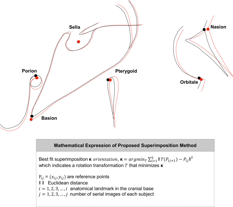
In the current study, six cranial base landmarks were included as reference points: Sella, Nasion, Porion, Orbitale, Basion, and Pterygoid, which can be thought of as adding four supplementary landmarks to the conventional SN superimposition method to reduce over-reliance on Sella and Nasion. For each lateral cephalometric radiograph and its associated cranial base landmarks, the second tracing was location shifted and rotation transformed until the sum of squared Euclidean distance measures ( ) in the cranial base landmarks between the first and second images were minimized while Sella position remained in an identical location (Figure 1). Mathematically, if reference points were expressed in the Cartesian coordinates as Pij = (xij,yij), where i is defined as the anatomical landmark in the cranial base (i = 1 – 5, each representing Nasion, Porion, Orbitale, Basion, and Pterygoid) and j indicates the number of serial images of each subject, then for a given jth image, there is a rotation transformation T that minimizes
  where || || stands for the Euclidean distance measure between T(Pij+1) and Pij.
Figure 1.
The proposed superimposition method included a total of 6 cephalometric landmarks: Sella, Nasion, Portion, Orbitale, Basion, and Pterygoid. For each cephalometric radiograph (black line), the second tracing (red line) was superimposed until the sum of squared Euclidean distances between the six cranial base landmarks were minimized while Sella position remained in an identical position. This method can be expanded to superimpose j multiple images.
Comparisons Between Two Superimposition Methods
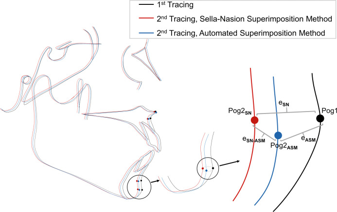
The first and second tracings by the first examiner were used for superimposition. For each of the 283 images, the second tracing was superimposed on the first tracing of the same radiograph by both the SN and automated superimposition methods. For a given cephalometric landmark P, the position of P in the first tracing was denoted as P1, and the position of P in the second tracing oriented by the SN superimposition method and the automated superimposition method were denoted as P2SN and P2ASM, respectively (Figure 2).
Figure 2.
For each of the 283 images, the second tracing was superimposed on the first tracing of the same radiograph by both the Sella-Nasion (SN) and automated superimposition methods. The figure illustrates focusing on the landmark Pogonion (Pog). For a given cephalometric landmark P, P1 = the position of P in the first tracing, P2SN = the position of P in the second tracing oriented by Sella Nasion method, P2ASM = the position of P in the second tracing oriented by the automated superimposition method. The Euclidean distance between P1 − P2SN, P1 − P2ASM, and P2SN − P2ASM were expressed as eSN, eASM, eSN-ASM, respectively.
Since the superimpositions were made on the same pair of X-ray images, the error of a given superimposition method was reported as Euclidean distance values of landmarks between the first tracing and second tracing. For each superimposition, the Euclidean distances between P1 and P2SN, P1 and P2ASM, P2SN and P2ASM were calculated in millimeters for five cephalometric landmarks: Anterior Nasal Spine (ANS), Point A, Point B, Pogonion, and Gonion. Those were chosen because they are known to be some of the most characteristic and representative variables in the maxilla and mandible.23 The superimposition error was evaluated based on whether the superimposition methods reduced or increased the resultant distance between the cephalometric landmarks located in the maxilla and mandible.
The distance between P1 and P2SN, the superimposition error by the SN method, was expressed as eSN. The distance between P1 and P2ASM, the superimposition error by the automated superimposition method, was expressed as eASM. The distance between P2SN and P2ASM, or the difference between the SN and automated superimposition methods, was denoted as eSN-ASM (Figure 2).
Statistical Analysis
The eSN and eASM were compared by paired t-test where the difference was considered statistically significant at P < .05. Regarding the clinical difference between the two superimposition methods, the t-test was performed to test the null hypothesis if eSN-ASM was less than 1 mm, which was considered clinically insignificant. The choice of 1 mm as a criterion for clinical significance was in accordance with previous studies.9,12 All statistical analyses were performed using Language R (Vienna, Austria).25
RESULTS
The errors of each superimposition method were assessed by comparing the superimposed second tracing to the first tracing. The means and standard deviations of the Euclidean distances between the position of landmarks in the first tracing and superimposed second tracing are shown for each cephalometric landmark in Table 1. For all four cephalometric landmarks, there was a significant difference in the superimposition error between the SN and automated superimposition methods.
Table 1. .
Comparison of Errors between the Two Superimposition Methods. Values are the Euclidean Distances (mm) Between the Position of a Landmark in the First Tracing and the Superimposed (Second) Tracing for a Given Cephalometric Landmark
| Sella-Nasion Method (P1 − P2SN)a |
Automated Method (P1 − P2ASM)a |
Mean Difference |
||||
| Mean |
SDb |
Mean |
SD |
P Valuec |
||
| ANS | 4.0 | 3.6 | 2.6 | 2.2 | 1.38 | <.0001 |
| Point A | 4.2 | 4.3 | 2.2 | 2.2 | 1.97 | <.0001 |
| Point B | 3.6 | 4.2 | 1.9 | 2.1 | 1.64 | <.0001 |
| Pogonion | 3.9 | 3.5 | 2.7 | 2.1 | 1.18 | <.0001 |
| Gonion | 3.7 | 3.6 | 2.4 | 2.2 | 1.27 | <.0001 |
Distance indicates Euclidean distance. P1 stands for the position of landmark P in the first tracing, P2SN the position of landmark P in the second tracing oriented by the Sella-Nasion superimposition method, and P2ASM stands for the position of landmark P in the second tracing oriented by the automated superimposition method, where P = Anterior Nasal Spine (ANS), Point A, Point B, Pogonion, and Gonion.
SD indicates standard deviation.
Results from the paired t-test.
In the comparison between the superimposition methods (Table 2), the null hypothesis that the distance between the landmarks located by the two superimposition methods would be less than 1 mm was rejected for all the cephalometric landmarks; clinically significant differences between the SN and new superimposition methods were found.
Table 2. .
Comparison of Two Superimposition Methods. Values are the Euclidean Distance (mm) Between the Positions of a Landmark in the Second Tracing, Which Is Oriented by Two Different Superimposition Methods for a Given Cephalometric Landmarka
| Difference Between Two Methods (eSN−ASM) |
H0: eSN−ASM < 1 mm |
||
| Mean |
SDb |
P Valuec |
|
| ANS | 3.3 | 3.8 | <.0001 |
| Point A | 4.0 | 4.7 | <.0001 |
| Point B | 3.6 | 4.2 | <.0001 |
| Pogonion | 2.8 | 3.3 | <.0001 |
| Gonion | 3.2 | 3.7 | <.0001 |
eSN−ASM stands for the difference between the SN and automated superimposition methods of landmark P, where P = Anterior Nasal Spine (ANS), Point A, Point B, Pogonion, and Gonion.
SD indicates standard deviation.
Results from t-tests under the null hypothesis if the error was less than 1 mm.
DISCUSSION
The purpose of this study was to evaluate a new superimposition method compatible with computer-aided cephalometrics. Additionally, superimposition error with this method was compared to that of the conventional SN superimposition method that has widely been used. When comparing the superimposition errors, statistically and clinically significant differences were found between the two methods. The proposed method demonstrated smaller superimposition errors than did the conventional SN superimposition method.
Errors in superimposition could be caused by errors in identification of landmarks and tracing, quality of images, the skill of the clinician, reproducibility of superimposition in the reference plane itself, and changes in the reference planes by remodeling and growth.11,13 It was illustrated by a number of investigators that the greatest source of error in cephalometrics originated from the landmark identification procedure.26,27 Although SN superimposition has been a popular method, accurate identification of both Sella and Nasion was important to the utmost for exact superimposition. Incorrect identification of Nasion in relation to Sella may lead to a false impression of facial growth even after the completion of growth.11
The results of this study demonstrated that the proposed method showed smaller superimposition errors than the SN superimposition method. Instead of using only two cranial base landmarks as in the SN superimposition method, the proposed superimposition method used six cranial landmarks: Sella, Nasion, Porion, Orbitale, Basion, and Pterygoid. In the SN superimposition method, it was likely that Nasion identification error led to the greatest superimposition error. The proposed superposition method, however, reduced over-reliance on Nasion by using additional landmarks. An identification error would have been inevitable for each of the cranial base landmarks even by the same examiner. The proposed method oriented the superimposed cephalometric image to a best fit position, which minimized the squared sum of errors at each point. Thus, landmark identification errors at each point could be averaged, minimized, reduced, and thus resulted in a more accurate superimposition.
The importance of accuracy in superimposition is indisputable, but the choice of superimposition method could also be affected by cost, time, and expediency.1 The proposed method may take more time and be laborious since more cranial base landmarks are necessary to be identified correctly. Additionally, mathematical computation is required, minimizing the sum of squared Euclidean distances, which might look more complex to perform than the conventional method. However, such possible drawbacks would hardly be a problem in computer-aided cephalometrics. In a recent study regarding fully automatic landmark identification by artificial intelligence (AI), it took only 0.05 seconds to identify 80 cephalometric landmarks per image.14,17 Likewise, the mathematical computation required in the proposed method can be solved without much difficulty.
When differences between/among various superimposition methods were examined previously,1,9–13 most studies used serial cephalometric radiographs of growing or non-growing subjects, before and after orthodontic or orthopedic treatment, to compare superimposition methods. In only one study,10 the same cephalometric image was traced multiple times to identify differences between superimposition methods. Similarly, in the present study, a single cephalometric image was traced twice and superimposed. An additional strength of the current study was that a large number of images were used compared to the study by Gliddon et al.,10 which investigated eight images. The present study greatly increased the number (n = 283) of images that were taken from various malocclusion patients.14,17 This was somewhat different from the methods used in most previous studies. Since the tracings of the same image were superimposed, any difference could indicate an error either because of tracing error or superimposition error. On the other hand, since the same radiographs were used, all the structures including the cranial base would not have changed, which made the circumstances similar to serial radiographs of non-growing subjects without undergoing any treatment. Tracing the same image twice would have resulted in fewer differences between the positions of identified cranial base landmarks than using successive images of the same subject. Therefore, if serial lateral cephalometric images were used, the difference between the two superimposition methods would have been greater because the difference between the cranial base landmarks would be greater in the serial images than between the same images.
By growth and remodeling, changes in the Sella, Nasion, and Basion landmarks were noted in previous studies.18,28 For growing patients, Björk's structural method using stable cranial base structures could be considered a reliable superimposition method. However, it has been reported that there was little difference between Björk's structural method and the SN superimposition method when treatment time was short but the difference became clinically relevant as the treatment time exceeded three years in growing patients.12 The proposed superimposition method allows the second image to have some freedom in rotation around Sella. To accurately superimpose the second image to the first image, several cranial base landmarks around Sella were selected to reduce over-reliance on a particular landmark. Then, to minimize Euclidean distance measures in the cranial base landmarks between the first and second images, performing location shift and rotation transformation using computer technology was necessary.
Further study is needed to determine how the proposed method would differ from the SN superimposition method or Björk's structural method when applied in growing patients. Since the present study assumed no growth by superimposing two tracings on the same image, the proposed method might be suitable for comparing post-surgery change or treatment outcomes for non-growing patients. The question as to whether this proposed method might also be suitable for observing long term growth changes should be tested. Björk's structural method, which was thought to be a reliable method in growing patients, necessitated locating stable cranial base structures and overlapping as many structures as possible, which might negate the strong points of computer-aided cephalometric workflow. The superimposition method, which would be most highly compatible with computer-aided cephalometrics in growing patients, has yet to be determined.
CONCLUSIONS
The proposed method was compatible with computer-aided cephalometrics and demonstrated smaller superimposition errors than the conventional SN superimposition method. It might be a clinically more reliable method for superimposing serial cephalometric images.
ACKNOWLEDGMENTS
We thank Mr. Youngsung Yu (DDH Inc., Seoul, Korea) for his development of user-friendly interfaces without which we could have never conducted this study's experimentation. We also thank Dr. Steven J. Lindauer at VCU School of Dentistry (Richmond, Virginia) for his invaluable efforts in editing and improving this manuscript.
REFERENCES
- 1. .Goel S, Bansal M, Kalra A. A preliminary assessment of cephalometric orthodontic superimposition. Eur J Orthod. 2004;26:217–222. doi: 10.1093/ejo/26.2.217. [DOI] [PubMed] [Google Scholar]
- 2. .Björk A, Skieller V. Normal and abnormal growth of the mandible. A synthesis of longitudinal cephalometric implant studies over a period of 25 years. Eur J Orthod. 1983;5:1–46. doi: 10.1093/ejo/5.1.1. [DOI] [PubMed] [Google Scholar]
- 3. .Ricketts RM. A four-step method to distinguish orthodontic changes from natural growth. J Clin Orthod. 1975;9:208–228. [PubMed] [Google Scholar]
- 4. .Steiner CC. Cephalometrics in clinical practice. Angle Orthod. 1959;29:8–29. [Google Scholar]
- 5. .Viazis A. The cranial base triangle. J Clin Orthod. 1991;25:565–570. [PubMed] [Google Scholar]
- 6. .Donatelli RE, Lee SJ. How to report reliability in orthodontic research: Part 1. Am J Orthod Dentofacial Orthop. 2013;144:156–161. doi: 10.1016/j.ajodo.2013.03.014. [DOI] [PubMed] [Google Scholar]
- 7. .Donatelli RE, Lee SJ. How to report reliability in orthodontic research: Part 2. Am J Orthod Dentofacial Orthop. 2013;144:315–318. doi: 10.1016/j.ajodo.2013.03.023. [DOI] [PubMed] [Google Scholar]
- 8. .Donatelli RE, Lee SJ. How to test validity in orthodontic research: a mixed dentition analysis example. Am J Orthod Dentofacial Orthop. 2015;147:272–279. doi: 10.1016/j.ajodo.2014.09.021. [DOI] [PubMed] [Google Scholar]
- 9. .Ghafari J, Engel FE, Laster LL. Cephalometric superimposition on the cranial base: a review and a comparison of four methods. Am J Orthod Dentofacial Orthop. 1987;91:403–413. doi: 10.1016/0889-5406(87)90393-3. [DOI] [PubMed] [Google Scholar]
- 10. .Gliddon MJ, Xia JJ, Gateno J, et al. The accuracy of cephalometric tracing superimposition. J Oral Maxillofac Surg. 2006;64:194–202. doi: 10.1016/j.joms.2005.10.028. [DOI] [PubMed] [Google Scholar]
- 11. .Houston W, Lee R. Accuracy of different methods of radiographic superimposition on cranial base structures. Eur J Orthod. 1985;7:127–135. doi: 10.1093/ejo/7.2.127. [DOI] [PubMed] [Google Scholar]
- 12. .Huja S, Grubaugh E, Rummel A, Fields H, Beck F. Comparison of hand-traced and computer-based cephalometric superimpositions. Angle Orthod. 2009;79:428–435. doi: 10.2319/052708-283.1. [DOI] [PubMed] [Google Scholar]
- 13. .You QL, Hagg U. A comparison of three superimposition methods. Eur J Orthod. 1999;21:717–725. doi: 10.1093/ejo/21.6.717. [DOI] [PubMed] [Google Scholar]
- 14. .Hwang HW, Park JH, Moon JH, et al. Automated identification of cephalometric landmarks: Part 2-Might it be better than human? Angle Orthod. 2020;90:69–76. doi: 10.2319/022019-129.1. [DOI] [PMC free article] [PubMed] [Google Scholar]
- 15. .Livas C, Delli K, Spijkervet FKL, Vissink A, Dijkstra PU. Concurrent validity and reliability of cephalometric analysis using smartphone apps and computer software. Angle Orthod. 2019;89:889–896. doi: 10.2319/021919-124.1. [DOI] [PMC free article] [PubMed] [Google Scholar]
- 16. .Moylan HB, Carrico CK, Lindauer SJ, Tufekci E. Accuracy of a smartphone-based orthodontic treatment-monitoring application: A pilot study. Angle Orthod. 2019;89:727–733. doi: 10.2319/100218-710.1. [DOI] [PMC free article] [PubMed] [Google Scholar]
- 17. .Park JH, Hwang HW, Moon JH, et al. Automated identification of cephalometric landmarks: Part 1-Comparisons between the latest deep-learning methods YOLOV3 and SSD. Angle Orthod. 2019;89:903–909. doi: 10.2319/022019-127.1. [DOI] [PMC free article] [PubMed] [Google Scholar]
- 18. .Arat ZM, Türkkahraman H, English JD, Gallerano RL, Boley JC. Longitudinal growth changes of the cranial base from puberty to adulthood: a comparison of different superimposition methods. Angle Orthod. 2010;80:725–732. doi: 10.2319/080709-447.1. [DOI] [PMC free article] [PubMed] [Google Scholar]
- 19. .Lee HJ, Suh HY, Lee YS, et al. A better statistical method of predicting postsurgery soft tissue response in Class II patients. Angle Orthod. 2014;84:322–328. doi: 10.2319/050313-338.1. [DOI] [PMC free article] [PubMed] [Google Scholar]
- 20. .Lee YS, Suh HY, Lee SJ, Donatelli RE. A more accurate soft-tissue prediction model for Class III 2-jaw surgeries. Am J Orthod Dentofacial Orthop. 2014;146:724–733. doi: 10.1016/j.ajodo.2014.08.010. [DOI] [PubMed] [Google Scholar]
- 21. .Suh HY, Lee SJ, Lee YS, et al. A more accurate method of predicting soft tissue changes after mandibular setback surgery. J Oral Maxillofac Surg. 2012;70:e553–562. doi: 10.1016/j.joms.2012.06.187. [DOI] [PubMed] [Google Scholar]
- 22. .Yoon KS, Lee HJ, Lee SJ, Donatelli RE. Testing a better method of predicting postsurgery soft tissue response in Class II patients: a prospective study and validity assessment. Angle Orthod. 2015;85:597–603. doi: 10.2319/052514-370.1. [DOI] [PMC free article] [PubMed] [Google Scholar]
- 23. .Kang TJ, Eo SH, Cho H, Donatelli RE, Lee SJ. A sparse principal component analysis of Class III malocclusions. Angle Orthod. 2019;89:768–774. doi: 10.2319/100518-717.1. [DOI] [PMC free article] [PubMed] [Google Scholar]
- 24. .Suh HY, Lee HJ, Lee YS, Eo SH, Donatelli RE, Lee SJ. Predicting soft tissue changes after orthognathic surgery: the sparse partial least squares method. Angle Orthod. 2019;89:910–916. doi: 10.2319/120518-851.1. [DOI] [PMC free article] [PubMed] [Google Scholar]
- 25. .R Development Core Team. R A language and environment for statistical computing. Vienna, Austria: R Foundation for Statistical Computing; 2019. [Google Scholar]
- 26. .Baumrind S, Frantz RC. The reliability of head film measurements: 1. Landmark identification. Am J Orthod. 1971;60:111–127. doi: 10.1016/0002-9416(71)90028-5. [DOI] [PubMed] [Google Scholar]
- 27. .Trpkova B, Major P, Prasad N, Nebbe B. Cephalometric landmarks identification and reproducibility: a meta analysis. Am J Orthod. 1997;112:165–170. doi: 10.1016/s0889-5406(97)70242-7. [DOI] [PubMed] [Google Scholar]
- 28. .Arat M, Köklü A, Özdiler E, Rübendüz M, Erdogan B. Craniofacial growth and skeletal maturation: a mixed longitudinal study. Eur J Orthod. 2001;23:355–361. doi: 10.1093/ejo/23.4.355. [DOI] [PubMed] [Google Scholar]


