



Abstract
Osteosarcoma occurs largely in children and adolescents and is the most common primary malignant tumour of bone. Although surgical advances and neoadjuvant chemotherapy have made great strides in recent years, rates of local recurrence and lung metastasis remain high, with a plateau in overall survival during the past decade. It is thus urgent to explore the pathogenesis of osteosarcoma and identify potential therapeutic targets. Parathyroid hormone receptor 1 (PTHR1) belongs to the broad family of G protein–coupled receptors, binding both parathyroid hormone (PTH) and parathyroid hormone–related peptide (PTHrP, a paracrine factor). Previous studies have shown that in tissues and cells of osteosarcoma, expression of PTHR1 is markedly increased, correlating with aggressive biologic behaviour and a poor prognosis. PTHR1 expression also correlates closely with epigenetic regulation, transcriptional regulation, post‐translational modification and protein interaction. Herein, we have summarized the latest research on the role played by PTHR1 in progression of osteosarcoma, assessing its clinical utility as a novel biomarker and its therapeutic ramifications.
Keywords: oncogene, osteosarcoma, prognosis, PTHR1, targeted therapy
1. INTRODUCTION
Osteosarcoma is a common and often rapidly progressing primary malignant tumour of adolescents and children. It tends to arise near the ends of limbs and long bones but may involve the iliac bone, spine and other skeletal parts. There is a proclivity for distant metastasis as well, and the generally low long‐term survival rate of such patients reflects its high degree of malignancy. 1 , 2 , 3 Surgery is the chief means of early treatment for osteosarcoma 4 , 5 (Table 1). However, this method has two clear disadvantages, namely the trauma entailed and the less than encouraging prospects of long‐term survival. 6 The 5‐year survival rate in patients treated exclusively by surgery is quite low, owing to pulmonary metastases, prognostic factors and matters of therapeutic compliance. 7 , 8
TABLE 1.
Potential therapeutic approaches of osteosarcoma
| Approach | Related drugs | Related gene |
|---|---|---|
| Surgery | ||
| Tumour resection | / | / |
| Limb function reconstruction | / | / |
| Chemotherapy | MTX, ADM, | / |
| DDP, IFO | / | |
| Radiotherapy | ||
| Three‐dimensional conformal radiation therapy (3D‐CRT) Intensity‐modulated radiation therapy (IMRT) TomoTherapy (TOMO) | / | / |
| Molecular targeted therapy | / | Ezrin, HER2, telomerase |
| Immunity therapy | ||
| DC therapy | / | / |
| Cytokine therapy | / | / |
| CAR‐T cell immunotherapy | / | / |
| Immune checkpoint block therapy | / | PD‐1/PD‐L1 |
| Gene therapy | / | p53, p16, p21 and Rb |
| Embolization therapy | ||
| Selective arterial embolization | / | / |
| Transcatheter arterial chemoembolization | / | / |
| Radiofrequency ablation therapy | ||
| High intensity energy focused ablation | / | / |
| Radiofrequency ablation | / | / |
| Cryoablation | / | / |
| Microwave ablation | / | / |
| Stem cell therapy | / | HH, NOTCH, |
| Wnt/p‐catenin and MAP | ||
More recently, molecular mechanisms fuelling the development and progression of osteosarcoma, particularly LncRNA DANCR promotion of ROCK1‐mediated proliferation and metastasis (via decoying of miR‐335‐5p and miR‐1972) or expression patterns of programmed death proteins (PD‐L1, PD‐L2 and PD‐1), have provided a crux for therapeutic targeting. 9 , 10 , 11 Still, an optimal approach to routine chemotherapy has yet to be devised. Combination treatments, such as gemcitabine/ docetaxel or doxorubicin plus a heart protectant, may effectively reduce individual toxicities while boosting responses, but these strategies are of limited utility. 12 , 13 The precise pathways contributing to occurrences and metastasis of osteosarcoma remain elusive and must be clarified in our search for better preventive and therapeutic measures.
Parathyroid hormone (PTH) is a polypeptide hormone secreted by parathyroid master cells. Through its action on osteoblasts and osteoclasts, via cyclic adenosine monophosphate (cAMP) and phospholipase C pathways, it serves to modulate blood calcium levels. Parathyroid hormone receptor 1 (PTHR1) belongs to the G protein–coupled cell membrane receptor family, widely distributed and of greatest import in canine and rat osteosarcoma. PTHR1 binds both PTH and PTH‐related peptide (PTHrP) and is primarily expressed in bone, kidney and cartilage. The vascular system, certain developmental organs and human MCF7 breast cancer cells also show high levels of expression. 14 , 15 It is encoded by 14 exon genes on chromosome 3 and plays a critical role in regulating serum and endochondral bone concentrations of calcium. 16
Along with other specifics (ie target cell type, molecular structure of binding ligand and homeostatic bodily conditions), PTHR1 has been implicated in a number of intracellular signalling pathways, the nature, degree and duration of which are decisive in the biologic responses induced. 17 Biochemical and cellular responses to PTHR1 activation may thus differ according to cell type. In primary failure of eruption (PFE), clinical and radiographic characteristics are highly specific for PTHR1 effects 18 ; and compared with responses in wild‐type (WT) mice, physiological responses to injected PTH ligands are acutely and severely disrupted in mice bearing the phosphorylation‐deficient (PD) PTHR1 knock‐in mutation. 19 In both WT and PD animals, PTH administration increases the volume and trabecular thickness of vertebral and distal femoral bones, but PTHR1 phosphorylation is not a major factor in anabolic actions of PTH. 20 PTH/PTHR1 and phytoestrogens have both performed positively in an animal model of bilateral ovariectomy. 21 Finally, activation of PTHR1 appears to modulate diverse molecular cascades through autocrine mechanisms. These cascades are involved in a variety of processes, including hormonal feedback control, receptor desensitization and catabolism, as well as removal of hormone‐ligand complexes from the circulation. 17 , 22 Many studies have underscored the impact of abnormal PTHR1 expression, showing a close association with occurrences and malignant progression of osteosarcoma (Figure 1).
FIGURE 1.
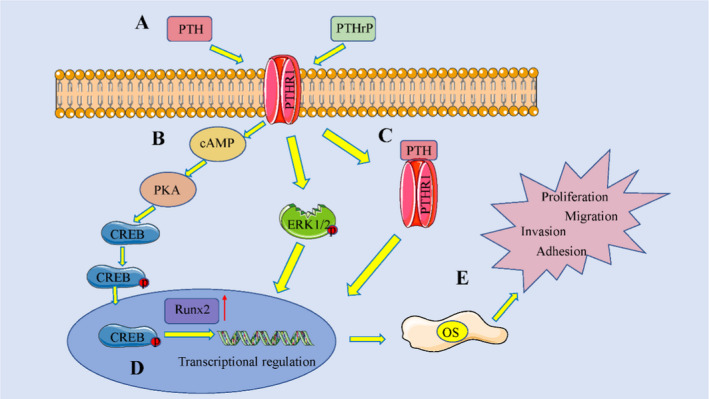
The effect of parathyroid hormone (PTH)/PTH‑related peptide (PTHrP)–dependent extracellular matrix (ECM) signalling on osteosarcoma (OS) cell functions. (A) PTHR1 activation; (B) receptor respective downstream signalling; (C) transcriptional regulation; (D) modulation of ECM‑correlated target genes; and (E) regulation of basic OS cell functions. PTHR1, PTH receptor 1; cAMP, cyclic adenosine monophosphate; CREB, cAMP response element–binding protein; and Runx‑2, runt‑related transcription factor 2
In this review, we have summarized present knowledge surrounding regulatory and functional aspects of PTHR1 in osteosarcoma. We have also probed the clinical implications of altering PTHR1 expression and the latest therapeutic strategies for targeting PTHR1 in this setting (Table 2, Figure 2).
TABLE 2.
The biological function of PTHR1 in osteosarcoma
| Researchers | Expression | Functional role | Related gene | References |
|---|---|---|---|---|
| Yang et al | Up‐regulated | Cell proliferation and invasion | TGF‐b1 and connective tissue growth factor | 32 |
| Ho et al | Up‐regulated | Cell invasion, growth and tumour differentiation | Wnt signal pathway and RANKL | 44 |
| Guan et al | / | / | ZFPM2, LEF1, NR4A2, HAS2 and RHOC | 43 |
| Li et al | / | / | Dkk1, Lef1, Agt‐CCR3 and Agt‐CCL9 | 17 |
| Li et al | Up‐regulated | Cell proliferation, invasion and tumorigenesis | MMP‐2/9 and Vmp1 | 40 |
| Li et al | Up‐regulated | Cell proliferation and tumour metastasis | MMP‐2/9 | 39 |
| Li et al | / | / | miR‐124‐3p‐AR‐Tgfb1i1, miR‐27a‐3p‐PPARG‐Abca1, and miR‐103/590‐3p‐AXIN2 | 42 |
| Wen et al | Cell proliferation and tumour metastasis | MMP‐2/9 | 41 | |
| Qu et al | Up‐regulated | Cell proliferation and apoptosis | LINC01278/ miR‐133a‐3p | 31 |
| Al‐Khan et al | Up‐regulated | Prognostic role | / | 14 |
FIGURE 2.
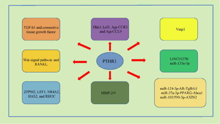
Potential mechanism and regulatory axis of PTHR1 in osteosarcoma. (A) Related to TGF‐b1 and connective tissue growth factor; (B) Wnt signal pathway and RANKL regulation; (C) potential downstream genes (ZFPM2, LEF1, NR4A2, HAS2 and RHOC); (D) potential downstream genes (DKK1, LEF1, Agt‐CCR3 and Agt‐CCL9); (E) MMP‐2/9 regulation; (F) VMP1 regulation; (G) regulated by LINC01278/ miR‐133a‐3p; and (H) potential regulatory axis (miR‐124‐3p‐AR‐Tgfb1i1, miR‐27a‐3p‐PPARG‐Abca1 and miR‐103/590‐3p‐AXIN2). RANKL, TNF superfamily member 11; ZFPM2, zinc finger protein, FOG family member 2; LEF1, lymphoid enhancer–binding factor 1; NR4A2, nuclear receptor subfamily 4 group A member 2; HAS2, hyaluronan synthase 2; RHOC, ras homolog family member C; DKK1, dickkopf WNT signalling pathway inhibitor 1; MMP‐2/9, matrix metallopeptidase 2/9; and VMP1, vacuole membrane protein 1
2. BIOLOGY OF PTHR1
PTHR1 is a class B G protein–coupled receptor that binds PTH and PTHrP. Although present in other tissues, it is primarily expressed in bone, kidney and cartilage. 23 , 24 Despite their shared signalling mechanisms, the biologic functions of PTH and PTHrP are actually quite different. 25 , 26 PTH modulates serum calcium through endocrine effects on bone and kidney cells, whereas PTHrP is a paracrine modulator of cell proliferation and differentiation at developmental sites, such as bony growth plates. 22 The biologic responses induced by PTHR1 activation generally reflect the nature, intensity and duration of signalling, in conjunction with other variables (ie target cell type, ligand structure and primary homeostatic conditions). Activation of PTHR1 thus triggers unique biochemical and cellular responses, depending on cell type.
In osteoblasts and chondrocytes, PTHR1 activation modulates proliferative and apoptotic efficiency and contributes to production of various signalling factors involved in bone and cartilage metabolism. 22 In renal tubules, PTHR1 activation modulates transmembrane transport of mineral ions through expression levels and functional activities of related proteins. The global response to PTHR1 activation is modulated by processes at several levels, including intracellular routes for receptor desensitization, systemic feedback loops for hormonal release, and steps in the destruction and metabolic clearance of peptide hormones from the circulation. Despite the many mechanisms that modulate PTHR1 activity, maladjustments may occur, imposing serious physiological consequences. 17 , 27 , 28
3. EXTENT AND CLINICAL RELEVANCE OF PTHR1 EXPRESSION IN OSTEOSARCOMA
In patients with osteosarcoma, PTHR1 overexpression has been linked to greater risk of metastasis and a poor prognosis. Using quantitative reverse transcription‐polymerase chain reaction (QRT‐PCR), Qu et al have recorded dramatically higher levels of PTHR1 expression in a 40‐patient sampling of osteosarcomas (vs normal control tissues), culminating in adverse clinical outcome. 29 Levels of PTHR1 mRNA expressed in metastases or recurrences of osteosarcoma have also proven much higher than those in primary tumours, conferring more aggressive phenotypes and microenvironments more conducive to malignancy. 30 A canine model of osteosarcoma constructed by Al‐khan and colleagues has demonstrated a poorer prognosis for dogs showing intensified PTHR1 expression. 31 Besides, PTHrP (1‐40) stimulates calcium uptake which transporters TRPV6 and CaBP‐D9k via PTHR1 receptor and PKC α/β signaling pathway in rat enterocytes. 32 As a whole, these findings indicate the important influence of PTHR1 on progression of osteosarcoma and its utility in predicting patient prognosis.
4. REGULATORY MECHANISMS OF PTHR1 IN OSTEOSARCOMA
Various sources have confirmed that PTHR1 actually modulates malignant progression of osteosarcoma through certain mechanisms involving matrix metalloproteinases, non‐coding RNA and genetic foci. The precise regulatory mechanisms and their potential for new therapeutic targets must be further studied and fully explored.
4.1. Matrix metalloproteinases and extracellular matrix regulation by PTHR1
The family of matrix metalloproteinases (MMPs), which variably relies on metal (ie, zinc) and calcium ions as cofactors, is now comprised of at least 26 different members assigned to five major substrate‐defined categories as follows: interstitial collagen enzyme, gelatin enzymes, stromelysin, metalloproteinases and other enzymes. Given their importance in tissue remodelling and organ development, abnormal MMP expression (especially MMP‐2, MMP‐9 and MMP‐14) is seen in a host of diseases, from autoimmune disorders to cancers, impacting tumour and immune‐cell microenvironments. 33 , 34 , 35 , 36
PTHR1 likely stimulates malignant progression of osteosarcoma by regulating MMP expression. In a past study of metastatic human osteosarcoma cells, quercetin clearly diminished expression of PTHR1 mRNA, thus attenuating expression levels of MMP‐2 and MMP‐9 mRNA and ultimately reducing cellular invasion, adhesion, proliferation and migration. 37 Li and colleagues have similarly witnessed attenuation of PTHR1 mRNA and protein expression in osteosarcoma cells incubated with β‐alanine, indicating its positive relation with tumour invasion and metastasis 38 ; and upon incubating Saos‐2 and U2OS human osteosarcoma cell lines in β‐alanine, Wen et al found expression levels of MMP‐2 and MMP‐9 mRNA notably reduced, whereas tissue inhibitors of metalloproteinase (TIMP)‐1 and TIMP‐2 were expressed at high levels. Osteosarcoma cells treated in mangiferin (a xanthone) have also shown substantial attenuation of PTHR1 mRNA and protein expression, along with growth inhibition and induction of apoptosis. 39 Nonetheless, the interplay of PTHR1 and MMP expression levels in progression of osteosarcoma requires greater scrutiny.
The extracellular matrix (ECM) is an extensive network of cellular secretions, composed of collagens, elastin, proteoglycans (PGs), glycosaminoglycans (GAGs), fibronectins and laminins. Relative to physiological bone, the ECM of osteosarcoma is extensively altered. At least one prior study has addressed the role of ECM role in regulating osteosarcoma cells in vivo and in vitro. 40 Fibroblast growth factor‐2 is known to modulate migration of MG‐63 osteosarcoma cells by regulating expression levels of ECM‐associated proteoglycans. 41 Downstream signalling pathways of the ECM components are distinctive 42 , 43 and have received growing attention for their interconnected mechanisms of carcinogenesis in osteosarcoma. Broader study is essential in this area to assess their potential as clinical biomarkers and therapeutic targets.
The microenvironment, including bone, stroma, vascular elements and immune cells, is also critical in regulating the growth and metastasis of osteosarcoma. 44 Mesenchymal stem cells (MSCs) within the microenvironment of bone are paramount in promoting tumour metastasis, and many signalling pathways, such as PI3K/Akt, Wnt/β‐Catenin, MAPK/ERK, Hedgehog and Notch, facilitate the transition of MSCs to osteosarcoma cells. In animal models, MSCs clearly contribute to pulmonary metastasis. 45 Immunotherapy has gained prominence as a clinical treatment, showing promise in patients with lung cancer, melanoma, and oesophageal cancer. Tumour immune cells are varied, consisting of mesenchymal cells, tumour‐infiltrating immune cells (TIICs), endothelial cells, ECM molecules and inflammatory mediators. Immune scores are associated with overall survival and gauge immune‐related risk in patients with osteosarcoma. 46
4.2. Non‐coding RNA and key genes implicated in PTHR1 regulation
Non‐coding RNA (ncRNA) chiefly includes microRNA (miRNA), long non‐coding RNA (lncRNA) and circular RNA (circRNA). Because the assumed region is a complementary base pair to the seed sequence of miRNA, located at its 3' untranslated region (UTR), PTHR1 may be influenced by regulating miRNAs. According to Li et al, PTHR1 may exert important influence on progression of osteosarcoma by activating miRNA genes miR‐124‐3‐p‐AR‐Tgfb1i1, miR‐a‐3p‐PPARG‐27 Abca1 and miR‐103/590‐3p‐AXIN2. 47 Qu et al have also noted that in osteosarcoma, LINC01278 is a competing endogenous RNA of PTHR1 (by sponging miR‐133a‐3p) and a likely point of tumour inhibition in osteosarcoma. 29
The vastness of bioinformatics applications has allowed researchers to trace many potential downstream targets of PTHR1. In particular, NR4A2, ZFPM2, RHOC, LEF1 and HAS2 may be potential targets of PTHR1 in osteosarcoma 48 ; and the pro‐malignant effects of PTHR1 are apparently mediated through Wnt, angiogenesis and fever pathways, modulating expression of pivotal enriched genes (Dkk1, Lef1, AGT‐CCR3 and AGT‐CCL9). 15 PTHR1 may thus regulate AGT‐CCL9 in osteosarcoma cells, impacting cellular viability, apoptosis, migration, invasion and colony formation. 49
5. FUNCTION OF PTHR1 IN OSTEOSARCOMA
As a crucial oncogenic gene, oncogenic properties of PTHR1 pertaining to osteosarcoma have been adequately chronicled. In this section, we describe the functions of PTHR1 as a pro‐oncogenic gene in this setting, serving to regulate PTHR1 expression.
5.1. Promoting cellular proliferation and growth
In the absence of added exogenous PTHrP, overexpression of PHTR1 intensifies cellular proliferation, motility and Matrigel invasion, as presumptive autocrine effects. PTHR1 overexpression is also associated with deferred osteoblastic differentiation and up‐regulation of genes involved in ECM production, inclusive of TGF‐b1 and connective tissue growth factor. 30 Ho and colleagues have used shRNA to diminish PTHR1 expression, observing a mild hindrance of cellular proliferation in vitro but markedly diminished collagen invasion and reduced expression of RANK ligand (RANKL). Although in vivo administration of PTH (1‐34) has no proliferative effect on osteosarcoma cells, knockdown of PTHR1 yields remarkable growth inhibition and intensifies tumour differentiation/mineralization. 50 In a study by Wen et al, mangiferin treatment served to greatly reduce expression of PTHR1 mRNA and protein in osteosarcoma cells, curbing their viability, proliferation, invasion, adhesion, and migration, and triggering apoptosis. 39 Al‐Khan et al have investigated expression levels of PTHR1 and PTHrP in canine osteosarcoma tissues as a means of prognostication. They found that overexpression of PTHR1 antigen correlated with poorer outcome, supporting its use as prognostic index. 31
5.2. Promoting cellular invasion and metastasis
Li et al have shown that β‐alanine inhibition of PTHR1 expression diminishes proliferation, invasion, migration and tumorigenesis in U2OS cells, signifying a positive relation between PTHR1 expression and tumour invasion/metastasis. 38 Quercetin‐inhibited proliferation and inhibition of osteosarcoma are otherwise enhanced by knockdown of PTHR1, underscoring its importance in this setting. 37
6. THERAPEUTIC STRATEGIES BASED ON PTHR1 IN OSTEOSARCOMA
As shown by Qu et al, LINC01278 (an lncRNA affiliate) promotes cellular proliferation and limits apoptosis in osteosarcoma cells. Mechanistic studies further indicate that LINC01278 is a competing endogenous RNA of PTHR1 (by sponging miR‐133a‐3p) and a likely point of tumour inhibition. Down‐regulation of PTHR1 serves to restore the inhibitory impact of miR‐133a‐3p. This carcinogenic effect of LINC01278, as a consequence of miR‐133a‐3p/PTHR1 signalling, represents a viable opportunity for therapeutic targeting. 29
7. CONCLUSION AND FUTURE PERSPECTIVES
Patients with advanced osteosarcoma are currently confined to systemic chemotherapy as treatment, although the limitations of present‐day regimens have become increasingly evident. 51 , 52 Finding new treatments is therefore a matter of urgency. The mTOR pathway inhibitor, rapamycin, mitigates mRNA translation and inhibits metastasis of osteosarcoma cells, 53 , 54 but prospects beyond this are seriously lacking. Although targeted therapy is the new paradigm in treating advanced malignant tumours, the research to date on targeted treatments for osteosarcoma remains insufficient. More studies and drug testing are needed to identify better and safer agents.
The G protein–coupled receptor PTHR1 is highly expressed in cartilage, kidney, bone, and other vascular and developmental tissues. It is encoded by 14 exon genes on chromosome 3 and is critical in regulating calcium concentrations of serum and endochondral bone. A number of intracellular signalling pathways are linked to PTHR1, the nature, intensity and duration of signalling determining subsequent biologic responses. There are ancillary variables as well, including target cell type, molecular structure of binding ligand and homeostatic conditions. 16 , 20 , 55 Biochemical and cellular responses thus differ according to cell type. 24 , 56 , 57 , 58
In osteoblasts and chondrocytes, activation of PTHR1 modulates proliferation, apoptosis and production of assorted signal transduction factors involved metabolism of bone and cartilage. In renal tubules, PTHR1 activation modulates the transmembrane transport of mineral ions by regulating expression levels and functions of related proteins. Through autocrine mechanisms, PTHR1 activation also modulates diverse molecular cascades involved in receptor desensitization, hormonal feedback loops, catabolism and removal of hormone‐ligand complexes from circulation.
Clinical data on patients with osteosarcoma indicate that PTHR1 overexpression carries greater risk of metastasis and a poor prognosis. Investigational studies have likewise shown that PTHR1 plays an important pro‐oncogenic role in tumour growth and distant metastasis, both in vitro and in vivo. Hence, a new therapeutic strategy for osteosarcoma, aimed at inhibiting PTHR1 expression or function, seems quite feasible. This may involve specific mechanisms under genetic and non‐genetic control, using epigenetic drugs and natural compounds known to target PTHR1 in vitro. Because specific mechanisms of PTHR1 activity have yet to be fully delineated in the context of osteosarcoma, our research efforts must intensify going forward, propelling routine therapeutics to new and more acceptable heights.
CONFLICT OF INTEREST
The authors declare no competing interests.
AUTHOR CONTRIBUTION
Original draft preparation,allocation, revision, supplement and edition, Chaonan Sun and Shenglong Li. All authors have read and agreed to the published version of the manuscript.
ACKNOWLEDGEMENTS
We thank the generous support by Liaoning Cancer Hospital & Institute (Shenyang).
Sun C, Li S. PTHR1 in osteosarcoma: Specific molecular mechanisms and comprehensive functional perspective. J Cell Mol Med. 2021;25:3175–3181. 10.1111/jcmm.16420
Funding information
This work was supported by Natural Science Foundation of Liaoning Province (2020‐MS‐058) and Shenyang Young and Middle‐aged Scientific and Technological Innovation Talent Support Plan (RC190456).
REFERENCES
- 1. Ługowska I, Pieńkowski A, Szumera‐Ciećkiewicz A, et al. The long‐term treatment outcomes of adult osteosarcoma. Pol Merkur Lekarski. 2017;250:158‐164. [PubMed] [Google Scholar]
- 2. Meazza C, Scanagatta P. Metastatic osteosarcoma: a challenging multidisciplinary treatment. Expert Rev Anticancer Ther. 2016;16(5):543‐556. [DOI] [PubMed] [Google Scholar]
- 3. Kerr DL, Dial BL, Lazarides AL, et al. Epidemiologic and survival trends in adult primary bone tumors of the spine. Spine J. 2019;19(12):1941‐1949. [DOI] [PubMed] [Google Scholar]
- 4. Suehara Y, Alex D, Bowman A, et al. Clinical genomic sequencing of pediatric and adult osteosarcoma reveals distinct molecular subsets with potentially targetable alterations. Clin Cancer Res. 2019;25:6346‐6356. [DOI] [PMC free article] [PubMed] [Google Scholar]
- 5. Wu Y, Xie Z, Chen J, et al. Circular RNA circTADA2A promotes osteosarcoma progression and metastasis by sponging miR‐203a‐3p and regulating CREB3 expression. Mol Cancer. 2019;18:73. [DOI] [PMC free article] [PubMed] [Google Scholar]
- 6. Nomura M, Rainusso N, Lee YC, et al. Tegavivint and the beta‐Catenin/ALDH axis in chemotherapy‐resistant and metastatic osteosarcoma. J Natl Cancer Inst. 2019;111:1216‐1227. [DOI] [PMC free article] [PubMed] [Google Scholar]
- 7. Faisham WI, Mat Saad AZ, Alsaigh LN, et al. Prognostic factors and survival rate of osteosarcoma: a single‐institution study. Asia Pac J Clin Oncol. 2017;13(2):104‐110. [DOI] [PubMed] [Google Scholar]
- 8. Pruksakorn D, Phanphaisarn A, Arpornchayanon O, Uttamo N, Leerapun T, Settakorn J. Survival rate and prognostic factors of conventional osteosarcoma in Northern Thailand: a series from Chiang Mai University Hospital. Cancer Epidemiol. 2015;39(6):956‐963. [DOI] [PubMed] [Google Scholar]
- 9. Wang Y, Zeng X, Wang N, et al. Long noncoding RNA DANCR working as a competitive endogenous RNA, promotes ROCK1‐mediated proliferation and metastasis via decoying of miR‐335‐5p and miR‐1972 in osteosarcoma. Mol Cancer. 2018;17:89. [DOI] [PMC free article] [PubMed] [Google Scholar]
- 10. Wagner F, Holzapfel BM, McGovern JA, et al. Humanization of bone and bone marrow in an orthotopic site reveals new potential therapeutic targets in osteosarcoma. Biomaterials. 2018;171:230‐246. [DOI] [PubMed] [Google Scholar]
- 11. Zheng B, Ren T, Huang Y, et al. PD‐1 axis expression in musculoskeletal tumors and antitumor effect of nivolumab in osteosarcoma model of humanized mouse. J Hematol Oncol. 2018;11:16. [DOI] [PMC free article] [PubMed] [Google Scholar]
- 12. Lin YH, Jewell BE, Gingold J, et al. Osteosarcoma: molecular pathogenesis and iPSC modeling. Trends Mol Med. 2017;23:737‐755. [DOI] [PMC free article] [PubMed] [Google Scholar]
- 13. Fernandez L, Metais JY, Escudero A, et al. Perez‐martinez.memory T Cells expressing an NKG2D‐CAR efficiently target osteosarcoma cells. Clin Cancer Res. 2017;23:5824‐5835. [DOI] [PubMed] [Google Scholar]
- 14. Johnson RW, Sun Y, Ho PWM, et al. Parathyroid hormone‐related protein negatively regulates tumor cell dormancy genes in a PTHR1/Cyclic AMP‐independent manner. Front Endocrinol (Lausanne). 2018;9:241. [DOI] [PMC free article] [PubMed] [Google Scholar]
- 15. Li S, Dong Y, Wang K, Wang Z, Zhang X. Transcriptomic analyses reveal the underlying pro‐malignant functions of PTHR1 for osteosarcoma via activation of Wnt and angiogenesis pathways. J Orthop Surg Res. 2017;12:168. [DOI] [PMC free article] [PubMed] [Google Scholar]
- 16. Mansoori MN, Shukla P, Singh D. Combination of PTH (1–34) with anti‐IL17 prevents bone loss by inhibiting IL‐17/N‐cadherin mediated disruption of PTHR1/LRP‐6 interaction. Bone. 2017;105:226‐236. [DOI] [PubMed] [Google Scholar]
- 17. Cheloha RW, Gellman SH, Vilardaga JP, Gardella TJ. PTH receptor‐1 signalling‐mechanistic insights and therapeutic prospects. Nat Rev Endocrinol. 2015;11:712‐724. [DOI] [PMC free article] [PubMed] [Google Scholar]
- 18. Pilz P, Meyer‐Marcotty P, Eigenthaler M, Roth H, Weber BH, Stellzig‐Eisenhauer A. Differential diagnosis of primary failure of eruption (PFE) with and without evidence of pathogenic mutations in the PTHR1 gene. J Orofac Orthop. 2014;75:226‐239. [DOI] [PubMed] [Google Scholar]
- 19. Maeda A, Okazaki M, Baron DM, et al. Critical role of parathyroid hormone (PTH) receptor‐1 phosphorylation in regulating acute responses to PTH. Proc Natl Acad Sci U S A. 2013;110:5864‐5869. [DOI] [PMC free article] [PubMed] [Google Scholar]
- 20. Datta NS, Samra TA, Abou‐Samra AB. Parathyroid hormone induces bone formation in phosphorylation‐deficient PTHR1 knockin mice. Am J Physiol Endocrinol Metab. 2012;302:1183‐1188. [DOI] [PMC free article] [PubMed] [Google Scholar]
- 21. Miao Q, Li JG, Miao S, et al. The bone‐protective effect of genistein in the animal model of bilateral ovariectomy: roles of phytoestrogens and PTH/PTHR1 against post‐menopausal osteoporosis. Int J Mol Sci. 2012;13:56‐70. [DOI] [PMC free article] [PubMed] [Google Scholar]
- 22. Lupp A, Klenk C, Rocken C, Evert M, Mawrin C, Schulz S. Immunohistochemical identification of the PTHR1 parathyroid hormone receptor in normal and neoplastic human tissues. Eur J Endocrinol. 2010;162:979‐986. [DOI] [PubMed] [Google Scholar]
- 23. Yavropoulou MP, Michopoulos A, Yovos JG. PTH and PTHR1 in osteocytes. New insights into old partners. Hormones (Athens). 2017;16150‐16160. [DOI] [PubMed] [Google Scholar]
- 24. Tamura T, Noda H, Joyashiki E, et al. Identification of an orally active small‐molecule PTHR1 agonist for the treatment of hypoparathyroidism. Nat Commun. 2016;7:13384. [DOI] [PMC free article] [PubMed] [Google Scholar]
- 25. Wang M, Nasiri AR, Broadus AE, Tommasini SM. Periosteal PTHrP regulates cortical bone remodeling during fracture healing. Bone. 2015;81:104‐111. [DOI] [PMC free article] [PubMed] [Google Scholar]
- 26. Filipovic N, Vrdoljak M, Vuica A, et al. Expression of PTHrP and PTH/PTHrP receptor 1 in the superior cervical ganglia of rats. Neuropeptides. 2014;48:353‐359. [DOI] [PubMed] [Google Scholar]
- 27. Hopyan S, Gokgoz N, Poon R, et al. A mutant PTH/PTHrP type I receptor in enchondromatosis. Nat Genet. 2002;30:306‐310. [DOI] [PubMed] [Google Scholar]
- 28. Liu S, Jean‐Alphonse FG, White AD, et al. Use of backbone modification to enlarge the spatiotemporal diversity of parathyroid hormone receptor‐1 signaling via biased agonism. J Am Chem Soc. 2019;141:14486‐14490. [DOI] [PMC free article] [PubMed] [Google Scholar]
- 29. Qu Z, Li S. Long noncoding RNA LINC01278 favors the progression of osteosarcoma via modulating miR‐133a‐3p/PTHR1 signaling. J Cell Physiol. 2020. 10.1002/jcp.29582. [Epub ahead of print]. [DOI] [PubMed] [Google Scholar]
- 30. Yang R, Hoang BH, Kubo T, et al. Over‐expression of parathyroid hormone Type 1 receptor confers an aggressive phenotype in osteosarcoma. Int J Cancer. 2007;121:943‐954. [DOI] [PubMed] [Google Scholar]
- 31. Al‐Khan AA, Nimmo JS, Tayebi M, et al. Parathyroid hormone receptor 1 (PTHR1) is a prognostic indicator in canine osteosarcoma. Sci Rep. 2020;10:1564. [DOI] [PMC free article] [PubMed] [Google Scholar]
- 32. Liu C, Shao G, Lu Y, et al. Parathyroid hormone‐related protein (1–40) enhances calcium uptake in rat enterocytes through PTHR1 receptor and protein kinase calpha/beta signaling. Cell Physiol Biochem. 2018;51:1695‐1709. [DOI] [PubMed] [Google Scholar]
- 33. Ye Y, Kuang X, Xie Z, et al. Small‐molecule MMP2/MMP9 inhibitor SB‐3CT modulates tumor immune surveillance by regulating PD‐L1. Genome Med. 2020;12:83. [DOI] [PMC free article] [PubMed] [Google Scholar]
- 34. Park SS, Lee DH, Lee SM, Lee CH, Kim SY. Single‐sided Deafness Leads to Changes in Vesicular Synaptic Transporters and Matrix Metalloproteinase 9 in the Primary Auditory Cortex. Neuroscience. 2020;449:189‐201. [DOI] [PubMed] [Google Scholar]
- 35. Ochieng JK, Kundu ST, Bajaj R, Leticia Rodriguez B, Fradette JJ, Gibbons DL. MBIP (MAP3K12 binding inhibitory protein) drives NSCLC metastasis by JNK‐dependent activation of MMPs. Oncogene. 2020;39(43):6719‐6732. [DOI] [PMC free article] [PubMed] [Google Scholar]
- 36. Mazzoni TS, Quagio‐Grassiotto I. Presence of the matrix metalloproteinases during the migration of the primordial germ cells in zebrafish gonadal ridge. Cell Tissue Res. 2021;383(2):707–722. [DOI] [PubMed] [Google Scholar]
- 37. Li S, Pei Y, Wang W, Liu F, Zheng K, Zhang X. Quercetin suppresses the proliferation and metastasis of metastatic osteosarcoma cells by inhibiting parathyroid hormone receptor 1. Biomed Pharmacother. 2019;114:108839. [DOI] [PubMed] [Google Scholar]
- 38. Li S, Chen P, Zheng K, et al. β‐Alanine mediated inhibition of PTHR1suppresses the proliferation, invasion and tumorigenesis in metastatic human osteosarcoma U2OS cells. Int J Biol Macromol. 2018;111:1255‐1263. [DOI] [PubMed] [Google Scholar]
- 39. Wen J, Qin Y, Li C, Dai X, Wu T, Yin W. Mangiferin suppresses human metastatic osteosarcoma cell growth by down‐regulating the expression of metalloproteinases‐1/2 and parathyroid hormone receptor 1. AMB Express. 2020;10:13. [DOI] [PMC free article] [PubMed] [Google Scholar]
- 40. Nikitovic D, Kavasi R‐M, Aikaterini Berdiaki DIONYSIOSJ, et al. Parathyroid hormone/parathyroid hormone‐related peptide regulate osteosarcoma cell functions: Focus on the extracellular matrix (Review). Oncol Rep. 2016;36(4):1787‐1792. [DOI] [PMC free article] [PubMed] [Google Scholar]
- 41. Datsis GA, Berdiaki A, Nikitovic D, et al. Parathyroid hormone affects the fibroblast growth factor‐proteoglycan signaling axis to regulate osteosarcoma cell migration. FEBS J. 2011;278(19):3782‐3792. [DOI] [PubMed] [Google Scholar]
- 42. Cui J, Dean D, Hornicek FJ, Chen Z, Duan Z. The role of extracelluar matrix in osteosarcoma progression and metastasis. J Exp Clin Cancer Res. 2020;39:178. [DOI] [PMC free article] [PubMed] [Google Scholar]
- 43. Ralph DS, Shyam KB, Israel V. Proteases and glycosidases on the surface of exosomes: newly discovered mechanisms for extracellular remodelling. Matrix Biol. 2019;75–76:160‐169. [DOI] [PMC free article] [PubMed] [Google Scholar]
- 44. Corre I, Verrecchia F, Crenn V, Redini F, Trichet V. The osteosarcoma microenvironment: a complex but targetable ecosystem. Cells. 2020;9(4):976. [DOI] [PMC free article] [PubMed] [Google Scholar]
- 45. Yang C, Tian Y, Zhao F, et al. Bone microenvironment and osteosarcoma metastasis. Int J Mol Sci. 2020;21(19):6985. [DOI] [PMC free article] [PubMed] [Google Scholar]
- 46. Zhang C, Zheng J‐H, Lin Z‐H, et al. Profiles of immune cell infiltration and immune‐related genes in the tumor microenvironment of osteosarcoma. Aging (Albany NY). 2020;12(4):3486‐3501. [DOI] [PMC free article] [PubMed] [Google Scholar]
- 47. Li S, Wu X, Pei Y, et al. PTHR1 may be involved in progression of osteosarcoma by regulating miR‐124‐3p‐AR‐Tgfb1i1, miR‐27a‐3p‐PPARG‐Abca1, and miR‐103/590‐3p‐AXIN2 Axes. DNA Cell Biol. 2019;38:1323‐1337. [DOI] [PubMed] [Google Scholar]
- 48. Guan D, Tian H. Integrated network analysis to explore the key genes regulated by parathyroid hormone receptor 1 in osteosarcoma. World J Surg Oncol. 2017;15:177. [DOI] [PMC free article] [PubMed] [Google Scholar]
- 49. Li S, Liu F, Pei YI, Dong Y, Shang Y. Parathyroid hormone type 1 receptor regulates osteosarcoma K7M2 Cell growth by interacting with angiotensinogen. J Cell Mol Med. 2021. 10.1111/jcmm.16314. [Epub ahead of print]. [DOI] [PMC free article] [PubMed] [Google Scholar]
- 50. Ho PW, Goradia A, Russell MR, et al. Knockdown of PTHR1 in osteosarcoma cells decreases invasion and growth and increases tumor differentiation in vivo. Oncogene. 2015;34:2922‐2933. [DOI] [PubMed] [Google Scholar]
- 51. Angulo P, Kaushik G, Subramaniam D, et al. Natural compounds targeting major cell signaling pathways: a novel paradigm for osteosarcoma therapy. J Hematol Oncol. 2017;10:10. [DOI] [PMC free article] [PubMed] [Google Scholar]
- 52. Baglio SR, Lagerweij T, Perez‐Lanzon M, et al. Blocking tumor‐educated MSC paracrine activity halts osteosarcoma progression. Clin Cancer Res. 2017;23:3721‐3733. [DOI] [PubMed] [Google Scholar]
- 53. Morrow JJ, Mendoza A, Koyen A, et al. mTOR inhibition mitigates enhanced mRNA translation associated with the metastatic phenotype of osteosarcoma cells in vivo. Clin Cancer Res. 2016;22:6129‐6141. [DOI] [PMC free article] [PubMed] [Google Scholar]
- 54. Ballatori SE, Hinds PW. Osteosarcoma: prognosis plateau warrants retinoblastoma pathway targeted therapy. Signal Transduct Target Ther. 2016;1:16001. [DOI] [PMC free article] [PubMed] [Google Scholar]
- 55. Bollerslev J, Pretorius M, Heck A. Parathyroid hormone independent hypercalcemia in adults. Best Pract Res Clin Endocrinol Metab. 2018;32:621‐638. [DOI] [PubMed] [Google Scholar]
- 56. Arai Y, Kiyotsuka Y, Shimada K, Oyama K, Izumi M. Discovery of novel PTHR1 antagonists: design, synthesis, and structure activity relationships. Bioorg Med Chem Lett. 2019;29:2613‐2616. [DOI] [PubMed] [Google Scholar]
- 57. Komori T. Regulation of proliferation, differentiation and functions of osteoblasts by Runx2. Int J Mol Sci. 2019;20(7):1694. [DOI] [PMC free article] [PubMed] [Google Scholar]
- 58. Hattersley G, Dean T, Corbin BA, Bahar H, Gardella TJ. Binding selectivity of abaloparatide for PTH‐Type‐1‐receptor conformations and effects on downstream signaling. Endocrinology. 2016;157:141‐149. [DOI] [PMC free article] [PubMed] [Google Scholar]