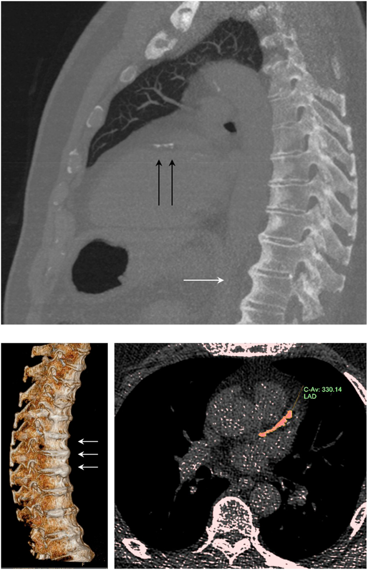
Fig. 1.

Illustration of a 68 year old male with DISH and CAC score category > 100–400.
(Upper panel) MIP 7 mm sagittal/oblique. White arrows: thoracic DISH; black arrows: calcifications in the left anterior descending coronary artery (LAD). (Lower left panel) 3D reconstruction of the spine showing DISH. (Lower right panel) LAD calcification quantification.
