PLOS ONE logoLink to PLOS ONE
. 2021 Apr 12;16(4):e0249179. doi: 10.1371/journal.pone.0249179

In vivo assessment of a delayed release formulation of larazotide acetate indicated for celiac disease using a porcine model

Hiroko Enomoto 1, James Yeatts 1, Liliana Carbajal 1, B Radha Krishnan 2, Jay P Madan 2, Sandeep Laumas 2, Anthony T Blikslager 1, Kristen M Messenger 1,*
Editor: Vivek Gupta3
PMCID: PMC8041193  PMID: 33844694

Abstract

There is no FDA approved therapy for the treatment of celiac disease (CeD), aside from avoidance of dietary gluten. Larazotide acetate (LA) is a first in class oral peptide developed as a tight junction regulator, which is a lead candidate for management of CeD. A delayed release formulation was tested in vitro and predicted release in the mid duodenum and jejunum, the target site of CeD. The aim of this study was to follow the concentration versus time profile of orally administered LA in the small intestine using a porcine model. A sensitive liquid chromatography/tandem mass spectrometry method was developed to quantify LA concentrations in porcine intestinal fluid samples. Oral dosing of LA (1 mg total) in overnight fasted pigs resulted in time dependent appearance of LA in the distal duodenum and proximal jejunum. Peak LA concentrations (0.32–1.76 μM) occurred at 1 hour in the duodenum and in proximal jejunum following oral dosing, with the continued presence of LA (0.02–0.47 μM) in the distal duodenum and in proximal jejunum (0.00–0.43 μM) from 2 to 4 hours following oral dosing. The data shows that LA is available in detectable concentrations at the site of CeD.

Introduction

Celiac disease (CeD) is one of the most common autoimmune disorders affecting around 1% of the population worldwide [1, 2]. There has been a notable rise in the prevalence of CeD in the last 50 years and a rise in the rate of diagnosis in the last 10 years [2]. CeD is defined as a chronic small intestinal immune mediated enteropathy that is precipitated by exposure to dietary gluten, which is broken down into immunologically active gliadin fragments in genetically predisposed individuals. Gliadin indirectly stimulates the secretion of zonulin from the lamina propria of the intestine into the intestinal lumen, which leads to the binding of zonulin to purported apical receptors of the enterocyte. This initiates a complex series of tight junction events that involves phosphorylation of tight junction proteins, which induces a loss of epithelial barrier function, exacerbating innate and adaptive immune responses [1, 3, 4]. Untreated and partially treated CeD is associated with an increased risk for multiple comorbidities, such as diarrhea, abdominal pain, infertility, osteoporosis, joint pain, arthritis, uveitis, cataracts, alopecia areata, neuropathies, and lymphomas [2, 47]. Currently, a gluten free diet (GFD) is the only available approach to manage symptoms in CeD patients, but it is not completely effective due to hidden gluten from food contamination in kitchens, restaurants, and during food processing [4]. Recurrent CeD signs and symptoms resulting from inadvertent or deliberate gluten exposure have been reported in approximately 70% of CeD patients on a GFD [8, 9]. Additionally, refractory celiac disease is unresponsive to the treatment even with a strict gluten free diet and no other effective treatment has been established [7, 10, 11]. There is therefore an unmet need for non-dietary therapies for the management of CeD.

Larazotide acetate (LA) is an orally administered, locally acting, synthetic eight amino acid peptide that is known to act as a tight junction regulator [12]. LA acts as a zonulin inhibitor, capable of closing leaky or open interepithelial junctions, thereby having the potential to prevent exposure to gliadin [13]. Presently, LA is being studied in phase 3 clinical trials using a delayed release formulation designed to reach the target site for treatment of CeD in the proximal small intestine [12, 1416].

No study has been conducted to show the presence of LA and/or its fragments (Fig 1A and 1B) in the gastrointestinal tract (GIT) upon dosing. While earlier studies attempted (unsuccessfully) to determine LA pharmacokinetics from plasma, the peptide is broken down in the small intestine and there is no systemic absorption of LA or the fragments, therefore collection of intestinal fluid directly from the intestinal tracts is ultimately necessary to determine the presence, concentrations, and duration of LA at its site of action [17]. However, several anatomic and physiologic characteristics of the intestinal tract make frequent sampling challenging. Repeated aspiration of the gastrointestinal fluid increases the risk of peritonitis. Furthermore, it is impossible to repeatedly aspirate from the same anatomic location due to GI motility, and abdominal aspiration is invasive and painful to the subject. The sacrifice of multiple animals for collection of intestinal fluid contents does not allow for repeated sampling in the same animal. In vivo ultrafiltration (UF) is a minimally invasive method to collect and determine protein unbound drug concentrations in animal models [18, 19]. The probe fibers are made of a semipermeable dialysis membrane allowing water, electrolytes and low weight molecules (<30,000 Daltons) to pass into the collection system. A vacutainer attached to the assembly passively aspirates fluid over time and allows for the collection of multiple samples without causing distress to the animal. UF probes were previously successfully implanted into the intestinal tract of calves, which allowed for safe and effective continuous sampling of intestinal fluid [20]. UF probes have been used to collect several drug compounds from various tissue sites in animals [2125], but to our knowledge, this technique has never been applied to the collection of LA and its fragments directly from the intestinal tract. The purpose of this study was to (1) evaluate the dissolution profile of LA in the simulated media and to quantify the concentrations of LA in the porcine small intestine, and to (2) evaluate the use of UF as a method for intestinal fluid collection in a porcine model. We hypothesized that UF could be used to evaluate concentrations of LA directly at the intestinal site of action.

Fig 1.

Fig 1

Amino acid sequence of larazotide acetate (a) and its fragments (b) Larazotide acetate: H-Gly-Gly-Val-Leu-Val-Gln-Pro-Gly-OH (a) Fragment 1: H-Gly-Val-Leu-Val-Gln-Pro-Gly-OH; Fragment 2: H-Val-Leu-Val-Gln-Pro-Gly-OH; Fragment 3: H-Gly-Gly-Val-Leu-Val-Gln-Pro-OH; Fragment 4: H-Val-Leu-Val-Gln-Pro-OH.

Materials and methods

In vitro dissolution experiment

1 mg LA capsules were dissolved in the simulated gastric media and simulated intestinal media, and percent dissolved LA were calculated at appropriate time points. The dissolution method was performed according to the current USP dissolution monograph chapter 711 [26]. The analytical method was validated following the FDA Bioanalytical Method Validation Guidelines for Industry [27]. The detailed methods are shown in S1 Text.

LA bead composition

In this study, a delayed release formulation of LA was tested. This is the formulation of LA that is being tested in the phase 3 clinical trials. The capsule contained a mixture of two types of beads (A and B) with two different thicknesses of enteric coating to trigger release in the mid duodenum and complete release within the proximal jejunum. The enteric coating included an enteric methacrylate polymer, eudragit, which is well known as an enteric coating polymer that delays the release of the active substance in the acidic surroundings of the stomach. The enteric coating was designed to release LA at pH level above pH 5, which would include release in the duodenum and jejunum over the course of about 3 hours.

In vivo ultrafiltration experiment

Animals and housing

This study was carried out in accordance with the recommendations in the Guide for the Care and Use of the Laboratory Animals [28] and approved by the Institutional Animal Care and Use Committee at North Carolina State University (protocol number: 18-154-B). Three female Yorkshire cross pigs weighing between 15–20 kg and 6–8 weeks of age were used in this study. Pigs were obtained from North Carolina State University’s commercial herd. All pigs were considered healthy on the basis of physical exams, which were performed upon delivery and after a minimum of 3 days’ acclimatization. The animals were housed individually in stainless steel pens to allow comprehensive observation of changes to the animal’s behavior, feed consumption, and volume or character of excreta. While housed in pens, each animal had free access to water, and commercial pig pelleted feed was provided twice daily. Housing was controlled at temperature 17.8–28.9°C, humidity 30–70% and an alternating 12 hour light/dark cycle was maintained and air flow was at least 10 air changes per hour with 100% fresh air. On the last day of the study, each pig was sedated with a xylazine/ketamine combination, then humanely euthanized by administration of an overdose of sodium pentobarbital intravenously. Once death was confirmed by lack of a heart beat and corneal reflex, a post mortem exam was performed to confirm the location of UF probes in the intestines during the duration of the study period.

Surgical procedure for ultrafiltration probe placement

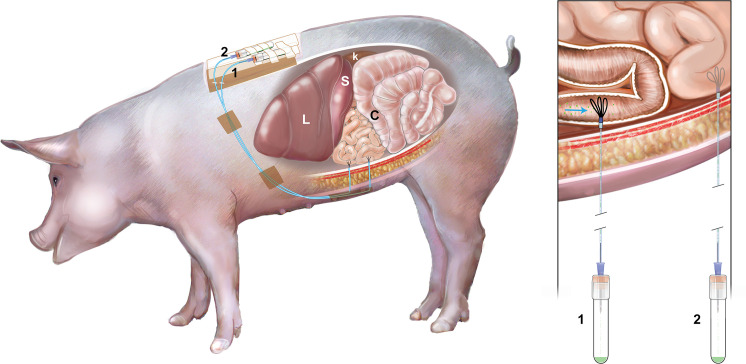
Food and water were withheld from pigs at least 12 hours prior to anesthesia and surgery. Pigs were sedated with injection of ketamine (11 mg/kg, IM; Vedco) and xylazine (0.25 mg/kg, IM; Akorn Animal Health). Intravenous administration of buprenorphine (0.04 mg/kg; Par Pharmaceutical) and intramuscular administration of flunixin meglumine (2.2 mg/kg; Norbrook) and ceftiofur sodium (Excede for Swine, 8mg/kg; Med Pharmex Inc) were given for analgesia and infection prophylaxis, respectively. Pigs were intubated and maintained under general anesthesia using isoflurane delivered in 100% oxygen during the surgery. The abdomen was approached via midline laparotomy, and the pylorus was located. The duodenum was traced from the pylorus to an accessible point on the left side of the abdomen approximately 15–18 cm distal to the pylorus (Fig 2). The intestine was lifted and an incision was made using a #11 scalpel blade, piercing the anti-mesenteric intestinal wall into the lumen. A guide needle was used to introduce the UF probe into the duodenum threading the probe proximally toward the pylorus. The guide needle was subsequently removed, taking care to leave the UF probe in place [20]. The three loops of the UF probe remained within the intestine, and each probe was secured in the intestine by using a Halstead pattern suture around the probe, followed by a finger trap suture pattern using 3–0 Vicryl (Henry Schein). The nonpermeable connection tubing of the UF probe was extended to the lateral body wall, paramedian to the incision, and retrieved through a guide needle in order to exteriorize the tubing. The tubing was secured outside the animal by suturing the tubing to the skin using 3–0 nylon suture (Fig 3). The tubing was attached to a needle inserted into a vacutainer for sample collection during the remainder of the experiment. A second UF probe was placed into the jejunum 20 cm distal to the duodenum using the same methodology (Fig 2). The abdomen was closed in a routine manner. The porcine gastrointestinal model shown in Fig 3. The pigs were monitored for assessment of pain each day of the experiment and given buprenorphine as an analgesic medication if necessary.

Fig 2. The location of implantation of ultrafiltration probes.
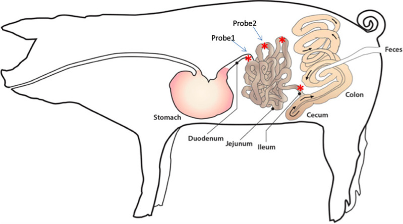
Fig 2

The location of the first probe placement was 15–18 cm distal to the pylorus, the distal duodenum. The location of the second probe placement was 20 cm distal to the duodenum, the proximal jejunum.

Fig 3. The porcine gastrointestinal model.

Fig 3

This model was implanted the ultrafiltration probes (UF) at duodenum and jejunum and the intestinal fluid was collected via UF probes continuously. L: liver, S: spleen, K: kidney, C: colon, 1: first UF probe, 2: second UF probe.

Drug formulation, administration and intestinal fluid collection

The delayed release formulation of LA was provided by Innovate Biopharmaceuticals Inc. (currently, 9Meters Biopharm Inc., Raleigh, NC, USA). This formulation was identical to the formulation tested in the in vitro experiment as previously described. The formulated beads were weighed and placed into gelatin capsules prior to dosing. Pigs were fasted overnight for approximately 14 hours; water was withheld for 2 hours prior to dosing. On the morning of each experiment, a single capsule (1 mg) of LA, approximate dose of 0.05 mg/kg, or a placebo capsule was administered to each animal by mouth, then chased with 120 mL water. The animals were observed to ensure swallowing of the entire capsule. After drug or placebo administration, water was withheld for a further 2 hours, and food for four hours. Intestinal fluid (S1 Fig) was collected into a 3mL vacutainer (Becton Dickinson) via the UF probes every 1 hour over a 4 hour period on each day of the study (Fig 4). Each vacutainer was prefilled with 100 μL of a quenching solution (5% trifluoroacetic acid (TFA) in 80% acetonitrile (ACN):15% water) to prevent degradation of LA by intestinal enzymes during the collection period. Following sampling, an additional amount of quenching solution was added to the sample if necessary based on the sample volume collected to maintain an approximately consistent ratio of intestinal fluid: quench solution (250: 40). Samples were centrifuged at 13,200 x g for 5 minutes at 4°C. The resulting supernatant was removed and stored at -80°C until analysis by ultra performance liquid chromatography and tandem mass spectrometry (UPLC-MS/MS).

Fig 4. A timeline of the study design.

Fig 4

Overnight fasted pigs received 1 mg larazotide acetate (or placebo) and the intestinal fluid was collected every hour for four hours via the ultrafiltration probe on each experimental day.

Chemicals and reagents

All reagents were of LC/MS grade. ACN, formic acid and TFA were purchased from Fisher Chemical (Raleigh, NC, USA). LA and its fragments (1, 2, 3 and 4) standards were provided by Innovate Biopharmaceuticals Inc. (Currently, 9Meters Biopharm Inc).

Instrumentation

Analysis of LA and fragments were carried out via ultra performance liquid chromatography (UPLC) and tandem mass spectrometric (MS/MS) detection (Waters Corporation, Milford, MA). The UPLC-MS/MS system consisted of a Waters Acquity UPLC I class Binary Solvent Manager, Acquity UPLC Sample Manager FTN and a Xevo TQD tandem mass spectrometer (Waters Corporation, Milford, MA).

Preparation of standard solutions

The 3.5, 7, 14, 35, 70, 140 and 350 μM standards of LA were prepared by dilution of mixed stock solutions of LA (700 μM) and its fragments 1–4 (750, 800, 750 and 850 μM respectively) with diluent of 1:1 ACN: water. The standards of fragment 1–4 were made from mixed stock solution at the range of 3.75–375, 4.0–400, 3.75–375 and 4.25–425 μM respectively.

Calibration standard preparation

Each standard solution (3.5, 7, 14, 35, 70, 140 and 350 μM) of LA was diluted with the blank intestinal fluid to give concentrations 0.035, 0.07, 0.14, 0.35, 0.7, 1.4 and 3.5 μM for the calibration curve. The range of the calibration curve for fragment 1–4 was 0.038–3.75, 0.04–4.0, 0.038–3.75, and 0.04–4.25 μM respectively. The blank intestinal fluid and dilution solvent were injected with every batch.

Sample preparation

The samples were centrifuged at 10,000 g for 5 minutes. The supernatant was filtered through a 0.2 μm polyvinylidene difluoride membrane filter directly into Waters maximum recovery sample vial (Waters Corporation, Milford, MA) prior to injection into the UPLC-MS/MS system. Quality control standards were injected with every batch.

UPLC- MS/MS conditions

Chromatographic separation was performed by a gradient elution on the ACQUITY UPLC BEH C18 1.7 μm column (2.1 mm x 50 mm) with VanGuard Pre-column (Waters Corporation, Milford, MA). The mobile phase solvents were 0.02% TFA in water (A) and 0.02% TFA in ACN (B) at a flow rate of 0.4 mL/min for 8 minutes. The gradient program mobile phase conditions were 95% of A and 5% of B for the first 5.5 minutes, then changed linearly to 10% of A and 90% of B from 5.5–6.5 minutes, then immediately back to 95% of A and 5% of B from 6.51–8 minutes to re-equilibrate at the initial conditions. The column temperature was 35°C, the autosampler temperature was maintained at 4°C and the injection volume was 7 μL. The positive electrospray ionization mode (ESI (+)) was used with the multiple reactions monitoring (MRM). The tune page source voltages were 2.0 kV and 47 V for the capillary and cone respectively. The source desolvation temperature was 600°C. The source desolvation gas flow was 750 L/hr and the cone gas was 50 L/hr. The source temperature was 150°C. The MS file cone voltage (V) settings for LA, fragments 1, 2, 3, and 4 were 44, 42, 42, 40, and 38 V with collision energy settings (V) of 60, 58, 54, 62, and 54 V respectively. Argon was used as the collision gas and nitrogen as the desolvation and cone gases. Quantification was performed using the transitions Parent (m/z) 726.65, 669.54, 612.58, 669.54 and 555.64 for LA, fragment 1, 2, 3 and 4 respectively and Daughter (m/z); 72.08, 86.14, 72.1, 72.1 and 72.1 for LA, fragment 1, 2, 3 and 4 respectively with the retention time 2.33, 2.32, 2.23, 2.43 and 2.35 minutes for LA, fragment 1, 2, 3 and 4 respectively (S1 Table). The chromatogram of LA is shown in S2 Fig.

The lower limit of quantification and the lower limit of detection

Lower limit of quantification (LLOQ) was defined as the lowest concentration that produced a peak area 5 times the blank solvent peak area, had an accuracy within 20% of the nominal value, and a precision of no more than 20% CV. The LLOQ of the LA was 0.07 μM (0.05 μg/mL). The LLOQ of the fragment 1, 2, 3 and 4 was 0.15, 0.16, 0.15 and 0.17 μM (0.1 μg/mL) respectively. The lower limit of detection (LLOD) was the lowest concentration that produced a peak area > 3 times blank solvent peak area. The LLOD of the LA was 0.0175 μM (0.0125 μg/mL). The LLOD of the fragment 1, 2, 3 and 4 was 0.0024, 0.0050, 0.0188 and 0.0213 μM (0.0016, 0.0031, 0.0012 and 0.0125 μg/mL) respectively.

Calibration curve

The calibration curves of LA and its fragment 1–4 were fit with a weighted (1/concentration) linear equation. The calibration range of 0.035–3.5 μM (LA), 0.038–3.75 μM (fragment 1 and 3), 0.04–4.0 μM (fragment 2), 0.04–4.25 μM (fragment 4) was linear with a coefficient of determination, R2, greater than or equal to 0.99. Each calibration standard concentration could be back calculated to within 15% of the true concentration (S3 Fig).

Precision and accuracy

A total 6 replicate samples at low, medium and high concentrations (0.21, 1.05 and 2.8 μM of LA, 0.45, 1.13 and 3 μM of fragment 1, 0.48, 1.2 and 3.2 μM of fragment 2, 0.45, 1.13 and 3 μM of fragment 3 and 0.51, 1.28 and 3.4 μM of fragment 4) were tested on 3 days and interday and intraday precision and accuracy were calculated. Mean of each concentration were within 15% of the nominal value and have a precision not exceeding 15% CV (S2 Table).

Pharmacokinetics

Non compartmental pharmacokinetic analyses of LA in the intestinal fluid was performed using commercially available software (Phoenix® WinNonlin® Software version 8.3, Certara, Princeton, NJ). The pharmacokinetic parameters estimated for LA in intestinal fluid after oral administration included the elimination rate constant (λz), elimination half life (HLλz), the area under the curve from time zero to the last time point (AUClast), the maximum concentration (Cmax), time to maximum concentration (Tmax), the mean drug residence time from time zero to the last time point (MRTlast), which were calculated using the linear log trapezoidal method.

Results

In vitro experiment

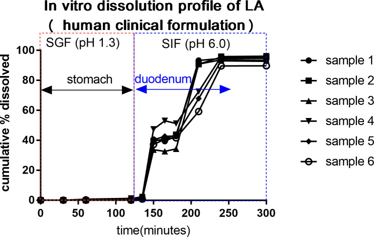
Data was collected from 6 capsules. Average (± SD) dissolution (%) are shown in Table 1 and Fig 5. The in vitro dissolution data predicted that the delayed release formulation of LA was not dissolved in the simulated gastric dissolution media for 2 hours. Dissolution was 39.7–42.8%, 79.1% and 93.7% at 30–60 minutes, 90 minutes and 120–180 minutes in intestinal dissolution media.

Table 1. In vitro dissolution data of 1 mg larazotide acetate of a delayed release formulation in 6 capsules.

Sampling time points (minutes) Time in GIT (minutes) Average (±SD) Dissolution (%) of 1 mg LA (n = 6)
Simulated gastric media (Stomach) 0 0 0
30 30 0
60 60 0.1 ± 300
120 120 0.7 ± 85.7
Simulated intestinal media (Small intestine) 15 135 1.4 ± 42.9
30 150 39.7 ± 11.4
45 165 41.2 ± 14.4
60 180 42.8 ± 12.6
90 210 79.1 ± 18.3
120 240 93.7 ± 2.4
180 300 93.7 ± 2.5

Times (minutes) in small intestine were back calculated by adding 120 minutes to sampling time point respectively. LA: larazotide acetate, GIT: gastrointestinal tract, SD: standard deviation

Fig 5. The in vitro dissolution profile of larazotide acetate (LA) of the delayed release formulation.

Fig 5

The in vitro dissolution data predicts the delayed release formulation of LA will begin releasing in the mid duodenum and complete release in the proximal jejunum. SGF: simulated gastric fluid, SIF: simulated intestinal fluid. Samples 1–6 were replicated and included 1 mg of LA respectively.

In vivo experiment

A pilot study was initially performed with one pig to determine the feasibility of intestinal fluid collection. Sample data collected from this pilot study was not pooled with the study data. Thus, two animals were included in the study with one dosing event per day per animal. Each animal was used for up to four dosing events (either LA or placebo) without any signs of adverse effects. In the two experimental animals, the UF probes continuously collected intestinal fluid over the entire study period. Average (± SD) volume of fluid collected each hour at the distal duodenum was 604 ±259, 835 ±72, 515 ±75 and 487 ±113 μL at 1, 2, 3, and 4 hour time points, respectively. Average (± SD) volume of fluid collected each hour at the proximal jejunum was 720 ±70, 575 ± 80, 765 ±190 and 556 ± 250 μL at the 1, 2, 3, and 4 hour time points respectively (Table 2 and S3 Table).

Table 2. Concentration data of larazotide acetate in the intestinal samples upon oral administration of 1mg larazotide acetate in a delayed release formulation in pigs.

Time (hour) Probe Location Average (±SD) Concentration of LA (μM) in intestinal fluid (n = 3) Average (±SD) Volume (mL) of intestinal fluid (n = 3)
Clinical formulation Distal duodenum
0 0.00 ± 0.00 2583 ± 93
1 0.74 ± 0.30 604 ± 259
2 0.27 ± 0.14 835 ± 72
3 0.12 ± 0.09 515 ± 75
4 0.03 ± 0.01 487 ± 113
Proximal jejunum
0 0.00 ± 0.00 2364 ± 621
1 1.10 ± 0.48 720 ± 70
2 0.25 ± 0.13 575 ± 80
3 0.09 ± 0.10 765 ± 190
4 0.03 ± 0.04 556 ± 250
Placebo control Distal duodenum
0 0.00 ± 0.00 1848 ± 929
1 0.00 ± 0.00 827 ± 113
Proximal jejunum
0 0.00 ± 0.00 1751 ± 373
1 0.00 ± 0.00 943 ± 148

1μM of larazotide acetate (LA) = 0.71 μg/ml, SD: standard deviation

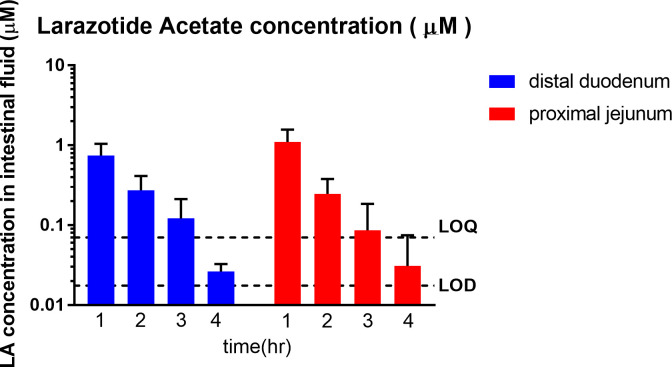
Oral dosing of the clinical formulation of LA (1mg) in overnight fasted pigs resulted in time dependent appearance of LA in the distal duodenum and the proximal jejunum. Peak LA concentrations ranged from 0.32–1.76 μM, and were noted at 1 hour in the distal duodenum and the proximal jejunum. The continued presence of LA was detected in distal duodenum for 2–4 hours (0.02–0.47 μM), and in proximal jejunum for 2–4 hours (0.00–0.43 μM). The LA concentrations were below LOD in one sample at the 3 hour time point and in 2 samples at the 4 hour time point in the proximal jejunum. The LA concentrations of these samples were reported as zero. The average (± standard deviation) concentrations of LA at 1, 2, 3 and 4 hours are shown in Fig 6, Table 2 and S4 Table.

Fig 6. Concentration v time profile of larazotide acetate (LA, μM) in the porcine intestinal fluid analyzed via UPLC-MS/MS (n = 3).

Fig 6

Time dependent appearance of LA in the distal duodenum and the proximal jejunum was observed. Peak LA concentrations ranged from 0.32 to 1.76 μM and were first observed at the 1 hour time point in the distal duodenum and the proximal jejunum. The highest concentration of LA was detected in the proximal jejunum at 1 hour. The continued presence of LA was detected in the distal duodenum (0.02–0.47 μM) and proximal jejunum (0.00–0.43 μM) from 2 to 4 hours post administration.

The LA was not detected in the placebo (control) group throughout the study (Table 2 and S4 Table).

The presence of LA fragments (1, 2, 3 and 4) were detected in duodenal and jejunal samples. However, all concentrations were below the LLOQ of 0.15, 0.16, 0.15 and 0.17 μM respectively.

The results of the pharmacokinetic analysis are presented in Table 3.

Table 3. The pharmacokinetic parameters for larazotide acetate in the intestinal fluid after oral larazotide acetate administration (1 mg total; approximate dose 0.05 mg / kg).

Distal duodenum Dose Replicate
Parameter (unit) # 1 # 2 # 3 Mean SD CV %
Λz (1/h) 0.79 1.23 1.41 1.14 0.32 28.1
HLλz (h) 0.88 0.56 0.49 0.65 0.21 32.2
Tmax (h) 1 1 1 1 0 0
Cmax (μg / mL) 0.23 0.70 0.67 0.53 0.26 49.3
AUClast (μg·h /mL) 0.36 0.83 1.13 0.77 0.39 50.2
MRTlast (h) 1.58 1.42 1.65 1.55 0.12 7.80
Proximal jejunum
Λz (1/h) 1.39 0.90 0.69 1.0 0.36 36.2
HLλz (h) 0.50 0.77 1 0.76 0.25 33.3
Tmax (h) 1 1 1 1 0 0
Cmax (μg / mL) 0.46 1.26 0.63 0.78 0.42 53.4
AUClast (μg·h /mL) 0.52 1.22 0.99 0.91 0.36 39.4
MRTlast (h) 1.31 1.26 1.53 1.37 0.14 10.3

λz: elimination rate constant, HLλz: elimination half life, Tmax: time to the maximum concentration, Cmax: maximum concentration, AUClast: area under the curve from time zero to the last time point, MRTlast: mean residence time from time zero to the last time point

1 μg / mL = 1.4 μM. SD: standard deviation, CV: coefficient of variation

Discussion and conclusions

This study was designed to assess the in vivo delivery and gastrointestinal transit profile of the delayed release formulation of LA intended for use in CeD patients using a porcine model. The results of the animal experiments show that concentrations of LA were present in the distal duodenum for the entire four hours and proximal jejunum for three hours (4 hours in one sample) following oral administration of the delayed release formulation in vivo. Since CeD patients have mucosal morphological changes localized to the upper small intestine [6, 7], this delayed release formulation is ideal to target diseased tissue in the duodenum and proximal jejunum. This is the first study to quantify the presence of LA in the small intestine in vivo over a recommended dosing interval, although numerous previous studies have confirmed pharmacodynamic effects [2, 4, 2931].

The GIT comprises rather complex biochemical and physiological process such as enzyme, luminal pH, body temperature, peristalsis movement, gastric and intestinal residence time and luminal composition; thus it is more complex than the in vitro static dissolution model [32]. An in vivo model is more ideal to study the true movement and release of a drug formulation. In this study, a porcine model was established for in vivo human oral drug assessments for several reasons. In particular, pigs have similar intestinal anatomy and physiology to that of human beings [33]. The porcine small intestinal pH has a similar range to that of humans [3336]. More specifically, the fasted pig’s gastric pH is 1.2–4.0 and the proximal small intestinal pH is 6.7. The fasted human gastric pH is 1.0–3.5 and 6.0–7.0 for the proximal small intestine [3336]. The variation in gastrointestinal volume, osmolality and intestinal transit time are other important determinants of drug release and absorption [36, 37]. The transit time from mouth through the small intestine is reported to be similar in pigs and humans, taking 3–4 hours in pigs as compared to 2–4 hours in humans in the fasted state [33].

Following oral administration, the parameters having the most influence on the drug dissolution are the physical and chemical characteristics of the dosing formulation [3841]. The enteric methacrylate polymer, eudragit, is derived from esters of acrylic and methacrylic acid by free radical polymerization. The use of eudragit for targeted drug release and eudragit’s stability in gastric fluid in the presence of digestive enzymes are well known [39, 42]. This in vitro study confirmed the stability of LA in the simulated gastric fluid (dissolved LA % was 0.7%) and dissolution of LA in simulated media in time dependent manner. LA solubility in the digestive fluid depends primarily on time and luminal pH [37]. The variability of drug transit time, luminal pH and other factors such as gastrointestinal fluid volume may have contributed to the variability of the LA concentrations in the small intestine.

UF probes were successfully placed in two different locations in the small intestine in all three pigs. Although unanticipated complications occurred in the initial pilot pig (loss of the UF probe from within the intestinal lumen), the UF probes were well tolerated at both duodenal and proximal jejunal sites in the remaining two pigs. Using the probes, intestinal fluid was collected over four hours, continuously from the exact same location, without invasive sampling or euthanasia, allowing for significant reduction in the number of experimental animals, and minimization of intra-animal variation in the data. The UF probes exclude all proteins and molecules larger than 30,000 Daltons and allows the collection of a “clean” ultrafiltrate that can be analyzed for drug concentrations or even biomarkers of drug efficacy. Warren et al., reported that intestinal fluid could be collected from the ileum and spiral colon of steers over 48 hours with an 88% success rate. Occasional absence of sample in the collection tube was noted as a possible complication, attributable to clogging of the pores on the fragile membrane of the UF or to damage of the membrane due to the motility of the intestinal tract and ingesta [20]. Although some samples were not obtained in the pilot pig, all samples at all time points were collected in the other two animals. In another study in horse hooves, 33% of the UF probe sites became infected, which was attributed to inadequate preparation of the probe placement site, insufficient maintenance of sterility during placement, or prolonged probe placement time [23]. In the present study, no signs of infection were observed; the probes were placed aseptically and tissues remained healthy over the 5 day study duration. Overall, it appears that UF probes can be used at multiple sites in the intestinal tract of pigs to collect gastrointestinal fluids. Continued refinement of optimal UF probe placement and maintenance techniques are necessary for future studies on gastrointestinal drug pharmacokinetics.

In vitro dissolution is the standard method used in the pharmaceutical industry to assess drug release from solid oral dosage forms and to predict the release profile in the GI tract by simulating the GI environment with appropriate buffer media. In vivo/ in vitro correlation analysis (IVIVC) was specifically designed to assess whether in vitro predictions match in vivo drug behavior. However, in this study IVIVC could not be performed for several reasons, including the lack of an injectable formulation, an immediate release and other multiple other extended release formulations [43] and the inability to quantify the total volume of the intestinal fluid as the UF probes only collected a small fraction of the entire volume. However, we described the pharmacokinetics of LA at two different sites in the small intestine in the pigs in this study. The pharmacokinetics of LA have not been described in people, despite the compound undergoing multiple clinical trials to date. Paterson et al., reported that oral LA (12mg) could be detected in the human plasma by HPLC-MS/MS, but all plasma concentrations were below LLOQ (0.5 ng/ml) [17]. Leffler et al., reported that the concentrations of LA and metabolites in human plasma were below the LOQ (0.5 ng/ml) in all groups administered 0.25, 1, 4 and 8 mg LA [16], thus LA pharmacokinetics could not be determined in human plasma. Since the present study only included the small number of pigs, data interpretation was limited. Additionally, our UPLC-MS/MS method was not sensitive enough to quantify the larazotide fragments that were identified. Lastly, healthy pigs were used in this study. Drug release and pharmacokinetics could be different in patients with CeD or other gastrointestinal pathology [44]. In spite of these limitations, the data showed that the LA was detectable in the small intestine after dosing, and an animal model was described to study drugs that act locally in the intestinal tract.

Despite the limitations discussed, this was the first study in which the UF probes were successfully placed in the intestinal tract of pigs to obtain concentrations of LA directly from the site of action. Our technique may be useful in future pharmacokinetic studies to analyze other formulations of LA or other locally acting drugs in the small intestine.

Supporting information

S1 Text. The in vitro dissolution test methods.

(PDF)

S1 Fig. Intestinal fluid collection system.

(TIF)

S2 Fig. A typical chromatogram of larazotide acetate at 1.38 μM.

(TIF)

S3 Fig. Calibration curves of larazotide acetate and its fragment 1–4.

(TIF)

S1 Table. Detection of larazotide acetate and its fragments in porcine intestinal fluid matrix by UPLC-MS/MS.

(PDF)

S2 Table. Analytical method suitability study data.

(PDF)

S3 Table. Individual intestinal fluid volume (μL) collected via ultrafiltration probes.

(PDF)

S4 Table. Individual concentration of larazotide acetate (μM) in the intestinal samples for the delayed release formulation and placebo administration.

(PDF)

Acknowledgments

We are grateful to Alice MacGregor Harvey for the medical illustrations.

Data Availability

All relevant data are within the manuscript and its Supporting Information files.

Funding Statement

The study was funded by Innovate Biopharmaceuticals (Jay Madan), which is now 9meters (https://9meters.com/). KMM and AB received the award (TSA #91690). Innovate Biopharmaceuticals (now 9meters) provided support in the form of salary for authors BRK, TPM, and SL. The funders had no role in study design, data collection and analysis, decision to publish, or preparation of the manuscript. The specific roles of these authors are articulated in the ‘author contributions’ section.

References

  • 1.Gonzalez LM, Moeser AJ, Blikslager AT Porcine models of digestive disease; the future of large animal translational research. Translational research. 2015. July;166 (1);12–27. 10.1016/j.trsl.2015.01.004 [DOI] [PMC free article] [PubMed] [Google Scholar]
  • 2.Alhassan E, Yadav A, Kelly CP, Mukherjee R. Novel non dietary therapies for celiac disease. Cellular and molecular gastroenterology and hepatology. 2019;8 (3):335–345. 10.1016/j.jcmgh.2019.04.017 [DOI] [PMC free article] [PubMed] [Google Scholar]
  • 3.Arrieta MC, Bistritz L, Meddings JB. Alterations in intestinal permeability. Gut. 2006. October;55 (10):1512–1520. 10.1136/gut.2005.085373 [DOI] [PMC free article] [PubMed] [Google Scholar]
  • 4.Khaleghi S, Ju JM, Lamba A, Murray JA. The potential utility of tight junction regulation in celiac disease: focus on larazotide acetate. Therapeutic Advances in gastroenterology. 2016;9 (1):37–49. 10.1177/1756283X15616576 [DOI] [PMC free article] [PubMed] [Google Scholar]
  • 5.Therrien A, Kelly CP, Silvester JA. Celiac disease: Extraintestinal Manifestations and associated Conditions. Journal of Clinical Gastroenterol. 2020. January; 54 (1); 8–21 10.1097/MCG.0000000000001267 [DOI] [PMC free article] [PubMed] [Google Scholar]
  • 6.Torborg L. Mayo Clinic Q and A: Diagnosing celiac disease not always a one- step process. 2015. Available from: http://newsnetwork.mayoclinic.org/discussion/mayo-clinic-q-and-a-diagnosing-celiac-disease-not-always-a-one-step-process [Google Scholar]
  • 7.Freeman HJ. Celiac disease: A disorder emerging from antiquity, its evolving classification and risk, and potential new treatment paradigms. Gut and liver. 2015. January;9(1):28–37. 10.5009/gnl14288 [DOI] [PMC free article] [PubMed] [Google Scholar]
  • 8.Leffler DA, Dennis M, Hyett B, Kelly E, Schuppan D, Kelly CP. Etiologies and predictors of diagnosis in nonresponsive celiac disease. Clin Gastroenterol Hepatol. 2007; 5:445–450 10.1016/j.cgh.2006.12.006 [DOI] [PubMed] [Google Scholar]
  • 9.Hall NJ, Rubin GP, Charnock A. Intentional and inadvertent non-adherence in adult coeliac disease. A crosssectional survey. Appetite 2013; 68:56–62. 10.1016/j.appet.2013.04.016 [DOI] [PubMed] [Google Scholar]
  • 10.Donnelly SC, Ellis HJ and Ciclitira RJ. Pharmacotherapy and management strategies for coeliac disease. Expert opinion on pharmacotherapy. 2011:12(11):1731–1744. 10.1517/14656566.2011.592140 [DOI] [PubMed] [Google Scholar]
  • 11.Vaquero L, Bernardo D, Leon F, Martin LR, Cuenllas BA, Vivas S. Challenges to drug discovery for celiac disease and approaches to overcome them. Expert opinion on drug discovery. 2019;14(10):957–968. 10.1080/17460441.2019.1642321 [DOI] [PubMed] [Google Scholar]
  • 12.Leffler DA, Kelly CP, Green PHR, Fedorak RN, DiMarino A and Perrow W. Larazotide acetate for persistent symptoms of celiac disease despite a gluten free diet: A randomized controlled trial. Gastroenterology. 2015. June;148:7:1311–1319 10.1053/j.gastro.2015.02.008 [DOI] [PMC free article] [PubMed] [Google Scholar]
  • 13.Valitutti F and Fasano A. Breaking down barriers: How understanding celiac disease pathogenesis informed the development of novel treatments. Digestive diseases and sciences. 2019; 64; 1748–1758. 10.1007/s10620-019-05646-y [DOI] [PMC free article] [PubMed] [Google Scholar]
  • 14.http://clinicaltrials.gov/ct2/show/NCT03569007, ClinicalTrials.gov, NIH U.S. National Library of Medicine
  • 15.Kelly CP, Green PHR, Murray JA, DiMarino A, Colatrella A and Leffler DA et al. Larazotide acetate in patients with coeliac disease undergoing a gluten challenge: a randomised placebo controlled study. Alimentary pharmacology and therapeutics. 2013:37:252–262 10.1111/apt.12147 [DOI] [PubMed] [Google Scholar]
  • 16.Leffler DA, Kelly CP, Abdallah HZ, Colatrella AM, Harris LA, Leon F et al. A randomized, double blind study of Larazotide acetate to prevent the activation of celiac disease during gluten challenge. The American journal of gastroenterology. 2012. October:107:1554–1562. 10.1038/ajg.2012.211 [DOI] [PMC free article] [PubMed] [Google Scholar]
  • 17.Paterson B M, Lammers K M, Arrieta M C, Fasano A, Meddings J B. The safety, tolerance, pharmacokinetic and pharmacodynamic effects of single doses of AT-1001 in coeliac disease subjects: a proof of concept study. Alimentary Pharmacology and Therapeutics. 2007; 26:757–766. 10.1111/j.1365-2036.2007.03413.x [DOI] [PubMed] [Google Scholar]
  • 18.Messenger KM, Papich MG, Blikslager AT. Distribution of enrofloxacin and its active metabolite, using an in-vivo ultrafiltration sampling technique after the injection of enrofloxacin to pigs. Journal of Veterinary Pharmacology and Therapeutics. 2011. September; 35:452–459. 10.1111/j.1365-2885.2011.01338.x [DOI] [PubMed] [Google Scholar]
  • 19.Papich MG, Davis JL, Floerchinger AM. Pharmacokinetics, protein binding, and tissue distribution of orally administered cefpodoxime proxetil and cephalexin in dogs. American journal of Veterinary Research. 2010. December;71: 1484–1491 10.2460/ajvr.71.12.1484 [DOI] [PubMed] [Google Scholar]
  • 20.Warren CD, Prange T, Campbell NB, Gerard MP, Martin LG, Jacob ME et al. Implantation of an ultrafiltration device in the ileum and spiral colon of steers to continuously collect intestinal fluid. Research in veterinary science. 2014; 97:611–615 10.1016/j.rvsc.2014.10.012 [DOI] [PubMed] [Google Scholar]
  • 21.Messenger KM, Papich MG, Blikslager AT. Distribution of enrofloxacin and its active metabolite, using an in-vivo ultrafiltration sampling technique after injection of enrofloxacin to pigs. Journal of veterinary pharmacology and therapeutics. 2012; 35:452–459. 10.1111/j.1365-2885.2011.01338.x [DOI] [PubMed] [Google Scholar]
  • 22.Messenger KM, Wofford JA, Papich MG. Carprofen pharmacokinetics in plasma and in control and inflamed canine tissue fluid using in-vivo ultrafiltration. Journal of veterinary pharmacology and therapeutics. 2015. 10.1111/jvp.12233 [DOI] [PubMed] [Google Scholar]
  • 23.Underwood C, Collins SN, Van Eps AW, Allavena RE, Torres CEM, Pollitt CC. Ultrafiltration of equine digital lamellar tissue. The veterinary journal. 2014; 202:314–322 10.1016/j.tvjl.2014.05.007 [DOI] [PubMed] [Google Scholar]
  • 24.Mzyk DA, Bublitz CM, Sylvester H, Mullen KAE, Hobgood GD, Baynes RE, et al. Short communication: Use of an ultrafiltration device in gland cistern for continuous sampling of healthy and mastitic quarters of lactating cattle for pharmacokinetic modeling. Journal of Dairy Science. 2018; 101:10414–10420 10.3168/jds.2018-14849 [DOI] [PubMed] [Google Scholar]
  • 25.Yan L, Xie S, Chen D, Pan Y, Tao Y, Qu W, et al. Pharmacokinetic and pharmacodynamic modeling of cyadox against clostridium perfringens in swine. Scientific reports. 2017; 7:4064. 10.1038/s41598-017-03970-9 [DOI] [PMC free article] [PubMed] [Google Scholar]
  • 26.<711> dissolution, USP 35, physical test: 5642–5648
  • 27.Bioanalytical method validation guidance for industry. U.S. Department of Health and Human Services Food and Drug Administration. 2018 [Google Scholar]
  • 28.Janet CG, Joseph TB, Leigh AC, John CD, Coenraad FMH, Wayne RB, et al. Guide for the Care and Use of Laboratory Animals: Eighth Edition [Google Scholar]
  • 29.Jin Y, Madan J, Laumas S, Radha Krishnan B, Blikslager AT. Larazotide protects the intestinal tight junction barrier during Anoxia/Reoxygenation injury via inhibition of myosin light chain kinase. Gastroenterology. 2019;156:S–487 [Google Scholar]
  • 30.Gopalakrishnan S, Tripathi A, Tamiz AP, Alkan SS, Pandey NB. Larazotide acetate promotes tight junction assembly in epithelial cells. Peptides. 2012;35: 95–101 10.1016/j.peptides.2012.02.016 [DOI] [PubMed] [Google Scholar]
  • 31.Gopalakrishnan S, Durai M, Kitchens K, Tamiz AP, Somerville R, Pandey NB, et al. Larazotide acetate regulates epithelial tight junctions in vitro and in vivo. Peptides. 2012; 35 (1): 86–94 10.1016/j.peptides.2012.02.015 [DOI] [PubMed] [Google Scholar]
  • 32.Ting Y, Zhao Q, Xia C and Huang Q. Using in vitro and in vivo models to evaluate the oral bioavailability of nutraceuticals. Journal of agricultural and food chemistry. 2015. January: 63:5:1332–1338 10.1021/jf5047464 [DOI] [PubMed] [Google Scholar]
  • 33.Henze LJ, Koehl NJ, O’Shea JP, Kostewicz ES, Holm R, Griffin BT. The pig as a preclinical model for predicting oral bioavailability and in-vivo performance of pharmaceutical oral dosage forms: a PEARRL review. Journal of pharmacy and pharmacology. 2019; 71:581–602 10.1111/jphp.12912 [DOI] [PubMed] [Google Scholar]
  • 34.Merchant HA, McConnell EL, Liu F, Ramaswamy C, Kulkarni RP, Basit AW, et al. Assessment of gastrointestinal pH, fluid and lymphoid tissue in the guinea pig, rabbit and pig, and implications for their use in drug development. European Journal of Pharmaceutical Sciences. 2011;42: 3–10, 10.1016/j.ejps.2010.09.019 [DOI] [PubMed] [Google Scholar]
  • 35.Merchant HA, Pereira FA, Rabbie SC, Youssef SA, Basit AW. Gastrointestinal characterisation and drug solubility determination in animals. Journal of Pharmacy and Pharmacology. 2014;67: 630–639, 10.1111/jphp.12361 [DOI] [PubMed] [Google Scholar]
  • 36.Ibekwe VC, Fadda HM, McConnell EL, Khela MK, Evans DF, Basit AW. Interplay Between Intestinal pH, Transit Time and Feed Status on the In-vivo Performance of pH Responsive Ileo-Colonic Release Systems. Pharmaceutical Research. 2008. August; 25 (8). 10.1007/s11095-008-9580-9 [DOI] [PubMed] [Google Scholar]
  • 37.Kararli TT. Comparison of the gastrointestinal anatomy, physiology, and Biochemistry of Humans and Commonly Used Laboratory Animals. Biopharmaceutics and Drug disposition. 1995; 16:351–380 10.1002/bdd.2510160502 [DOI] [PubMed] [Google Scholar]
  • 38.Hosny KM, Alhakamy NA, Almodhwahi MA, Kurakula M, Almehmady AM and Elgebaly SS. Self-nanoemulsifying system loaded with sildenafil citrate and incorporated within oral lyophilized flash tablets: preparation, optimization, and in vivo evaluation. Pharmaceutics. 2020:12:1124. 10.3390/pharmaceutics12111124 [DOI] [PMC free article] [PubMed] [Google Scholar]
  • 39.Alhakamy NA, Ahmed OAA, Kurakula M, Caruso G, Caraci F and Asfour HZ. Chitosan based microparticles enhance ellagic acid’s colon targeting and proapoptotic activity. Pharmaceutics. 2020;12:652. 10.3390/pharmaceutics12070652 [DOI] [PMC free article] [PubMed] [Google Scholar]
  • 40.Yadav S, Ahuja M, Kumar A and Kaur H. Gellan-thioglycolic acid conjugate: Synthesis, characterization and evaluation as mucoadhesive polymer. Carbohydrate polymers. 2014;99:601–607. 10.1016/j.carbpol.2013.08.068 [DOI] [PubMed] [Google Scholar]
  • 41.Naveen NR, Gopinath C and Kurakula M. Okra-thioglycolic acid conjugate- synthesis, characterization, and evaluation as a mucoadhesive polymer. Processes. 2020;8:316. 10.3390/pr8030316 [DOI] [Google Scholar]
  • 42.Naiserova M, Kubova K, Vyslouzil J, Bernatoniene J, Brokalakis I and Vetchy D. (Meth)acrylate copolymers of Eudragit type in oral tablet technology. Ceska a slovenska farmacie. 2019:68:183–197 [PubMed] [Google Scholar]
  • 43.Guidance for industry. Extended release oral dosage forms: development, evaluation, and application of in vitro/ in vivo correlations. U.S. Department of Health and Human Services. Food and Drug Administration, 1997. September:BP2:1–24. [Google Scholar]
  • 44.Nugent S, Kumar D, Rampton D and Evans D. Intestinal luminal pH in inflammatory bowel disease: possible determinants and implications for therapy with aminosalicylates and other drugs. Gut. 2001. April:48(4):571–577. 10.1136/gut.48.4.571 [DOI] [PMC free article] [PubMed] [Google Scholar]

Decision Letter 0

Vivek Gupta

18 Aug 2020

PONE-D-20-20338

In-vivo assessment of a delayed-release clinical formulation of larazotide acetate indicated

for celiac disease using a novel porcine model

PLOS ONE

Dear Dr. Messenger,

Thank you for submitting your manuscript to PLOS ONE. After careful consideration, we feel that it has merit but does not fully meet PLOS ONE’s publication criteria as it currently stands. Therefore, we invite you to submit a revised version of the manuscript that addresses the points raised during the review process.

Please submit your revised manuscript by Oct 02 2020 11:59PM. If you will need more time than this to complete your revisions, please reply to this message or contact the journal office at plosone@plos.org. When you're ready to submit your revision, log on to https://www.editorialmanager.com/pone/ and select the 'Submissions Needing Revision' folder to locate your manuscript file.

Please include the following items when submitting your revised manuscript:

  • A rebuttal letter that responds to each point raised by the academic editor and reviewer(s). You should upload this letter as a separate file labeled 'Response to Reviewers'.

  • A marked-up copy of your manuscript that highlights changes made to the original version. You should upload this as a separate file labeled 'Revised Manuscript with Track Changes'.

  • An unmarked version of your revised paper without tracked changes. You should upload this as a separate file labeled 'Manuscript'.

If you would like to make changes to your financial disclosure, please include your updated statement in your cover letter. Guidelines for resubmitting your figure files are available below the reviewer comments at the end of this letter.

If applicable, we recommend that you deposit your laboratory protocols in protocols.io to enhance the reproducibility of your results. Protocols.io assigns your protocol its own identifier (DOI) so that it can be cited independently in the future. For instructions see: http://journals.plos.org/plosone/s/submission-guidelines#loc-laboratory-protocols

We look forward to receiving your revised manuscript.

Kind regards,

Vivek Gupta

Academic Editor

PLOS ONE

Journal Requirements:

When submitting your revision, we need you to address these additional requirements.

1. Please ensure that your manuscript meets PLOS ONE's style requirements, including those for file naming. The PLOS ONE style templates can be found at

https://journals.plos.org/plosone/s/file?id=wjVg/PLOSOne_formatting_sample_main_body.pdf and

https://journals.plos.org/plosone/s/file?id=ba62/PLOSOne_formatting_sample_title_authors_affiliations.pdf

2. Thank you for stating the following in the Financial Disclosure section:

"The study was funded by Innovate Biopharmaceuticals (Jay Madan), which is now 9meters (https://9meters.com/). KMM and AB received the award (TSA #91690). Yes, the funder played a role in the study: Study design,review of the data and the manuscript. "

We note that one or more of the authors have an affiliation to the commercial funders of this research study : Innovate Biopharmaceuticals.

2.1. Please provide an amended Funding Statement declaring this commercial affiliation, as well as a statement regarding the Role of Funders in your study. If the funding organization did not play a role in the study design, data collection and analysis, decision to publish, or preparation of the manuscript and only provided financial support in the form of authors' salaries and/or research materials, please review your statements relating to the author contributions, and ensure you have specifically and accurately indicated the role(s) that these authors had in your study. You can update author roles in the Author Contributions section of the online submission form.

Please also include the following statement within your amended Funding Statement.

“The funder provided support in the form of salaries for authors [insert relevant initials], but did not have any additional role in the study design, data collection and analysis, decision to publish, or preparation of the manuscript. The specific roles of these authors are articulated in the ‘author contributions’ section.”

If your commercial affiliation did play a role in your study, please state and explain this role within your updated Funding Statement.

2.2. Please also provide an updated Competing Interests Statement declaring this commercial affiliation along with any other relevant declarations relating to employment, consultancy, patents, products in development, or marketed products, etc.  

Within your Competing Interests Statement, please confirm that this commercial affiliation does not alter your adherence to all PLOS ONE policies on sharing data and materials by including the following statement: "This does not alter our adherence to  PLOS ONE policies on sharing data and materials.” (as detailed online in our guide for authors http://journals.plos.org/plosone/s/competing-interests). If this adherence statement is not accurate and  there are restrictions on sharing of data and/or materials, please state these. Please note that we cannot proceed with consideration of your article until this information has been declared.

Please include both an updated Funding Statement and Competing Interests Statement in your cover letter. We will change the online submission form on your behalf.

Please know it is PLOS ONE policy for corresponding authors to declare, on behalf of all authors, all potential competing interests for the purposes of transparency. PLOS defines a competing interest as anything that interferes with, or could reasonably be perceived as interfering with, the full and objective presentation, peer review, editorial decision-making, or publication of research or non-research articles submitted to one of the journals. Competing interests can be financial or non-financial, professional, or personal. Competing interests can arise in relationship to an organization or another person. Please follow this link to our website for more details on competing interests: http://journals.plos.org/plosone/s/competing-interests

3. We note that you have indicated that data from this study are available upon request. PLOS only allows data to be available upon request if there are legal or ethical restrictions on sharing data publicly. For information on unacceptable data access restrictions, please see http://journals.plos.org/plosone/s/data-availability#loc-unacceptable-data-access-restrictions.

In your revised cover letter, please address the following prompts:

a) If there are ethical or legal restrictions on sharing a de-identified data set, please explain them in detail (e.g., data contain potentially identifying or sensitive patient information) and who has imposed them (e.g., an ethics committee). Please also provide contact information for a data access committee, ethics committee, or other institutional body to which data requests may be sent.

b) If there are no restrictions, please upload the minimal anonymized data set necessary to replicate your study findings as either Supporting Information files or to a stable, public repository and provide us with the relevant URLs, DOIs, or accession numbers. Please see http://www.bmj.com/content/340/bmj.c181.long for guidelines on how to de-identify and prepare clinical data for publication. For a list of acceptable repositories, please see http://journals.plos.org/plosone/s/data-availability#loc-recommended-repositories.

We will update your Data Availability statement on your behalf to reflect the information you provide.

[Note: HTML markup is below. Please do not edit.]

Reviewers' comments:

Reviewer's Responses to Questions

Comments to the Author

1. Is the manuscript technically sound, and do the data support the conclusions?

The manuscript must describe a technically sound piece of scientific research with data that supports the conclusions. Experiments must have been conducted rigorously, with appropriate controls, replication, and sample sizes. The conclusions must be drawn appropriately based on the data presented.

Reviewer #1: No

Reviewer #2: Partly

**********

2. Has the statistical analysis been performed appropriately and rigorously?

Reviewer #1: No

Reviewer #2: I Don't Know

**********

3. Have the authors made all data underlying the findings in their manuscript fully available?

The PLOS Data policy requires authors to make all data underlying the findings described in their manuscript fully available without restriction, with rare exception (please refer to the Data Availability Statement in the manuscript PDF file). The data should be provided as part of the manuscript or its supporting information, or deposited to a public repository. For example, in addition to summary statistics, the data points behind means, medians and variance measures should be available. If there are restrictions on publicly sharing data—e.g. participant privacy or use of data from a third party—those must be specified.

Reviewer #1: Yes

Reviewer #2: No

**********

4. Is the manuscript presented in an intelligible fashion and written in standard English?

PLOS ONE does not copyedit accepted manuscripts, so the language in submitted articles must be clear, correct, and unambiguous. Any typographical or grammatical errors should be corrected at revision, so please note any specific errors here.

Reviewer #1: No

Reviewer #2: No

**********

5. Review Comments to the Author

Please use the space provided to explain your answers to the questions above. You may also include additional comments for the author, including concerns about dual publication, research ethics, or publication ethics. (Please upload your review as an attachment if it exceeds 20,000 characters)

Reviewer #1: The manuscript is not very well articulated. Needs extensive English language revision and making sentences meaningful, short, and formal. Lacks scientific correlation and does not follow or state the regulatory guidelines followed throughout the study. The title does not correlate to the study performed and reported. No proper references are cited. The aims and objectives are poorly defined. A major revision is required and cannot be accepted in the current state. The following are the recommendation the authors need to address and make suitable changes.

1. The abstract seems too descriptive; the authors are advised to make the abstract short and concise.

2. The term “novel’ for the porcine model used is ambiguous, the literature review does show other porcine models for this type of study. The authors need to show ceratin validation of this “novel” method developed or used in this study. This is “novel” accounted if the authors have independently published this porcine protocol and got approval from regulatory agencies or cited by fellow researchers after using this reported method. A clear justification is requested. If the authors want to report only the novel porcine model and analytical method developed then the title has to be changed suitably.

3. The authors are advised not to define what methodologies were followed or developed in the abstract making it verbose, instead, those can be reported as results are highly recommended. Mere mentioning about first development or use of any method in abstract does not increase the scientific quality or acceptance.

4. Including abstract the entire manuscript is recommended for extensive English grammar revision, making sentences formal and direct and not either way.

5. From the abstract, the readers would get confused, whether the author's would like to elevate the porcine model or absorbance of delayed LA formulation in s.intestine- contradicts with aim defined in the abstract. Kindly rephrase.

6. The conclusion in the abstract is poorly defined. Kindly rewrite.

7. Keywords need to be rechecked.

8. The sentence LA is being studied in phase 3 clinical trials using a clinical formulation designed to reach the target site for treatment of CeD in the small intestine [1]”- the reference cited seems no connection to this sentence. Kindly recheck and cite appropriately.

9. The introduction is poorly constructed. The objective defined at the end again contradicts with title and abstract written. How can we use a non-validated animal model for IVIVC? As per which guidelines the authors have performed the comparison? Kindly cite or remove the phrase. Complete revision is highly recommended.

10. The surgical procedures lack citing the references from where at least the method was inspired or developed.

11. The drug formulation lacks to disclose the type of polymers or excipients used to delay or make a trigger release in the duodenum. In-vitro dissolution procedure is not reported or cited. The authors need to carefully address this lacuna.

12. Authors have failed to mention or cite using or as per which regulatory guidelines the analytical method was developed and validated. If validated why all parameters were disclosed.

13. Apart from indicating concentration at distal or proximal duodenum by using analytical method, there is no substantial evidence provided by authors on explaining why the formulation could have restricted and had triggered at a specific site. The authors could have mentioned the polymer or excipient used for mucoadhesiveness, formulate into tablet and take x-ray images to locate and see the presence of the beads and then quantify the LA. This seems to be a more justified approach. The authors need to clarify on this.

14. There are various contamination and infections borne while using probes. The authors did not mention how they have taken measures to minimize this. Authors need to address this.

15. The study has only two studies- one analytical method to quantify LA and porcine model to collect sampling. The authors have failed to constructively frame the IVIVC. The authors re advised to follow regulatory guidelines while drawing a comparison study.

Reviewer #2: In this manuscript, the authors report an in vivo procedure for real-time sampling of intestinal fluid from pigs to profile the concentration of larazotide acetate (LA) for the first time. They developed a sophisticated and sensitive UPLC method for LA quantification at biorelevant concentrations. The in vivo procedure and analytical methods are well-described.

While this in vivo sampling technique holds multiple advantages, the authors need to better elaborate on the correlation of their findings with conventionally used in vitro dissolution studies. This study would be a good addition to the existing literature, following few major revisions as listed below:

Introduction:

1. Along with the publications [14-17] already cited by the authors, it may be worthwhile to include another reference utilizing a very similar in vivo study for intestinal tract measurements in pigs for a different orally administered drug:

Yan, L., Xie, S., Chen, D. et al. Pharmacokinetic and pharmacodynamic modeling of cyadox against Clostridium perfringens in swine. Sci Rep 7, 4064 (2017). https://doi.org/10.1038/s41598-017-03970-9

2. While the introduction provides a good overview of the need for a minimally invasive, real-time sampling method for in vivo GI profiling of an oral drug, it does not provide a concrete conclusion about the stated hypothesis. It will be helpful to add 2-3 sentences describing how (qualitatively and/or quantitatively) the in vitro-in vivo data correlated based on the demonstrated results.

Materials and methods:

1. Line 125: butterfly ‘valve (is missing)’

2. Line 153-154: For reproducibility, it will be helpful to mention the value of consistent ratio maintained for the intestinal fluid and quench solution

3. System suitability:

- Need to rephrase this section as the sentences are confusing due to repeated occurrences of the phrase ‘system suitability’.

- For complete definition of method, please include the standard concentration that was selected for these studies

-What quantitative criteria were set to determine the system suitability/ repeatability?

Results:

1. Reorganize Table 1 to include the volume of intestinal fluid collected from duodenum and jejunum at different time points, respectively and the corresponding LA concentrations

2. Table 1/Fig. 9: It is unclear how n=3 is obtained, if only 2 pigs were used for the study. Are these biological replicates or technical replicates?

Discussion and conclusions:

1. The authors need to revise the discussion sections to improve the flow of concepts and connectivity.

For e.g. The statement in line 262 ‘It has been reported…’ fits better in lines 310/311

2. Line 260: The use of adjective ‘meaningful’ is very vague. It is unclear if they want to imply bio-relevance or agreement with qualitative in vivo release profile, etc.

3. While the authors repeatedly state that the in vivo concentrations correlate well with the in vitro dissolution results, there is no explicit description on how these in vitro results were captured, interpreted and (qualitatively) compared/correlated. It will be helpful to provide a detailed discussion on Fig 3 and its implications for Fig 9.

Figures:

Figure 1 and 2 can be clubbed together

Figure 3:

- Is the in vitro dissolution data published in previous literature, or generated for this study? If reusing from previous literature, please cite the source. If the study was performed for this manuscript, include the methodology and result discussion for the same.

- Edit axes titles to be more descriptive of the units shown: e.g. X axis- time (minutes), Y-axis- cumulative % dissolved

- It is unclear how samples 1-6 are related to each other. Are they replicates? Please edit the legend appropriately.

Figure 5: Elaborate this caption to describe the model and provide legend for notations used (L, S, k, 1,2 etc.)

Figure 6: can be clubbed with Fig. 5 or moved to supplementary information

Figure 7: Multiple spelling errors

Figure 8: can be moved to supplementary information

Figure 9:

- Edit axes titles to be more descriptive of the units shown: e.g. X axis- time (hr), Y-axis- LA concentration in intestinal fluid (μM)

- (Optional) Can also include a similar dotted line to identify LOD

Supplementary information:

(Optional) Include calibration curves and method suitability study data

Copyediting recommendations:

- Need to standardize (italicize and/or remove hyphen) the format of words like ‘in vitro’ and ‘in vivo’, ‘et al’ throughout the document.

- Avoid unnecessary capitalization (e.g. line 135: In-vitro, line 461: Pig)

- Maintain a single space between values and their unit of measurements for standardization (e.g. line 29: ‘1 mg’ instead of ‘1mg’) throughout the manuscript

- Line 91: Capitalization of the letter ‘I’ in the word- institutional

- Line 215: Explicitly define ‘CV’ (as coefficient of variation?)

**********

6. PLOS authors have the option to publish the peer review history of their article (what does this mean?). If published, this will include your full peer review and any attached files.

If you choose “no”, your identity will remain anonymous but your review may still be made public.

Do you want your identity to be public for this peer review? For information about this choice, including consent withdrawal, please see our Privacy Policy.

Reviewer #1: No

Reviewer #2: No

[NOTE: If reviewer comments were submitted as an attachment file, they will be attached to this email and accessible via the submission site. Please log into your account, locate the manuscript record, and check for the action link "View Attachments". If this link does not appear, there are no attachment files.]

While revising your submission, please upload your figure files to the Preflight Analysis and Conversion Engine (PACE) digital diagnostic tool, https://pacev2.apexcovantage.com/. PACE helps ensure that figures meet PLOS requirements. To use PACE, you must first register as a user. Registration is free. Then, login and navigate to the UPLOAD tab, where you will find detailed instructions on how to use the tool. If you encounter any issues or have any questions when using PACE, please email PLOS at figures@plos.org. Please note that Supporting Information files do not need this step.

PLoS One. 2021 Apr 12;16(4):e0249179. doi: 10.1371/journal.pone.0249179.r002

Author response to Decision Letter 0


10 Dec 2020

Point-by-point Rebuttal PONE-D-20-20338

“In-vivo assessment of a delayed release formulation of larazotide acetate indicated for celiac disease using a porcine model”

We thank both reviewers for their time and constructive comments to strengthen our manuscript. This document addresses each Reviewer’s concerns. The revised manuscript contains highlighted text for the significant additions to the manuscript and used “track changes” to show deleted text. We look forward to your response.

Sincerely,

Kristen Messenger

2.1. Please provide an amended Funding Statement declaring this commercial affiliation, as well as a statement regarding the Role of Funders in your study. If the funding organization did not play a role in the study design, data collection and analysis, decision to publish, or preparation of the manuscript and only provided financial support in the form of authors' salaries and/or research materials, please review your statements relating to the author contributions, and ensure you have specifically and accurately indicated the role(s) that these authors had in your study. You can update author roles in the Author Contributions section of the online submission form.

Answer : We have amended this section and included the information in our Cover Letter. This study was funded by Innovate Biopharmaceuticals Inc., which is now 9meters Biopharma. Funding included salary support for authors (KM and AB). RK, JM, and SL were employees of Innovate Biopharmaceuticals and are now either employees or consultants for 9meters. Innovate Biopharmaceuticals (RK, JM, and SL) was involved in the study design, data collection, data analysis, decision to publish, and preparation of the manuscript.

Please also include the following statement within your amended Funding Statement.

“The funder provided support in the form of salaries for authors [insert relevant initials], but did not have any additional role in the study design, data collection and analysis, decision to publish, or preparation of the manuscript. The specific roles of these authors are articulated in the ‘author contributions’ section.”

If your commercial affiliation did play a role in your study, please state and explain this role within your updated Funding Statement.

Answer: See above.

2.2. Please also provide an updated Competing Interests Statement declaring this commercial affiliation along with any other relevant declarations relating to employment, consultancy, patents, products in development, or marketed products, etc.

Within your Competing Interests Statement, please confirm that this commercial affiliation does not alter your adherence to all PLOS ONE policies on sharing data and materials by including the following statement: "This does not alter our adherence to PLOS ONE policies on sharing data and materials.” (as detailed online in our guide for authors http://journals.plos.org/plosone/s/competing-interests). If this adherence statement is not accurate and there are restrictions on sharing of data and/or materials, please state these. Please note that we cannot proceed with consideration of your article until this information has been declared.

Please include both an updated Funding Statement and Competing Interests Statement in your cover letter. We will change the online submission form on your behalf.

Please know it is PLOS ONE policy for corresponding authors to declare, on behalf of all authors, all potential competing interests for the purposes of transparency. PLOS defines a competing interest as anything that interferes with, or could reasonably be perceived as interfering with, the full and objective presentation, peer review, editorial decision-making, or publication of research or non-research articles submitted to one of the journals. Competing interests can be financial or non-financial, professional, or personal. Competing interests can arise in relationship to an organization or another person. Please follow this link to our website for more details on competing interests: http://journals.plos.org/plosone/s/competing-interests

Answer: Updated and included in the Cover Letter

3. We note that you have indicated that data from this study are available upon request. PLOS only allows data to be available upon request if there are legal or ethical restrictions on sharing data publicly. For information on unacceptable data access restrictions, please see http://journals.plos.org/plosone/s/data-availability#loc-unacceptable-data-access-restrictions.

Answer: We have provided all the data in Supplementary Files.

In your revised cover letter, please address the following prompts:

a) If there are ethical or legal restrictions on sharing a de-identified data set, please explain them in detail (e.g., data contain potentially identifying or sensitive patient information) and who has imposed them (e.g., an ethics committee). Please also provide contact information for a data access committee, ethics committee, or other institutional body to which data requests may be sent.

b) If there are no restrictions, please upload the minimal anonymized data set necessary to replicate your study findings as either Supporting Information files or to a stable, public repository and provide us with the relevant URLs, DOIs, or accession numbers. Please see http://www.bmj.com/content/340/bmj.c181.long for guidelines on how to de-identify and prepare clinical data for publication. For a list of acceptable repositories, please see http://journals.plos.org/plosone/s/data-availability#loc-recommended-repositories.

We will update your Data Availability statement on your behalf to reflect the information you provide.

Reviewer 1.

1. The abstract seems too descriptive; the authors are advised to make the abstract short and concise.

Answer: Thank you very much for your advice. We shortened the abstract.

2. The term “novel’ for the porcine model used is ambiguous, the literature review does show other porcine models for this type of study. The authors need to show ceratin validation of this “novel” method developed or used in this study. This is “novel” accounted if the authors have independently published this porcine protocol and got approval from regulatory agencies or cited by fellow researchers after using this reported method. A clear justification is requested. If the authors want to report only the novel porcine model and analytical method developed then the title has to be changed suitably.

Answer: We have removed “novel” from the model description and title.

3. The authors are advised not to define what methodologies were followed or developed in the abstract making it verbose, instead, those can be reported as results are highly recommended. Mere mentioning about first development or use of any method in abstract does not increase the scientific quality or acceptance.

Answer: The abstract has been revised as suggested.

4. Including abstract the entire manuscript is recommended for extensive English grammar revision, making sentences formal and direct and not either way.

Answer: All has been revised.

5. From the abstract, the readers would get confused, whether the author's would like to elevate the porcine model or absorbance of delayed LA formulation in. intestine- contradicts with aim defined in the abstract. Kindly rephrase.

Answer: Abstract has been revised.

6. The conclusion in the abstract is poorly defined. Kindly rewrite.

Answer: Revised.

7. Keywords need to be rechecked.

Answer: We checked and revised them. Key words are now celiac disease, larazotide acetate, porcine, ultrafiltration

8. The sentence LA is being studied in phase 3 clinical trials using a clinical formulation designed to reach the target site for treatment of CeD in the small intestine [1]”- the reference cited seems no connection to this sentence. Kindly recheck and cite appropriately.

Answer: The citation has been updated.

9. The introduction is poorly constructed. The objective defined at the end again contradicts with title and abstract written. How can we use a non-validated animal model for IVIVC? As per which guidelines the authors have performed the comparison? Kindly cite or remove the phrase. Complete revision is highly recommended.

Answer: We removed the phrase of in vivo/ in vitro comparison and revised the conclusion, and revised the entire introduction. As you will read in the Discussion, IVIVC could not be performed for this study since only a single formulation was tested and there is no completely dissolved formulation of LA to compare with, although future studies could focus on different formulations and IVIVC could be more useful.

10. The surgical procedures lack citing the references from where at least the method was inspired or developed.

Answer: We added a citation on a bovine GI model from which the method was inspired.

11. The drug formulation lacks to disclose the type of polymers or excipients used to delay or make a trigger release in the duodenum. In-vitro dissolution procedure is not reported or cited. The authors need to carefully address this lacuna.

Answer: The manuscript has been updated. For the reviewer, the strategy for the manufacturing of the larazotide acetate delayed release capsules involved four layers applied over inert sugar cores. The first layer applied is a drug layer and the second layer applied is a sub coat layer. The third layer applied is an enteric coated layer and the fourth layer is a topcoat layer.

Enteric coat layer

This layer includes an enteric methacrylate polymer, eudragit, which is well known as an enteric coating polymer that delays the release of the active substance in the acidic surroundings of the stomach. The enteric coat layer enables to release Larazotide at pH level above pH 5 thus allow release in the duodenum as well as jejunum and ileum over the course of about 3 hours when the product exposed to simulated intestinal fluid.

The release rate is tested in vitro by an in house dissolution test method simulating the gastric fluid and the intestinal fluid. The development of the dissolution test method was based on the physicochemical in vitro and in vivo characteristics of the product considering the mechanism of release.

The in vitro dissolution test for Larazotide is capable of:

● discriminating between batches with respect to critical process parameters which may have an impact on the desired bioavailability.

● testing for batch to batch consistency of pivotal clinical, bioavailability and routine production batches.

● determining stability of the relevant release characteristics of the product over the proposed shelf life and storage conditions.

The specification parameters set for dissolution testing are:

In simulated gastric fluid:

● After 120 minutes not more than 10 % of the labeled amount of Larazotide should dissolve in gastric fluid.

In simulated intestinal fluid

● Not less than 85 % of the labeled amount of Larazotide should dissolve in 120 minutes.

12. Authors have failed to mention or cite using or as per which regulatory guidelines the analytical method was developed and validated. If validated why all parameters were disclosed.

Answer: The method was validated by showing intraday and interday precision and accuracy according to the FDA Bioanalytical Method Validation Guidelines for Industry. We uploaded the method suitability study data to the supplementary file.

13. Apart from indicating concentration at distal or proximal duodenum by using analytical method, there is no substantial evidence provided by authors on explaining why the formulation could have restricted and had triggered at a specific site. The authors could have mentioned the polymer or excipient used for mucoadhesiveness, formulate into tablet and take x-ray images to locate and see the presence of the beads and then quantify the LA. This seems to be a more justified approach. The authors need to clarify on this.

Answer: We have now included the in vitro dissolution test developed to serve as a surrogate marker for in vivo behavior and thereby confirm consistent therapeutic performance of batches from routine production. We don’t think that a pig abdominal radiograph would have sufficient resolution to see the beads, although this is a consideration for future studies. We think that measuring the concentration of larazotide acetate directly from different sites in the intestine is important to see larazotide on the surface of the intestinal membrane at the target site in dynamic environment.

14. There are various contamination and infections borne while using probes. The authors did not mention how they have taken measures to minimize this. Authors need to address this.

Answer: We performed the routine surgical preparation of the abdomen by using chlorhexidine/alcohol alternating scrubs, preoperative administration of antibiotics (ceftiofur sodium 8.0 mg/kg), used sterile ultrafiltration probes and close observation of the incision daily for signs of infection, including heat, pain, swelling or discharge from the surgical incision. Our lab has extensive experience using these probes and to date we have not had any serious issues with contamination or infections while using the probes as directed.

15. The study has only two studies- one analytical method to quantify LA and porcine model to collect sampling. The authors have failed to constructively frame the IVIVC. The authors are advised to follow regulatory guidelines while drawing a comparison study.

Answer: We could not perform the IVIVC because this study did not have the immediate release formulation and/or other multiple formulations for testing and validation of the model. This study design was not enough to compare in vivo data to in vitro data. Thus, we removed the phrase “in vivo data corresponded to in vitro data”.

Reviewer 2.

Introduction

1. Along with the publications [14-17] already cited by the authors, it may be worthwhile to include another reference utilizing a very similar in vivo study for intestinal tract measurements in pigs for a different orally administered drug:

Yan, L., Xie, S., Chen, D. et al. Pharmacokinetic and pharmacodynamic modeling of cyadox against Clostridium perfringens in swine. Sci Rep 7, 4064 (2017). https://doi.org/10.1038/s41598-017-03970-9

Answer: Thank you very much for your advice. We added this paper to the reference although it is important to note that this reference refers to ex-vivo model.

2. While the introduction provides a good overview of the need for a minimally invasive, real-time sampling method for in vivo GI profiling of an oral drug, it does not provide a concrete conclusion about the stated hypothesis. It will be helpful to add 2-3 sentences describing how (qualitatively and/or quantitatively) the in vitro-in vivo data correlated based on the demonstrated results.

Answer: Thank you for the advice. We discussed IVIVC with our coauthors, and discovered we could not perform the IVIVC because this study did not have the immediate release formulation and/or other multiple formulations for testing. This study design was not enough to compare in vivo data to in vitro data. We removed the phrase “in vivo data corresponded to in vitro data”.

Material and methods

1. Line 125: butterfly ‘valve (is missing)’

Answer: We revised the description of how we secured the outer tubing to the animal.

2. Line 153-154: For reproducibility, it will be helpful to mention the value of consistent ratio maintained for the intestinal fluid and quench solution

Answer: We added the ratio for the intestinal fluid and quenching solution (250:40).

3. System suitability:

-Need to rephrase this section as the sentences are confusing due to repeated occurrences of the phrase ‘system suitability’.

- For complete definition of method, please include the standard concentration that was selected for these studies.

- What quantitative criteria were set to determine the system suitability/ repeatability?

Answer: This section has been revised. It now reads: A total 6 replicate samples at low, medium and high concentrations (0.21, 1.05 and 2.8 µM of LA, 0.45, 1.13 and 3 µM of fragment 1, 0.48, 1.2 and 3.2 µM of fragment 2, 0.45, 1.13 and 3 µM of fragment 3 and 0.51, 1.28 and 3.4 µM of fragment 4) were tested on 3 days and interday and intraday precision and accuracy were calculated. Mean of each concentration were within 15 % of the nominal value and have a precision not exceeding 15 % CV (Table S2).

We uploaded method suitability study data to the supplementary files.

Results

1. Reorganize Table 1 to include the volume of intestinal fluid collected from duodenum and jejunum at different time points, respectively and the corresponding LA concentrations

Answer: We added the volume of intestinal fluid collected from duodenum and jejunum at each time points respectively. In addition, we added the raw data to the supplementary file.

2. Table 1/Fig. 9: It is unclear how n=3 is obtained, if only 2 pigs were used for the study. Are these biological replicates or technical replicates?

Answer: We dosed one pig twice with 48 hours washout (n=2). The second pig had a single dose (n=1).

Discussion and conclusions:

1. The authors need to revise the discussion sections to improve the flow of concepts and connectivity.

For e.g. The statement in line 262 ‘It has been reported…’ fits better in lines 310/311

Answer: We revised the discussion.

2. Line 260: The use of adjective ‘meaningful’ is very vague. It is unclear if they want to imply bio-relevance or agreement with qualitative in vivo release profile, etc.

Answer: We removed this adjective.

3. While the authors repeatedly state that the in vivo concentrations correlate well with the in vitro dissolution results, there is no explicit description on how these in vitro results were captured, interpreted and (qualitatively) compared/correlated. It will be helpful to provide a detailed discussion on Fig 3 and its implications for Fig 9.

Answer: See previous explanations on why IVIVC was not performed, which we also included in the revised Discussion.

Figures

Figure 1 and 2 can be clubbed together

Answer: Since figure 1 shows amino acid sequence of LA and fragments and figure 2 shows in vivo ultrafiltration probe, we want to keep these figures separately.

Figure 3:

Is the in vitro dissolution data published in previous literature, or generated for this study? If reusing from previous literature, please cite the source. If the study was performed for this manuscript, include the methodology and result discussion for the same as.

Answer: We added in vitro methodology, results and discussion.

Edit axes titles to be more descriptive of the units shown: e.g. X axis- time (minutes), Y-axis- cumulative % dissolved

Answer: We changed the description of the title of X axis and Y axis.

It is unclear how samples 1-6 are related to each other. Are they replicates? Please edit the legend appropriately.

Answer: The six capsules are replicated.

Figure 5: Elaborate this caption to describe the model and provide legend for notations used (L, S, k, 1, 2 etc.)

Answer: We added the description of this model and legend for all notations.

Figure 6: can be clubbed with Fig. 5 or moved to supplementary information

Answer: We moved figure 6 to the supplementary information.

Figure 7: Multiple spelling errors

Answer: We corrected the mistakes such as acclimation and intestinal.

Figure 8: can be moved to supplementary information

Answer: We moved figure 8 to the supplementary information.

Figure 9:

-Edit axes titles to be more descriptive of the units shown: e.g. X axis-time (hr), Y-axis- LA concentration in intestinal fluid (µM)

- (Optional) Can also include a similar dotted line to identify LOD

Answer: We changed the description of the title of X axis and Y axis and added an LOD line.

Supplementary information:

(Optional) Include calibration curves and method suitability study data

Answer: We added calibration curves (S3 figure) and method suitability study data (S2 table) to supplementary information.

Copyediting recommendations:

- Need to standardize (italicize and/or remove hyphen) the format of words like ‘in vitro’ and ‘in vivo’, ‘et al’ throughout the document.

- Avoid unnecessary capitalization (e.g. line 135: In-vitro, line 461: Pig)

- Maintain a single space between values and their unit of measurements for standardization (e.g. line 29: ‘1 mg’ instead of ‘1mg’) throughout the manuscript

- Line 91: Capitalization of the letter ‘I’ in the word- institutional

- Line 215: Explicitly define ‘CV’ (as coefficient of variation?)

Answer: All recommendations have been accepted and corrected.

CV stands for coefficient of variation.

Attachment

Submitted filename: Enomoto et al., Response to reviewers 11 30 2020.docx

Decision Letter 1

Vivek Gupta

13 Jan 2021

PONE-D-20-20338R1

In vivo assessment of a delayed release formulation of larazotide acetate indicated for celiac disease using a porcine model

PLOS ONE

Dear Dr. Messenger,

Thank you for submitting your manuscript to PLOS ONE. After careful consideration, we feel that it has merit but does not fully meet PLOS ONE’s publication criteria as it currently stands. Therefore, we invite you to submit a revised version of the manuscript that addresses the points raised during the review process.

Please submit your revised manuscript by Feb 27 2021 11:59PM. If you will need more time than this to complete your revisions, please reply to this message or contact the journal office at plosone@plos.org. When you're ready to submit your revision, log on to https://www.editorialmanager.com/pone/ and select the 'Submissions Needing Revision' folder to locate your manuscript file.

Please include the following items when submitting your revised manuscript:

  • A rebuttal letter that responds to each point raised by the academic editor and reviewer(s). You should upload this letter as a separate file labeled 'Response to Reviewers'.

  • A marked-up copy of your manuscript that highlights changes made to the original version. You should upload this as a separate file labeled 'Revised Manuscript with Track Changes'.

  • An unmarked version of your revised paper without tracked changes. You should upload this as a separate file labeled 'Manuscript'.

If you would like to make changes to your financial disclosure, please include your updated statement in your cover letter. Guidelines for resubmitting your figure files are available below the reviewer comments at the end of this letter.

If applicable, we recommend that you deposit your laboratory protocols in protocols.io to enhance the reproducibility of your results. Protocols.io assigns your protocol its own identifier (DOI) so that it can be cited independently in the future. For instructions see: http://journals.plos.org/plosone/s/submission-guidelines#loc-laboratory-protocols

We look forward to receiving your revised manuscript.

Kind regards,

Vivek Gupta

Academic Editor

PLOS ONE

[Note: HTML markup is below. Please do not edit.]

Reviewers' comments:

Reviewer's Responses to Questions

Comments to the Author

1. If the authors have adequately addressed your comments raised in a previous round of review and you feel that this manuscript is now acceptable for publication, you may indicate that here to bypass the “Comments to the Author” section, enter your conflict of interest statement in the “Confidential to Editor” section, and submit your "Accept" recommendation.

Reviewer #1: All comments have been addressed

Reviewer #2: All comments have been addressed

**********

2. Is the manuscript technically sound, and do the data support the conclusions?

The manuscript must describe a technically sound piece of scientific research with data that supports the conclusions. Experiments must have been conducted rigorously, with appropriate controls, replication, and sample sizes. The conclusions must be drawn appropriately based on the data presented.

Reviewer #1: Partly

Reviewer #2: Yes

**********

3. Has the statistical analysis been performed appropriately and rigorously?

Reviewer #1: Yes

Reviewer #2: I Don't Know

**********

4. Have the authors made all data underlying the findings in their manuscript fully available?

The PLOS Data policy requires authors to make all data underlying the findings described in their manuscript fully available without restriction, with rare exception (please refer to the Data Availability Statement in the manuscript PDF file). The data should be provided as part of the manuscript or its supporting information, or deposited to a public repository. For example, in addition to summary statistics, the data points behind means, medians and variance measures should be available. If there are restrictions on publicly sharing data—e.g. participant privacy or use of data from a third party—those must be specified.

Reviewer #1: Yes

Reviewer #2: Yes

**********

5. Is the manuscript presented in an intelligible fashion and written in standard English?

PLOS ONE does not copyedit accepted manuscripts, so the language in submitted articles must be clear, correct, and unambiguous. Any typographical or grammatical errors should be corrected at revision, so please note any specific errors here.

Reviewer #1: Yes

Reviewer #2: Yes

**********

6. Review Comments to the Author

Please use the space provided to explain your answers to the questions above. You may also include additional comments for the author, including concerns about dual publication, research ethics, or publication ethics. (Please upload your review as an attachment if it exceeds 20,000 characters)

Reviewer #1: The following recent works should be cited and discussed in the results & discussion, this would elevate the importance of the study.

Expert opinion on drug discovery 14, no. 10 (2019): 957-968.

Pharmaceutics 12, no. 11 (2020): 1124.

Gut and liver 9, no. 1 (2015): 28.

Pharmaceutics 12, no. 7 (2020): 652.

Expert Opinion on Pharmacotherapy 12, no. 11 (2011): 1731-1744.

Processes 8, no. 3 (2020): 316.

Gastroenterology 148, no. 7 (2015): 1311-1319.

Peptides 35, no. 1 (2012): 86-94.

Reviewer #2: (No Response)

**********

7. PLOS authors have the option to publish the peer review history of their article (what does this mean?). If published, this will include your full peer review and any attached files.

If you choose “no”, your identity will remain anonymous but your review may still be made public.

Do you want your identity to be public for this peer review? For information about this choice, including consent withdrawal, please see our Privacy Policy.

Reviewer #1: No

Reviewer #2: No

[NOTE: If reviewer comments were submitted as an attachment file, they will be attached to this email and accessible via the submission site. Please log into your account, locate the manuscript record, and check for the action link "View Attachments". If this link does not appear, there are no attachment files.]

While revising your submission, please upload your figure files to the Preflight Analysis and Conversion Engine (PACE) digital diagnostic tool, https://pacev2.apexcovantage.com/. PACE helps ensure that figures meet PLOS requirements. To use PACE, you must first register as a user. Registration is free. Then, login and navigate to the UPLOAD tab, where you will find detailed instructions on how to use the tool. If you encounter any issues or have any questions when using PACE, please email PLOS at figures@plos.org. Please note that Supporting Information files do not need this step.

PLoS One. 2021 Apr 12;16(4):e0249179. doi: 10.1371/journal.pone.0249179.r004

Author response to Decision Letter 1


27 Feb 2021

We thank the Reviewer for the additional reference suggestions and have included them in our Introduction and Discussion sections, where relevant. The references have been included in our Bibliography.

Decision Letter 2

Vivek Gupta

15 Mar 2021

In vivo assessment of a delayed release formulation of larazotide acetate indicated for celiac disease using a porcine model

PONE-D-20-20338R2

Dear Dr. Messenger,

We’re pleased to inform you that your manuscript has been judged scientifically suitable for publication and will be formally accepted for publication once it meets all outstanding technical requirements.

Within one week, you’ll receive an e-mail detailing the required amendments. When these have been addressed, you’ll receive a formal acceptance letter and your manuscript will be scheduled for publication.

An invoice for payment will follow shortly after the formal acceptance. To ensure an efficient process, please log into Editorial Manager at http://www.editorialmanager.com/pone/, click the 'Update My Information' link at the top of the page, and double check that your user information is up-to-date. If you have any billing related questions, please contact our Author Billing department directly at authorbilling@plos.org.

If your institution or institutions have a press office, please notify them about your upcoming paper to help maximize its impact. If they’ll be preparing press materials, please inform our press team as soon as possible -- no later than 48 hours after receiving the formal acceptance. Your manuscript will remain under strict press embargo until 2 pm Eastern Time on the date of publication. For more information, please contact onepress@plos.org.

Kind regards,

Vivek Gupta

Academic Editor

PLOS ONE

Acceptance letter

Vivek Gupta

30 Mar 2021

PONE-D-20-20338R2

In vivo assessment of a delayed release formulation of larazotide acetate indicated for celiac disease using a porcine model

Dear Dr. Messenger:

I'm pleased to inform you that your manuscript has been deemed suitable for publication in PLOS ONE. Congratulations! Your manuscript is now with our production department.

If your institution or institutions have a press office, please let them know about your upcoming paper now to help maximize its impact. If they'll be preparing press materials, please inform our press team within the next 48 hours. Your manuscript will remain under strict press embargo until 2 pm Eastern Time on the date of publication. For more information please contact onepress@plos.org.

If we can help with anything else, please email us at plosone@plos.org.

Thank you for submitting your work to PLOS ONE and supporting open access.

Kind regards,

PLOS ONE Editorial Office Staff

on behalf of

Dr. Vivek Gupta

Academic Editor

PLOS ONE

Associated Data

    This section collects any data citations, data availability statements, or supplementary materials included in this article.

    Supplementary Materials

    S1 Text. The in vitro dissolution test methods.

    (PDF)

    S1 Fig. Intestinal fluid collection system.

    (TIF)

    S2 Fig. A typical chromatogram of larazotide acetate at 1.38 μM.

    (TIF)

    S3 Fig. Calibration curves of larazotide acetate and its fragment 1–4.

    (TIF)

    S1 Table. Detection of larazotide acetate and its fragments in porcine intestinal fluid matrix by UPLC-MS/MS.

    (PDF)

    S2 Table. Analytical method suitability study data.

    (PDF)

    S3 Table. Individual intestinal fluid volume (μL) collected via ultrafiltration probes.

    (PDF)

    S4 Table. Individual concentration of larazotide acetate (μM) in the intestinal samples for the delayed release formulation and placebo administration.

    (PDF)

    Attachment

    Submitted filename: Enomoto et al., Response to reviewers 11 30 2020.docx

    Data Availability Statement

    All relevant data are within the manuscript and its Supporting Information files.


    Articles from PLoS ONE are provided here courtesy of PLOS

    RESOURCES