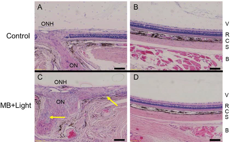
Figure 5:
Histological sections of optic nerve head (ONH) and peripheral retina (> 2 mm from ONH) from rats euthanized 10 days after unilateral treatment with targeted MB photocrosslinking were used to assess safety after photocrosslinking. Paraffin-embedded sections were stained with hematoxylin and eosin. While local tissue responses varied in eyes after photocrosslinking, we show the most severe damage seen among the three treated eyes from 3 rats examined. Control eye (retrobulbar injection of HBSS) showing healthy optic nerve and healthy retina both A) adjacent to the ONH, and B) in the periphery. C) A treated eye shows inflammatory infiltration near the ON (lower arrow) and retinal damage (upper arrow) near the ON. This damage was localized to the region adjacent to the optic nerve, since retina in the D) periphery appeared normal. Abbreviations: optic nerve head (ONH), optic nerve (ON), vitreous (V), retina (R), choroid (C), sclera (S), and retrobulbar tissue (B). Scale bars = 100 μm.

