Abstract
Cells have a regulatory mechanism known as heat shock (HS) response, which induces the expression of HS genes and proteins in response to heat and other cellular stresses. Exposure to moderate HS results in beneficial effects, such as thermotolerance and promotes survival, whereas excessive HS causes cell death. The effect of HS on cells depends on both exogenous factors, including the temperature and duration of heat application, and endogenous factors, such as the degree of cell differentiation. Neural stem cells (NSCs) can self-renew and differentiate into neurons and glial cells, but the changes in the HS response of symmetrically proliferating NSCs in culture are unclear. We evaluated the HS response of homogeneous proliferating NSCs derived from mouse embryonic stem cells during the proliferative phase and its effect on survival and cell death in vitro. The number of adherent cells and the expression ratios of HS protein (Hsp)40 and Hsp70 genes after exposure to HS for 20 min at temperatures above 43°C significantly increased with the extension of the culture period before exposure to HS. In contrast, caspase activity was significantly decreased by extension of the culture period before exposure to HS and suppressed the decrease in cell viability. These results suggest that the culture period before HS remarkably affects the HS response, influencing the expression of HS genes and cell survival of proliferating NSCs in culture.
Introduction
Cells possess a regulatory mechanism, termed heat shock (HS) response, which allows cells to respond to heat and other cellular stresses. This response includes the induction of expression of HS proteins (HSPs), including Hsp27, Hsp40, Hsp70, and Hsp90 [1, 2]. HS responses in cells under stress are extremely dependent on external factors, including the extent and duration of the temperature elevation and the timing of exposure, with exposure to excess stress potentially inducing cell death. For example, exposure of rat primary cortical and hippocampal neurons to HS at 44 or 45°C for 30 min can induce apoptotic cell death [3]. In contrast, exposure to moderate HS stress enhances thermotolerance by inducing HSP expression, which promotes cell survival. HSP expression induced by moderate heat exposure has been found to have a neuroprotective effect in cultured hippocampal neurons vulnerable to glutamate [4]. Other studies performed under different HS conditions found that prior exposure to mild heat stress protects primary neurons from more severe stress [5, 6].
HS responses also depend on internal factors, including the degree of neural cell differentiation. In the central nervous system, neural stem cells (NSCs) divide symmetrically until a sufficient number is achieved; then, they start to divide asymmetrically to produce neurons, astrocytes, and oligodendrocytes [7, 8]. NSCs are not only observed in neural tissues during early development, but can also be observed in some regions of the fetal and adult brains [9]. Isolated NSCs can be grown in vitro in medium supplemented with epidermal growth factor and/or fibroblast growth factor 2 (FGF2), forming spherical cell clusters known as neurospheres [10]. Although previous studies have examined the in vivo and in vitro effects of HS on the properties of NSCs and neural stem/precursor cells (NS/PCs), how the effects of HS vary depending on the time point of NSCs proliferation and differentiation is unknown. Previous studies have shown that exposure of NSCs to high temperatures during the proliferation phase in the early development arrested cell proliferation and induced cell death in cultured guinea pig [11–13], rat [14], and mouse embryos in vivo [15]. Moreover, severe heat stress exposure of NPCs reduced the cell viability of neurospheres [16], whereas mild heat exposure increased NS/PC proliferation in heat-acclimated rats [17]. These findings suggest that exposure to HS affects the survival and proliferation of NSCs and NS/PCs, both in vivo and in vitro. Although these in vivo studies tested the effects of HS on NSCs during the transition from symmetric to asymmetric division, and in vitro studies have tested the effects of HS on the properties of NS/PCs in neurospheres, none have examined the effects of HS specifically on symmetrically dividing NSCs in culture.
NSCs can be derived from pluripotent stem cells, such as embryonic stem (ES) cells [18–23] and induced pluripotent stem cells [24–26], and serve as a source of transplantation therapy for some central nervous system disorders. In particular, the neural stem sphere (NSS) method is a simple and efficient method for generating large numbers of homogeneous multipotent NSCs from mouse ES cells [27, 28], which can differentiate into neurons and glia [28–30]. In addition, NSCs prepared using the NSS method are able to maintain their properties in adherent monolayer cultures in the presence of FGF2, making them a suitable in vitro model to address the direct effects of exposure to HS [31] and other external stresses [32, 33] on proliferating NSCs. Therefore, in the current study, we evaluated the effects of brief exposure to HS on the response of proliferating NSCs derived from mouse ES cells and the resulting cell survival in different culture periods. We found that the culture period of proliferating NSCs prior to exposure to HS significantly altered the HS response, including HSP expression, altering both cell survival and cell death, which provides insight for determining the effective degree of exposure to HS to NSCs for cell therapy.
Materials and methods
Preparation of NSCs using the NSS method
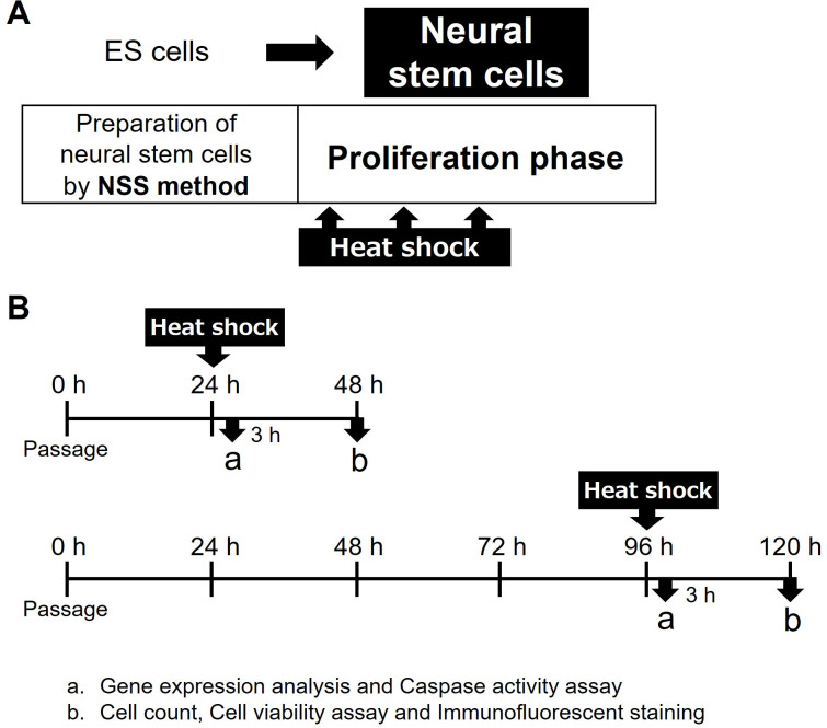
Mouse NSCs were prepared from undifferentiated mouse ES cells using the NSS method (Fig 1A), as previously described [28, 29, 31]. The mouse ES cell line (Cell No. RBRC-AES0140) was provided by the RIKEN BRC through the National Bio-Resource Project of MEXT, Japan. NSSs were formed by culturing the cells in neuron culture medium (FUJIFILM Wako, Tokyo, Japan) under free-floating conditions. Adherent cells were cultured in the presence of 20 ng/mL FGF2 (R&D Systems, Minneapolis, MN), resulting in large numbers of homogeneous NSCs migrating from the NSSs. The migrating NSCs were collected using conventional trypsin digestion to remove attached NSSs and plated in the presence of FGF2. The proliferating NSCs were suspended in culture media containing 10% dimethyl sulfoxide and stored at -80°C or in liquid nitrogen.
Fig 1. Experimental design and preparation of neural stem cells by the neural stem sphere (NSS) method.
(A) Preparation of mouse neural stem cells (NSCs) by NSS method and the times of exposure to heat shock (HS). Large numbers of NSCs were produced by the NSS method directly from mouse ES cells. These cells were cultured on dishes as monolayers and expanded in medium supplemented with FGF2. NSCs in the proliferation phase were exposed to HS. (B) Experimental schedule. NSCs were exposed to HS by immersing the culture dishes in thermostat baths at 37°C, 42°C, 43°C or 44°C after culture at 37°C under an atmosphere of 5% CO2 for 24 h (day 0) and 96 h (day 3). 3 h after HS, gene expression and caspase activities were assessed; after 24 h, the numbers of adherent cells, cell viability were measured and immunofluorescence staining was performed.
NSC culture
To prepare NSCs, the cryopreserved cells were thawed and expanded on proliferative monolayer cultures in NSC medium, consisting of neurobasal medium (Gibco, Carlsbad, CA) augmented with 2% B27 supplement (Gibco) and 20 ng/mL FGF2 (Fig 1A). NSCs were cultured at 37°C and 5% CO2, and the culture medium was replaced every 48 h.
Exposure to HS
NSCs were cultured in NSC medium on 35-mm culture dishes (BD Falcon; Becton Dickinson, Franklin Lakes, NJ, USA) for 24 h (time point considered day 0) or 96 h (day 3), tightly sealed with parafilm, and exposed to HS by immersion in thermostat baths (Thermominder TAITEC Co. Ltd., Saitama, Japan) maintained at 37°C, 42°C, 43°C, or 44°C for 20 min (Fig 1B). The cells were allowed to recover by culturing at 37°C and 5% CO2 for 24 h.
Measurement of cell numbers after HS
After the recovery period of 24 h, the number of adherent cells on culture dishes was counted in five cell-containing areas per dish using an inverted microscope equipped with a phase-contrast objective (DIAPHOT; Nikon, Tokyo, Japan).
Quantitative real-time RT-PCR analysis
After the recovery period of 3 h, mRNA was extracted from NSCs after HS at 24 h and 96 h, along with cells exposed to 37°C. mRNA extraction was performed using the QuickPrep™ Micro mRNA Purification Kit (GE Healthcare Bio-Sciences Corp., NJ, USA), according to the manufacturer’s instructions, although the DNase treatment step was not applied. Changes in the levels of gene expression were determined using real-time RT-PCR analysis with a 7300 Real-Time PCR System (Applied Biosystems, Foster City, CA, USA), Power SYBR®GREEN PCR Master Mix (Applied Biosystems), and the corresponding primers. The primer pairs were designed using Primer Express software (Applied Biosystems). Their details are mentioned in Tables 1 and S1. The potential reference genes glyceraldehyde-3-phosphate dehydrogenase (GAPDH) and ribosomal protein S29 (RPS29) were compared within the reference gene ranking [34]. As RPS29 expression showed less change after HS, it was chosen as the reference gene. The standard curve method was used to calculate the relative expression of each target gene. Expression values of each gene were normalized relative to the expression levels of RPS29.
Table 1. Primers used in quantitative real-time RT-PCR.
| Symbols | Gene Names | Synonyms | GenBank Accession Numbers |
|---|---|---|---|
| Gapdh | glyceraldehydes-3-phosphate dehydrogenase | Gapd | NM_008084 |
| Rps29 | ribosomal protein S29 | S29 ribosomal protein | NM_009093 |
| Hspa1b | heat shock protein 1B | Hsp70 | NM_010478 |
| Dnajb1 | DnaJ heat shock protein family (Hsp40) member B1 | Hsp40 | NM_018808 |
| Hsp90aa1 | heat shock protein 90, alpha (cytosolic), class A member 1 | Hsp90 | NM_010480 |
| Hspb2 | heat shock protein 2 | Hsp27 | NM_024441 |
| Cycs | cytochrome c, somatic | Cytochrome c | NM_007808 |
| Apaf-1 | apoptotic peptidase activating factor 1 | Apaf-1 | NM_009684 |
| Casp9 | caspase 9 | Caspase-9 | NM_015733 |
| Casp3 | caspase 3 | Caspase-3 | NM_009810 |
| Casp7 | caspase 7 | Caspase-7 | NM_007611 |
| Nes | nestin | ESTM46 | NM_016701 |
Cell viability assay
After NSCs were exposed to HS at 43°C or 44°C for 20 min and allowed to recover for 24 h at 37°C, along with cells exposed to 37°C, the cell viabilities were assessed. The cells were lysed in lysis buffer (Promega, Madison, WI, USA) for 5 min and were subjected to luminescence-based assays using CellTiter Glo reagent (Promega) according to the manufacturer’s instructions. Luminescence intensities were quantified using a luminometer (TD-20e; Turner Biosystems). The ratio of cell viability after HS was calculated relative to 37°C.
Caspase activity assay
NSCs were exposed to HS at 43°C or 44°C for 20 min and allowed to recover for 3 h at 37°C. Afterwards, HS-exposed cells along with cells exposed to 37°C were incubated with lysis buffer (Promega, Madison, WI) for 5 min and were subjected to luminescence-based assays with CellTiter Glo reagent (Promega) to assess cell viability. Luminescence intensities were quantified using a luminometer (TD-20e; Turner Biosystems). To assess caspase 3/7 activity, cells were incubated with Caspase-Glo 3/7 reagent (Promega), according to the manufacturer’s instructions. Caspase activity levels were normalized by cell viability levels. The ratio of caspase activity after HS was calculated relative to 37°C.
Immunofluorescence analysis
NSCs exposed to HS and subsequently allowed to recover, along with cells exposed to 37°C, were evaluated using immunofluorescence staining. Cells were fixed with 4% paraformaldehyde in phosphate-buffered saline (PBS) at room temperature, treated with 0.5% Triton X-100 in PBS, blocked with 10% bovine serum albumin (BSA) in PBS for 60 min at room temperature, and washed 3 times for 5 minutes with PBS. The cells were incubated with the primary antibody for 60 min and washed with PBS. The primary antibody used was anti-nestin (Rat-401, 1:100, Developmental Studies Hybridoma Bank, Iowa City, IA, USA), a marker of NSCs. The cells were subsequently incubated with Alexa Fluor546-labeled secondary antibodies (1:200, Molecular Probes, Eugene, OR, USA) for 30 min. The nuclei were counterstained with 4′,6-diamidine-2′-phenylindole dihydrochloride. Cells were viewed under a fluorescence microscope (BH-2; Nikon) and photos were analyzed with Photoshop Elements 12 (Adobe, CA, USA).
Statistical analysis
Data are expressed as mean ± SEM. Differences between two groups were analyzed using the Student’s t-test or Welch’s t-test. Statistical significance was set at p < 0.05. All statistical analyses were performed using SPSS 26 Statistics Base (IBM Japan, Tokyo, Japan).
Results
Effects of culture period before HS on the numbers of proliferating NSCs after HS treatment
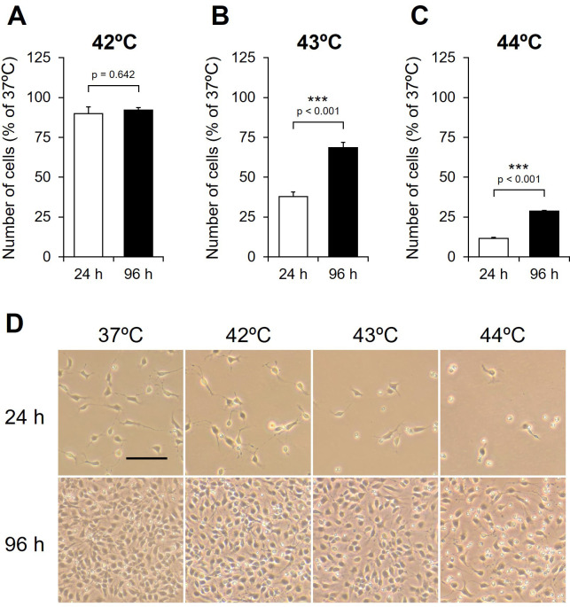
To determine the effects of HS on the number of NSCs, the cells were exposed to HS for 20 min at 42°C, 43°C, or 44°C after culture at 37°C for 24 and 96 h (Fig 1B), and the number of adherent cells was counted and compared to number of cells exposed to 37°C. The ratios of adherent cells exposed to HS after 24 h decreased in a temperature-dependent manner (Fig 2A–2C), with the ratio being significantly lower after exposure to HS at 43°C (24 h, 37.88 ± 2.74%; 96 h, 68.54 ± 3.35%) and 44°C (24 h, 11.59 ± 0.56%; 96 h, 28.57 ± 0.64%) (Fig 2B and 2C), along with an increase in the number of floating cells (Fig 2D). The ratio of adherent cells exposed to HS at 42°C did not differ significantly between 24 h and 96 h (Fig 2A). In contrast, the ratios of adherent cells exposed to HS at 43°C and 44°C after culture at 37°C for 96 h were significantly higher than those exposed after culture for 24 h (43 and 44°C, p < 0.001) (Fig 2B and 2C). Cell morphology after HS was observed using phase-contrast microscopy. The edges of NSCs exposed to HS at 43°C and 44°C after culture at 37°C for 24 h were unclear and some cells lacked neurites (S1 Fig). In contrast, the edges of NSCs exposed to HS after culture at 37°C for 96 h were clear with all cells having neurites (S1 Fig). This indicates that cell morphology is dependent on both the temperature of HS and the culture period before exposure to HS. These results indicate that the culture period before HS differentially affects the remaining adherent cells of proliferating NSCs, even with HS exposure at the same temperature and duration.
Fig 2. Effect of exposure to heat shock on the numbers of NSCs.
NSCs were exposed to heat shock (HS) at (A) 42°C, (B) 43°C, and (C) 44°C for 20 min. (A-C) Numbers of adherent cells, 24 h after HS performed after culture at 37°C for 24 h (day 0; white) and 96 h (day 3; black). The values are presented as mean ± SEM percentages of controls (n = 6). ***p < 0.001. (D) Phase-contrast micrographs of 37°C cells and HS-exposed cells, 24 h after HS was performed and after culture at 37°C for 24 h (day 0; upper) and 96 h (day 3; lower).
Effects of HS on expression of HSP genes in proliferating NSCs
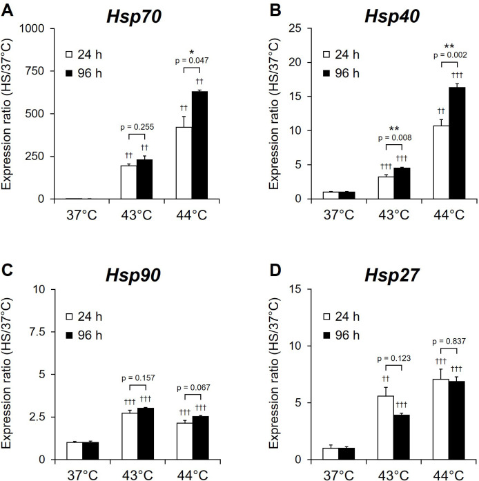
To evaluate changes in HS responses in proliferating NSCs, the ratios of HSP gene expression 3 h after the HS were analyzed using real-time RT-PCR. The ratios of expression of all four HSP genes were significantly higher in NSCs exposed to HS at 43°C and 44°C (p < 0.01, p < 0.001) than in NSCs exposed to 37°C at both timepoints (24 and 96 h) (Fig 3A–3D). The increase in the ratios of Hsp27, Hsp40, and Hsp70 was temperature-dependent (Fig 3A, 3B and 3D). HS at 24 and 96 h timepoints induced a 190- and 630-fold increase of Hsp70 expression, respectively. Expression of Hsp70 when the HS was delivered at 96 h was significantly higher than at 24 h in the case of HS at 44°C and 43°C (p = 0.047 and p = 0.008, respectively) (Fig 3A). Hsp40 expression was significantly increased after HS at 44°C for 96 h compared to that with 24°C (p = 0.002) (Fig 3B). These results demonstrate that the HSP expression increased with the extension of the culture period. In addition, these expression changes correlate with the change in the number of NSCs at these two time points in the culture (Fig 2B and 2C).
Fig 3. HSP gene expression 3 h after exposure of NSCs to heat shock.
NSCs cultured for 24 h (day 0; white) or 96 h (day 3; black) under proliferation conditions were exposed to heat shock (HS) at 43°C or 44°C and were then allowed to recover for 3 h. The expression of (A) Hsp70, (B) Hsp40, (C) Hsp90, and (D) Hsp27 was assayed by real-time RT-PCR and normalized by that of RPS29. Results are presented as means ± SEM (n = 4) (*P <0.05, **P <0.01 between 24 h and 96 h, ††P <0.01, †††P <0.001 between 37°C and 43°C or 44°C).
Effects of HS on expression of apoptosis-related genes in proliferating NSCs
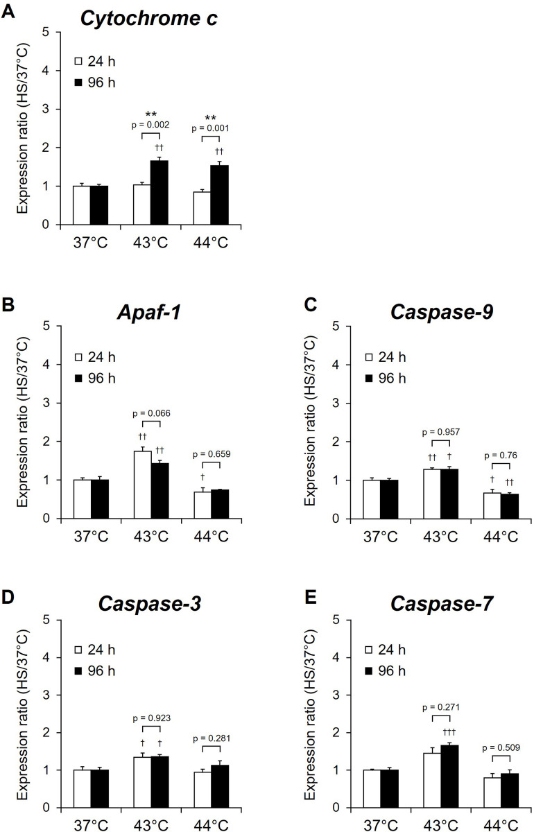
Expression of several HSPs induced by external stresses enhance cell survival by inhibiting the mitochondrial apoptotic pathways [35]. Therefore, to assess the effects on apoptotic signals of the increase of HSPs expression after HS in proliferating NSCs, the relative expression of the apoptosis-related genes cytochrome c, Apaf-1, caspase-9, caspase-3, and caspase-7 were analyzed using real-time RT-PCR 3 h after cells were exposed to HS at 43°C and 44°C after culturing for 24 and 96 h. The relative expression of Apaf-1 (24 h and 96 h; p < 0.01), caspase-9 (24 h; p < 0.01, 96 h; p < 0.05), caspase-3 (24 h and 96 h; p < 0.05), and caspase-7 (96 h; p < 0.001) in NSCs exposed to HS at 43°C were significantly higher than those in cells exposed to 37°C (Fig 4B–4E). In contrast, the relative expression of Apaf-1 (24 h; p < 0.05) and caspase-9 (24 h; p < 0.05, 96 h; p < 0.01) was significantly lower than in the cells exposed to 37°C (Fig 4B and 4C). The relative expression of caspase-9, Apaf-1, caspase-3, and caspase-7 did not differ significantly in cells exposed to the same temperature but at different timepoints (24 and 96 h) (Fig 4B–4E); however, cytochrome c expression was significantly higher than those exposed after culture for 24 h (43°C: p = 0.002; 44°C: p = 0.001) (Fig 4A).
Fig 4. Expression of apoptosis-related genes 3 h after exposure of NSCs to heat shock.
NSCs cultured for 24 h (day 0; white) or 96 h (day 3; black) under proliferation conditions were exposed to heat shock (HS) at 43°C or 44°C and were then allowed to recover for 3 h. The expression of (A) cytochrome c, (B) Apaf-1, (C) caspase-9, (D) caspase-3 and (E) caspase-7 was assayed by real-time RT-PCR and normalized by that of RPS29. Results are presented as means ± SEM (n = 4). (**P <0.01 between 24 h and 96 h, †P <0.05, ††P <0.01, †††P <0.001 between 37°C and 43°C or 44°C).
Regulations of cell viability and caspase activity in proliferating NSCs after HS
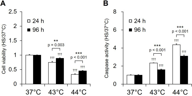
To examine the factors leading to changes in cell number between 24 h and 96 h in proliferating NSCs after exposure to HS, cell viability assay and caspase activity assay, defined as the ratio of caspase 3/7 activity/cell viability, were performed. Cell viability was assessed at 24 h after HS, the same time period in which cell counts were performed. Cell viability after HS at 44°C was maintained for 3 h, but gradually decreased until 12 h (S2A Fig), and Caspase 3/7 activities increased within 20 min after HS at 44°C for 20 min and were maintained until 12 h (S2B Fig). Therefore, caspase activity in NSCs was analyzed 3 h after HS. After recovery by incubation at 37°C for 24 h after HS, cell viability of cells exposed to HS at 43°C and 44°C after culture at 37°C for 96 h were significantly higher than those exposed after culture for 24 h (43°C: p = 0.003; 44°C: p = 0.001) (Fig 5A). Caspase activity was significantly lower in cells exposed to HS at 43°C and 44°C after 96 h than after 24 h (43 and 44°C; p < 0.001) (Fig 5B). These results suggest that extension of the culture period prior to HS could improve cell viability after HS by inducing an increase in HSP expression and suppressing the increase in caspase activity.
Fig 5. Caspase activities of NSCs exposed to heat shock.
(A) Cell viability was measured 3 h after exposure to heat shock (HS) using CellTiter Glo reagent (Promega), with results expressed as mean ± SEM (n = 8) ratios to the 37°C sample. (**P <0.01 for comparisons between cells exposed to HS after culture for 24 h (day 0; white) and 96 h (day 3; black); †††P <0.01 for comparisons between 37°C and 43°C or 44°C). (B) Caspase activity was measured 3 h after exposure to heat shock (HS) using the caspase-Glo 3/7 reagent (Promega) and normalized to cell viability measured using CellTiter Glo reagent (Promega), with results expressed as mean ± SEM (n = 8) ratios to the 37°C sample. The values are presented as the in the graph. (**P <0.01 for comparisons between cells exposed to HS after culture for 24 h (day 0; white) and 96 h (day 3; black); †††P <0.01 for comparisons between 37°C and 43°C or 44°C).
Maintenance of NSCs marker expressions in NSCs in culture
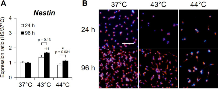
To evaluate the maintenance of the neural stem cells properties, the expression of NSC markers was evaluated using real-time RT-PCR analysis and immunofluorescence staining in NSCs at 3 h after at 37°C incubation or at 24 after 43°C and 44°C HS treatment. Real-time RT-PCR analysis indicated that the nestin gene, a marker that is specifically expressed in NSCs, was expressed in NSCs after HS at 37°C, 43°C, and 44°C for both 24 h and 96 h under proliferative conditions, and its expression at 96 h was significantly increased compared to that at 24 h (44°C: p = 0.031) (Fig 6A). Immunofluorescence staining demonstrated that NSCs after HS at 37°C were positive for nestin at both 24 h and 96 h under proliferative conditions, and similar results were observed for adherent cells after HS at 43°C and 44°C (Fig 6B). These results indicate that NSCs in culture maintain their properties as NSCs even after HS exposure and are preserved as NSCs although the culture period is extended.
Fig 6. Expressions of the gene and the protein of the NSC marker after heat shock.
(A) NSCs cultured for 24 h (day 0; white) or 96 h (day 3; black) under proliferation conditions were exposed to heat shock (HS) at 43°C or 44°C and were then allowed to recover for 3 h. The expression of Nestin was assayed by real-time RT-PCR and normalized by that of RPS29. Results are presented as means ± SEM (n = 4). (*P <0.05 between 24 h and 96 h, †††P <0.001 between 37°C and 43°C or 44°C). (B) NSCs cultured for 24 h (day 0; white) or 96 h (day 3; black) under proliferation conditions were exposed to heat shock (HS) at 43°C or 44°C and were then allowed to recover for 24 h. Immunofluorescence staining was performed on adherent cells after HS by using Nestin antibody. Fluorescence microscope images of Nestin (red) with DAPI counterstaining for nuclei (blue). Bars: 100 μm.
Discussion
We have previously shown that HS exposure above 43°C for 20 min before seeding in culture dishes inhibited the proliferation of ES cell-derived mouse NSCs [32]; however, the effect of the culture period prior to exposure to HS on the proliferating NSCs during culture under proliferative conditions was unknown. The current study shows that the culture period of proliferating homogeneous NSCs prior to HS exposure, even at the same temperature and for the same duration, profoundly affects the HS response, including the expression of HSP genes. Although HS above 43°C reduced the number of NSCs, the extension of the culture period prior to exposure to HS caused a relative increase in HSP expression, which resulted in a decrease in caspase activity and suppression of decreased cell viability. Several HSP genes whose expression is induced by HS help to limit the damage caused by stress and modulate apoptosis by different pathways [2]. Moreover, the relative expression levels of HS-induced HSPs are correlated with cell viability [5, 36–38]. The results presented in this study demonstrate that different HS conditions induce distinct HSP expression profiles, which effect the cell viability of a homogeneous population of proliferating NSCs cultured in proliferative conditions.
HSPs function as molecular chaperones, which prevent the aggregation of denatured proteins and promote protein refolding, and they also play a role in the primary resistance machinery to protect vulnerable cells [39] under both stress [40] and physiological [41] conditions. Hsp70 and Hsp27 have been reported to be the most strongly induced HSPs after stress, and they exert powerful neuroprotective effects [40, 42, 43]. Hsp40 also functions in the refolding of heat-denatured proteins by collaborating with Hsp70 [44]. Hsp90 is one of the most abundant proteins in cells in physiological conditions [45], with a function similar to that of Hsp70 and Hsp27 [46, 47]. Hsp27 and Hsp90 are also involved in regulating the cytoskeleton of neural cells; Hsp27 stabilizes major components of the cytoskeleton, including neurofilaments, actin, and microtubules [48] and is involved in neurite growth and/or axonal regeneration [49]. Hsp90 protects tubulin against thermal denaturation and maintains it in a state compatible with microtubule polymerization [50]. In the current study, we show that the soma and neurites morphology and the HSPs expression profile of NSCs exposed to HS at 43°C and 44°C were maintained by extension of the culture period prior to exposure to HS. Therefore, increased HSP expression may be protective in proliferating NSCs, maybe due to their function as molecular chaperones that refold denatured proteins, favoring cell viability.
We hypothesized that extension of the culture period before HS would not only affect HSP expression, but also the expression of apoptotic genes. In the mitochondrial apoptotic pathway, the leakage of cytochrome c from mitochondria activated of a downstream signal transduction cascade that includes complex formation by Apaf-1, recruitment of caspase 9, and activation of caspases 3 and 7, finally triggering apoptosis [35]. Hsp70 inhibits the release of cytochrome c from mitochondria [51] and together with Hsp90 interferes with Apaf-1 activity [51–53]. Hsp27 has also been reported to inhibit the release of cytochrome c [54, 55]. Inhibition of cytochrome c and Apaf-1 prevents the downstream activation of caspase 9, inhibiting apoptosis [51]. In the current study, there were no differences in the expression of apoptosis-related genes between 24 h and 96 h after HS except for cytochrome c. The absence of difference in HSP expression at 44°C compared to 37°C suggested that an increase in HSP expression is involved in the suppression of apoptosis-related genes. Because there were no changes in apoptosis-related genes during the culture period, we hypothesized that caspase 3/7 activity, which is located downstream of the apoptotic signaling pathway, and cell viability might be altered. Indeed, we found that caspase 3/7 activities were further inhibited by extension of the culture period; in contrast, cell viability improved. Altogether, our results suggest that expression of HSPs in NSCs induced by extension of the culture period before HS may have resulted in an anti-apoptotic effect by inhibiting the apoptotic signaling.
The survival of NS/PCs with mitotic capability has been reported to be affected by the extracellular matrix (ECM), which regulates the HS response and cell differentiation [56, 57]. Laminin, the main component of Matrigel® Matrix, has been reported to maintain the stem cell properties of hippocampal NPCs, thereby enhancing their survival [58]. In addition, NS/PC differentiation caused a reduced HS response and suppression of Hsp70 expression, resulting in a high risk of vulnerability to stress [56]. In the current study, NSCs were positive for the NSC marker nestin at both the gene and protein level (Fig 6) and maintained the properties of NSCs with proliferative capacity in culture conditions using Matrigel® matrix. These results indicate that maintaining properties under proliferative conditions and enhancing the HS response by extension of the culture period may modulate the survival of NSCs after exposure to severe HS stress.
In conclusion, the results of the present study demonstrate that mouse ES-derived NSCs under proliferative conditions show altered HSP expression and cell viability due to changes in the HS response and the properties of NSCs with the extension of the culture period prior to the HS. To the best of our knowledge, this is the first report to demonstrate that changes in the effects of HS on homogeneous NSCs occur in monolayer cultures under proliferative conditions. Stem cell therapies for regenerative medicine require to improve the viability of NS/NP cells after transplantation. The viability of transplanted cells may depend on the environment surrounding the foci as well as the condition of the donor cells [59]. Therefore, we believe that regulating the environment of NSCs before and after transplantation, including during culture, is critical for improving the viability of NSCs. Although the conditions in the current study were severe HS causing cell death, moderate HS could be a useful intervention to enhance cell survival, similar to what occurs in other neural cells. Studying HS conditions that are beneficial for the survival of stem cell-derived NSCs in culture or after transplantation may provide important insights to improve cell therapy.
Supporting information
Phase-contrast micrographs of control cells (left) and HS-exposed cells (43°C: middle, 44°C: right), 24 h after HS performed after culture at 37°C for 24 h (day 0; upper) and 96 h (day 3; lower).
(TIF)
(A) Viability of NSCs 0–12 h after HS was assessed using CellTiter Glo reagent (Promega), with the results presented as the means ± SEM (n = 3). **P <0.01 for comparisons between control cells (circles) and cells exposed to HS at 44°C (triangles). Control cells were cultured in the 5% CO2 incubator at 37°C. (B) Caspase activities of NSCs 0–12 h after HS. Caspase activity of NSCs 0–12 h after HS was assessed using Caspase-Glo 3/7 reagent (Promega), normalized to cell viability using CellTiter Glo reagent (Promega). The results are presented as the means ± SEM (n = 3). **P <0.01 for comparisons between control cells (white) and cells exposed to HS at 44°C (black). Control cells were cultured in the 5% CO2 incubator at 37°C.
(TIF)
(DOCX)
Acknowledgments
We are highly thankful to Drs. Takashi Nakayama and Nobuo Inoue, who developed the neural stem sphere method, for technical assistance and supportive comments on the manuscript. We are also grateful to Dr. Mitsuyo Makita for providing the instruments and reagents for the experiments.
Data Availability
All relevant data are within the paper and its Supporting Information files.
Funding Statement
This research was supported by JSPS KAKENHI Grant Number JP18K17685 to HO. Braizon Therapeutics Inc. provided support in the form of salaries for authors (MO). The funders had no role in study design, data collection and analysis, decision to publish, or preparation of the manuscript. Braizon Therapeutics Inc. provided support in the form of salaries for authors (MO) but did not have any additional role in the study design, data collection and analysis, decision to publish, or preparation of the manuscript. The specific role of each author is articulated in the ‘author contributions’ section.
References
- 1.Lindquist S, Craig EA. The heat-shock proteins. Annu Rev Genet. 1988; 22:631–677. 10.1146/annurev.ge.22.120188.003215 [DOI] [PubMed] [Google Scholar]
- 2.Arya R, Mallik M, Lakhotia SC. Heat shock genes—integrating cell survival and death. J Biosci. 2007; 32(3):595–610. 10.1007/s12038-007-0059-3 [DOI] [PubMed] [Google Scholar]
- 3.Vogel P, Dux E, Wiessner C. Evidence of apoptosis in primary neuronal cultures after heat shock. Brain Res. 1997; 764(1–2):205–213. 10.1016/s0006-8993(97)00458-7 [DOI] [PubMed] [Google Scholar]
- 4.Sato K, Matsuki N. A 72 kDa heat shock protein is protective against the selective vulnerability of CA1 neurons and is essential for the tolerance exhibited by CA3 neurons in the hippocampus. Neuroscience. 2002; 109(4):745–756. 10.1016/s0306-4522(01)00494-8 [DOI] [PubMed] [Google Scholar]
- 5.Mailhos C, Howard MK, Latchman DS. Heat shock proteins hsp90 and hsp70 protect neuronal cells from thermal stress but not from programmed cell death. J Neurochem. 1994; 63(5):1787–1795. 10.1046/j.1471-4159.1994.63051787.x [DOI] [PubMed] [Google Scholar]
- 6.Fink SL, Chang LK, Ho DY, Sapolsky RM. Defective herpes simplex virus vectors expressing the rat brain stress- inducible heat shock protein 72 protect cultured neurons from severe heat shock. J Neurochem. 1997; 68(3): 961–969. 10.1046/j.1471-4159.1997.68030961.x [DOI] [PubMed] [Google Scholar]
- 7.Temple S. The development of neural stem cells. Nature. 2001; 414(6859):112–117. 10.1038/35102174 [DOI] [PubMed] [Google Scholar]
- 8.Merkle FT, Alvarez-Buylla A. Neural stem cells in mammalian development. Curr Opin Cell Biol. 2006; 18(6):704–709. 10.1016/j.ceb.2006.09.008 [DOI] [PubMed] [Google Scholar]
- 9.Gage FH. Mammalian neural stem cells. Science. 2000; 287(5457):1433–1438. 10.1126/science.287.5457.1433 [DOI] [PubMed] [Google Scholar]
- 10.Reynolds BA, Weiss S. Generation of neurons and astrocytes from isolated cells of the adult mammalian central nervous system. Science. 1992; 255(5052):1707–1710. 10.1126/science.1553558 [DOI] [PubMed] [Google Scholar]
- 11.Edwards MJ, Mulley R, Ring S, Wanner RA. Mitotic cell death and delay of mitotic activity in guinea pig embryos following brief maternal hyperthermia. J Embryol Exp Morphol. 1974; 32(3):593–602. [PubMed] [Google Scholar]
- 12.Wanner RA, Edwards MJ, Wright RG. The effect of hyperthermia on the neuroepithelium of the 21-day guinea-pig foetus: histologic and ultrastructural study. J. Pathol. 1976; 118(4):235–244. 10.1002/path.1711180406 [DOI] [PubMed] [Google Scholar]
- 13.Edwards MJ. Review: Hyperthermia and fever during pregnancy. Birth Defects Res Part A—Clin Mol Teratol. 2006; 76(7):507–516. 10.1002/bdra.20277 [DOI] [PubMed] [Google Scholar]
- 14.Edwards MJ, Walsh DA. Hyperthermia, teratogenesis and the heat shock response in mammalian embryos in culture. Int. J. Dev. Biol. 1997; 41(2):345–358. [PubMed] [Google Scholar]
- 15.Shiota K. Induction of neural tube defects and skeletal malformations in micefollowing brief hyperthermia in utero. Biol. Neonat. 1988; 53(2):86–97. 10.1159/000242767 [DOI] [PubMed] [Google Scholar]
- 16.Vishwakarma SK, Bardia A, Fathima N, Chandrakala L, Rahamathulla S, Raju N, et al. Protective role of hypothermia against heat stress in differentiated and undifferentiated human neural precursor cells: A differential approach for the treatment of traumatic brain injury. Basic Clin Neurosci. 2017; 8(6):453–466. 10.29252/NIRP.BCN.8.6.453 [DOI] [PMC free article] [PubMed] [Google Scholar]
- 17.Hossain ME, Matsuzaki K, Katakura M, Sugimoto N, Mamun A Al, Islam R, et al. Direct exposure to mild heat promotes proliferation and neuronal differentiation of neural stem/progenitor cells in vitro. PLoS One. 2017; 12(12): e0190356. 10.1371/journal.pone.0190356 [DOI] [PMC free article] [PubMed] [Google Scholar]
- 18.Kawasaki H, Mizuseki K, Nishikawa S, Kaneko S, Kuwana Y, Nakanishi S, et al. Induction of midbrain dopaminergic neurons from ES cells by stromal cell-derived inducing activity. Neuron. 2000; 28(1) 31–40. 10.1016/s0896-6273(00)00083-0 [DOI] [PubMed] [Google Scholar]
- 19.Ying QL, Smith AG. Defined conditions for neural commitment and differentiation. Methods Enzymol. 2003; 365:327–341. 10.1016/s0076-6879(03)65023-8 [DOI] [PubMed] [Google Scholar]
- 20.Watanabe K, Kamiya D, Nishiyama A, Katayama T, Nozaki S, Kawasaki H, et al. Directed differentiation of telencephalic precursors from embryonic stem cells. Nat Neurosci. 2005; 8(3):288–296. 10.1038/nn1402 [DOI] [PubMed] [Google Scholar]
- 21.Smukler SR, Runciman SB, Xu S, van der Kooy D. Embryonic stem cells assume a primitive neural stem cell fate in the absence of extrinsic influences. J Cell Biol. 2006; 172(1):79–90. 10.1083/jcb.200508085 [DOI] [PMC free article] [PubMed] [Google Scholar]
- 22.Chambers SM, Fasano CA, Papapetrou EP, Tomishima M, Sadelain M, Studer L. Highly efficient neural conversion of human ES and iPS cells by dual inhibition of SMAD signaling. Nat Biotechnol. 2009; 27(3):275–280. 10.1038/nbt.1529 [DOI] [PMC free article] [PubMed] [Google Scholar]
- 23.Kamiya D, Banno S, Sasai N, Ohgushi M, Inomata H, Watanabe K, et al. Intrinsic transition of embryonic stem-cell differentiation into neural progenitors. Nature. 2011; 470(7335):503–509. 10.1038/nature09726 [DOI] [PubMed] [Google Scholar]
- 24.Kim J, Efe JA, Zhu S, Talantova M, Yuan X, Wang S, et al. Direct reprogramming of mouse fibroblasts to neural progenitors. Proc Natl Acad Sci U S A. 2011; 108(19):7838–7843. 10.1073/pnas.1103113108 [DOI] [PMC free article] [PubMed] [Google Scholar]
- 25.Matsui T, Takano M, Yoshida K, Ono S, Fujisaki C, Matsuzaki Y, et al. Neural stem cells directly differentiated from partially reprogrammed fibroblasts rapidly acquire gliogenic competency. Stem Cells. 2012; 30(6):1109–1119. 10.1002/stem.1091 [DOI] [PubMed] [Google Scholar]
- 26.Thier M, Wörsdörfer P, Lakes YB, Gorris R, Herms S, Opitz T, et al. Direct conversion of fibroblasts into stably expandable neural stem cells. Cell Stem Cell. 2012; 10(4):473–479. 10.1016/j.stem.2012.03.003 [DOI] [PubMed] [Google Scholar]
- 27.Nakayama T, Momoki-Soga T, Inoue N. Astrocyte-derived factors instruct differentiation of embryonic stem cells into neurons. Neurosci Res. 2003; 46(2):241–249. 10.1016/s0168-0102(03)00063-4 [DOI] [PubMed] [Google Scholar]
- 28.Nakayama T, Inoue N. Neural stem sphere method: induction of neural stem cells and neurons by astrocyte-derived factors in embryonic stem cells in vitro. Methods Mol Biol. 2006; 330:1–13. 10.1385/1-59745-036-7:001 [DOI] [PubMed] [Google Scholar]
- 29.Nakayama T, Momoki-Soga T, Yamaguchi K, Inoue N. Efficient production of neural stem cells and neurons from embryonic stem cells. Neuroreport 2004; 15(3):487–491. 10.1097/00001756-200403010-00021 [DOI] [PubMed] [Google Scholar]
- 30.Otsu M, Sai T, Nakayama T, Murakami K, Inoue N. Uni-directional differentiation of mouse embryonic stem cells into neurons by the neural stem sphere method. Neurosci Res. 2011; 69(4):314–321. 10.1016/j.neures.2010.12.014 [DOI] [PubMed] [Google Scholar]
- 31.Omori H, Otsu M, Suzuki A, Nakayama T, Akama K, Watanabe M, et al. Effects of heat shock on survival, proliferation and differentiation of mouse neural stem cells. Neurosci Res. 2014;79: 13–21. 10.1016/j.neures.2013.11.005 [DOI] [PubMed] [Google Scholar]
- 32.Isono M, Otsu M, Konishi T, Matsubara K, Tanabe T, Nakayama T, et al. Proliferation and differentiation of neural stem cells irradiated with X-rays in logarithmic growth phase. Neurosci Res. 2012; 73(3):263–268. 10.1016/j.neures.2012.04.005 [DOI] [PubMed] [Google Scholar]
- 33.Shibata M, Otsu M, Omori H, Kobayashi H, Suzuki A et al. Effects of continuous exposure of mouse primitive neural stem cells to methylmercury in proliferation and differentiation stages. 2016; 18(4)1–34. 10.24531/jhsaiih.18.4_187 [DOI] [Google Scholar]
- 34.de Jonge HJ, Fehrmann RS, Bont ES, Hofstra RM, Gerbens F, Kamps WA, et al., Evidence based selection of housekeeping genes. PLoS One. 2007; 2(9):e898 10.1371/journal.pone.0000898 [DOI] [PMC free article] [PubMed] [Google Scholar]
- 35.Parcellier A, Gurbuxani S, Schmitt E, Solary E, Garrido C. Heat shock proteins, cellular chaperones that modulate mitochondrial cell death pathways. Biochem Biophys Res Commun. 2003; 304(3):505–512. 10.1016/s0006-291x(03)00623-5 [DOI] [PubMed] [Google Scholar]
- 36.Mailhos C, Howard MK, Latchman DS. Heat shock protects neuronal cells from programmed cell death by apoptosis. Neuroscience. 1993; 55(3):621–627. 10.1016/0306-4522(93)90428-i [DOI] [PubMed] [Google Scholar]
- 37.Kitagawa K, Matsumoto M, Tagaya M, Hata R, Ueda H, Niinobe M, et al. “Ischemic tolerance” phenomenon found in the brain. Brain Res. 1990; 528(1): 21–24. 10.1016/0006-8993(90)90189-i [DOI] [PubMed] [Google Scholar]
- 38.Ohtsuka K, Suzuki T. Roles of molecular chaperones in the nervous system. Brain Res Bull. 2000; 53(2):141–146. 10.1016/s0361-9230(00)00325-7 [DOI] [PubMed] [Google Scholar]
- 39.San Gil R, Ooi L, Yerbury JJ, Ecroyd H. The heat shock response in neurons and astroglia and its role in neurodegenerative diseases. Mol Neurodegener. 2017; 12(1):65. 10.1186/s13024-017-0208-6 [DOI] [PMC free article] [PubMed] [Google Scholar]
- 40.O’Reilly AM, Currie RW, Clarke DB. HspB1 (Hsp 27) expression and neuroprotection in the retina. Mol Neurobiol. 2010; 42(2):124–132. 10.1007/s12035-010-8143-3 [DOI] [PubMed] [Google Scholar]
- 41.Zhao H, Michaelis ML, Blagg BSJ. Hsp90 modulation for the treatment of Alzheimer’s disease. Adv Pharmacol. 2012; 64:1–25. 10.1016/B978-0-12-394816-8.00001-5 [DOI] [PubMed] [Google Scholar]
- 42.Bukau B, Horwich AL. The Hsp70 and Hsp60 chaperone machines. Cell. 1998; 92(3):351–366. 10.1016/s0092-8674(00)80928-9 [DOI] [PubMed] [Google Scholar]
- 43.Nollen EAA, Brunsting JF, Roelofsen H, Weber LA, Kampinga HH. In vivo chaperone activity of heat shock protein 70 and thermotolerance. Mol Cell Biol. 1999; 19(3):2069–2079. 10.1128/mcb.19.3.2069 [DOI] [PMC free article] [PubMed] [Google Scholar]
- 44.Minami Y, Höhfeld J, Ohtsuka K, Hartl FU. Regulation of the heat-shock protein 70 reaction cycle by the mammalian DnaJ homolog, Hsp40. J Biol Chem. 1996; 271(32):19617–19624. 10.1074/jbc.271.32.19617 [DOI] [PubMed] [Google Scholar]
- 45.Wegele H, Müller L, Buchner J. Hsp70 and Hsp90—a relay team for protein folding. Rev Physiol Biochem Pharmacol. 2004; 151:1–44. 10.1007/s10254-003-0021-1 [DOI] [PubMed] [Google Scholar]
- 46.Samakovli D, Thanou A, Valmas C, Hatzopoulos P. Hsp90 canalizes developmental perturbation. J Exp Bot. 2007; 58(13):3513–3524. 10.1093/jxb/erm191 [DOI] [PubMed] [Google Scholar]
- 47.Specchia V, Piacentini L, Tritto P, Fanti L, Dalessandro R, Palumbo G, et al. Hsp90 prevents phenotypic variation by suppressing the mutagenic activity of transposons. Nature. 2010; 463(7281):662–665. 10.1038/nature08739 [DOI] [PubMed] [Google Scholar]
- 48.Walsh D, Li Z, Wu Y, Nagata K. Heat shock and the role of the HSPs during neural plate induction in early mammalian CNS and brain development. Cell Mol Life Sci. 1997; 53(2):198–211. 10.1007/pl00000592 [DOI] [PMC free article] [PubMed] [Google Scholar]
- 49.Williams KL, Rahimtula M, Mearow KM. Hsp27 and axonal growth in adult sensory neurons in vitro. BMC Neurosci. 2005; 6:24. 10.1186/1471-2202-6-24 [DOI] [PMC free article] [PubMed] [Google Scholar]
- 50.Weis F, Moullintraffort L, Heichette C, Chrétien D, Garnier C. The 90-kDa heat shock protein Hsp90 protects tubulin against thermal denaturation. J Biol Chem. 2010; 285(13):9525–9534. 10.1074/jbc.M109.096586 [DOI] [PMC free article] [PubMed] [Google Scholar]
- 51.Mosser DD, Caron AW, Bourget L, Meriin AB, Sherman MY, Morimoto RI, et al. The chaperone function of hsp70 is required for protection against stress-induced apoptosis. Mol Cell Biol. 2000; 20(19):7146–7159. 10.1128/mcb.20.19.7146-7159.2000 [DOI] [PMC free article] [PubMed] [Google Scholar]
- 52.Beere HM, Wolf BB, Cain K, Mosser DD, Mahboubi A, Kuwana T, et al. Heat-shock protein 70 inhibits apoptosis by preventing recruitment of procaspase-9 to the Apaf-1 apoptosome. Nat Cell Biol. 2000; 2(8):469–475. 10.1038/35019501 [DOI] [PubMed] [Google Scholar]
- 53.Saleh A, Srinivasula SM, Balkir L, Robbins PD, Alnemri ES. Negative regulation of the Apaf-1 apoptosome by Hsp70. Nat. Cell Biol. 2000; 2(8):476–483. 10.1038/35019510 [DOI] [PubMed] [Google Scholar]
- 54.Samali A, Robertson JD, Peterson E, Manero F, Van Zeijl L, Paul C, et al. Hsp27 protects mitochondria of thermotolerant cells against apoptotic stimuli. Cell Stress Chaperones. 2001; 6(1):49–58. [DOI] [PMC free article] [PubMed] [Google Scholar]
- 55.Paul C, Manero F, Gonin S, Kretz-remy C, Virot S, Arrigo AP. Hsp27 as a negative regulator of cytochrome c release. Mol Cell Biol. 2002; 22(3):816–834. 10.1128/mcb.22.3.816-834.2002 [DOI] [PMC free article] [PubMed] [Google Scholar]
- 56.Yang J, Oza J, Bridges K, Chen KY, Liu AY. Neural differentiation and the attenuated heat shock response. Brain Res. 2008; 1203: 39–50. 10.1016/j.brainres.2008.01.082 [DOI] [PubMed] [Google Scholar]
- 57.Stabenfeldt SE, Munglani G, García AJ, Laplaca MC. Biomimetic microenvironment modulates neural stem cell survival, migration, and differentiation. Tissue Eng—Part A. 2010; 16(12):3747–3758. 10.1089/ten.TEA.2009.0837 [DOI] [PMC free article] [PubMed] [Google Scholar]
- 58.Brooker SM, Bond AM, Peng CY, Kessler JA. beta1-integrin restricts astrocytic differentiation of adult hippocampal neural stem cells. Glia. 2016; 64(7):1235–1251. 10.1002/glia.22996 [DOI] [PMC free article] [PubMed] [Google Scholar]
- 59.Nishimura K, Murayama S, Takahashi J. Identification of neurexophilin 3 as a novel supportive factor for survival of induced pluripotent stem cell-derived dopaminergic progenitors. Stem Cells Transl Med. 2015; 4(8):932–944. 10.5966/sctm.2014-0197 [DOI] [PMC free article] [PubMed] [Google Scholar]
Associated Data
This section collects any data citations, data availability statements, or supplementary materials included in this article.
Supplementary Materials
Phase-contrast micrographs of control cells (left) and HS-exposed cells (43°C: middle, 44°C: right), 24 h after HS performed after culture at 37°C for 24 h (day 0; upper) and 96 h (day 3; lower).
(TIF)
(A) Viability of NSCs 0–12 h after HS was assessed using CellTiter Glo reagent (Promega), with the results presented as the means ± SEM (n = 3). **P <0.01 for comparisons between control cells (circles) and cells exposed to HS at 44°C (triangles). Control cells were cultured in the 5% CO2 incubator at 37°C. (B) Caspase activities of NSCs 0–12 h after HS. Caspase activity of NSCs 0–12 h after HS was assessed using Caspase-Glo 3/7 reagent (Promega), normalized to cell viability using CellTiter Glo reagent (Promega). The results are presented as the means ± SEM (n = 3). **P <0.01 for comparisons between control cells (white) and cells exposed to HS at 44°C (black). Control cells were cultured in the 5% CO2 incubator at 37°C.
(TIF)
(DOCX)
Data Availability Statement
All relevant data are within the paper and its Supporting Information files.


