

Abstract
Background
Knee stiffness after trauma, fracture fixation, arthroscopic surgery, infection, and knee arthroplasty is a known complication, which is challenging to manage and causes significant disability to the patients.
Methods
We did a comprehensive search on the stiff knees, in the last week of May 2020, from the search engines of PubMed, SCOPUS, Google Scholar, and Research Gates using the appropriate keywords.
Results
We found two types of articles related to knees stiffness: (a) following trauma, internal and external fixation of fractures and arthroscopic surgery, and (b) following total knee arthroplasty. Arthroscopic surgery was found to be a favored mode of management of stiff knees in both of the above groups. The Manipulation under Anesthesia (MUA) was also found effective if done carefully and in the early course of the stiffness.
Conclusion
Knee stiffness due to any cause is a trouble proposition to both patients and treating surgeons. Various methods of management have been described to deal with knee stiffness. Amongst the operative treatment, MUA and arthroscopic surgery were found to be the most effective. Arthroscopic surgery offers a good option of release stiff knees in the majority of cases, and it is most valuable and effective if done earlier in the course of the stiffness (preferably between 3 and 6 months).
Electronic supplementary material
The online version of this article (10.1007/s43465-020-00287-0) contains supplementary material, which is available to authorized users.
Keywords: Stiff knees, Ankylosis, Arthrolysis, Arthroscopy, Injuries, Arthroplasty
Introduction
Knee stiffness or ankylosis is not an uncommonly seen condition in clinical practice. The reported incidence of intra-articular fibrosis of the knee varies from 4 to 35% [1]. It is common (14.5%) after traumatic knee injuries (of the knee) and external fixation of the fractures [2]. The problem of knee stiffness has been studied extensively after the knee surgery like anterior cruciate ligament (ACL) reconstruction and total knee arthroplasty (TKA). It may or may not be associated with pain and is usually a significant cause of disability to the patient and challenging to treat.
A full range of motion is required for a normal gait and function of the knee, and hence the knee stiffness due to any cause could lead to pain and functional disability [3]. The common underlying causes of knee stiffness are injuries, infection, and surgery, on and around the knee joint (Table 1).
Table 1.
Causes of knee stiffness
| (A) Post-traumatic (fractures in and around the knee joint) |
| (B) Post-inflammatory and infective joint disease |
| (C) After cast immobilization |
| (D) Scarred skin (post-burn contractures, post-traumatic) |
| (D) After excessive massage (e.g., by quacks) |
| (E) Postoperative: |
| Open reduction and internal fixation (ORIF) |
| Arthroscopic procedures |
| Arthrotomy |
| Total knee arthroplasty |
The site and pathology of lesions in these knees could be either proximal to the knee or within the knee [4]. There could be fibrosis or shortening of the quadriceps muscles (e.g., rectus femoris and vastus intermedius), adhesion of the vastus lateralis to the femoral condyle, or intra-articular adhesions in the patello-femoral or tibio-femoral joint. The knee stiffness could result in a loss in extension or in flexion or combined [5]. The extension loss is usually due to adhesions in the intercondylar notch or tibio-femoral compartments. In contrast, the loss of flexion is mostly due to adhesions in the suprapatellar pouch or the patello-femoral joint, and fibrosis or adhesion of the quadriceps muscles [6]. Jackson and Schaefer [7] were the first ones to describe the “cyclops lesion” (Fig. 1), which are due to a fibro-proliferative scar formation in the inter-condylar notch. These may result in the loss extension and restriction of flexion (Table 2).
Fig. 1.

Cyclops lesion in the intercondylar notch (marked with an arrow), causing knee stiffness
Table 2.
Components of knee stiffness (5)
| S. no. | Intra-articular components | Extra-articular components |
|---|---|---|
| 1 | Intra-articular adhesions | Quadriceps muscle adhesions to femur bone, aponeurosis, and inter muscular septum |
| 2 | Excessive proliferation of fibrous tissue scar | Retraction of muscle due to scan formation |
| 3 | Retraction of per-articular soft tissues | Adhesion of skin in the deeper layers |
| 4 | Bone impingement due to intra-articular mal union |
The knee injuries, subsequent immobilization, and surgery are the major contributory factors in causing knee stiffness. McNamara et al. [8], in a meta-analysis, identified several risk factors for motion loss after the knee injuries. These included fracture severity, external fixation, malreduction, soft-tissue injury, surgical timing, and postoperative immobilization. On the contrary, a well-performed surgery with achieving anatomic fracture reduction and stable internal fixation, and early range of motion(ROM) is crucial in decreasing the risk of arthrofibrosis.
Several ways could treat the knee stiffness due to arthrofibrosis. These include physiotherapy, manipulation under anesthesia, arthroscopic surgical release, and open release with or without quadricepsplasty (Table 3). Each technique has pros and cons. In the last couple of decades, arthroscopic knee release has found favor of the surgeons for the management of knee stiffness, which are not amenable to conservative treatment [3, 4, 9, 10].
Table 3.
Treatment options for stiff knees
| S. no. | Treatment option | Treatment modality |
|---|---|---|
| 1. | Medicines | Analgesics, non-steroidal anti-inflammatory drugs (NSAIDs), muscle relaxants |
| 2. | Intra-articular injections | Local anesthetics, steroids, hyaluronic acid (HA) |
| 3. | Physiotherapy | Knee exercises, physiotherapy modalities (e.g., ultrasonic, TENS, wax, etc.), continuous passive motion (CPM) |
| 4. | Manipulation under anesthesia (MUA) | Alone or along with arthroscopic and open surgery |
| 5. | Arthroscopic surgery | Adhesiolysis |
| 6. | Open surgical release | Excision of scar tissue, the release of tethered quadriceps from the femur |
| 7. | Quadricepsplasty | Alone or with arthroscopic or open procedures |
In this review, we shall discuss the common causes and remedies for knee stiffness and discuss in detail the role of arthroscopic surgery.
Methods
We did a comprehensive search on the stiff knees, in the last week of May 2020, from the search engines of PubMed, SCOPUS, Google Scholar, and Research Gates using the appropriate keywords: “Stiff knees” OR “Knee Ankylosis” AND “Arthrolysis” OR “Arthroscopy” OR “Injuries” OR “Arthroplasty”.
Authors’ Preferred Technique of Arthroscopic Release of a Stiff Knee
Pre-procedure
Clinical assessment of the stiff knee involves documentation of the active and passive ROM of the knee before the anesthesia and after anesthesia. If there is no significant difference in these two findings, it would suggest a resistant and severe type of knee contracture. The muscle wasting around the knee, presence of scar or contractures, patellar mobility must be noted. The main investigations required in these cases include plain radiographs, and Magnetic Resonance Imaging (MRI). These imaging can demonstrate an injuries and fractures around the knee, the state of the articular cartilage and any other coincidental finding. Computed Tomography (CT) is needed if there is a need to assess any bony defect or deformity.
A supine position and a general or spinal/epidural anesthesia are preferred. For the postoperative pain relief, an epidural catheter may be left, or patient-controlled analgesia (PCA) and regional nerve blocks can be used. A pneumatic tourniquet may or may not be used, depending on the patients’ condition and the choice of a surgeon.
A preoperative ROM is recorded with the patient prior to and after the anesthesia (Fig. 1). A video recording and photographs of the ROM prior and after the procedure are useful for hospital documentation and patient awareness (Supplementary Video 1) (Fig. 2).
Fig. 2.

Clinical photograph of a pre- and post-arthroscopic release of the knee
Arthroscopic Procedure
-
Position of portals:
An anterolateral (AL), anteromedial (AM), superomedial (SM), and superolateral (SL) portals are used for both working portals and outflow cannula. The posteromedial (PM) and posterolateral (PL) portals are rarely required, especially when a posterior capsular release is required for severe flexion deformities.
- The sequence of arthroscopic release: (Fig. 3)
-
(i)Supra-patellar release: It is best done from the SL portal as the viewing portal and the SM portal as the working portal. It is not always possible to begin the arthroscopy traditionally from the AL portal, as in most cases, the knee cannot be flexed more than 90°.
-
(i)
Fig. 3.

Steps of procedure for an arthroscopic release of the stiff knee
First, debridement of the patella-femoral and supra-patellar region is done, using conventional 30° arthroscope (Fig. 4). Flow irrigation using an arthroscopy pump or pressure bags on saline solution helps in the visualization and lysis of the adhesions. Some arthroscopic hand instruments that are useful are scissors and sickle knife (Fig. 5) and the commonly used arthroscopic power shavers (of 4–5 mm) and a radiofrequency(RF) ablation device are recommended to debride the patello-femoral joint and the supra-patellar pouch, keeping the knee in extension to release all the adhesions, hypertrophied synovium, and intra-articular bands [11]. It is important to realize that the extent of the supra-patellar pouch is about a hand width or three inches proximal to the patella. After the debridement and release, the patella should be felt mobile.
-
(ii)
Clearance of gutters: The medial and lateral gutter is then freed from any adhesions.
-
(iii)
Medial and lateral capsular release. Lateral retinacular release is helpful in further releasing of the tethered patella. Similarly, the medial capsular release may also bee required.
-
(iv)
Anterior interval release: Kukreja et al. [11] have elaborately described the release of the anterior interval of the knee between the anterior tibial plateau and the patellar tendon. First, the hypertrophic and scarred infrapatellar (Hoffa) fat pad and adhesions in the pre-tibial recess is released.
-
(v)
Intercondylar notch debridement: All the scar tissue and hypertrophic synovium are excised, taking care of not damaging the cruciate ligaments. A notchplasty may also be done if required [12].
-
(vi)
Clearance of tibio-femoral compartment: Any obstructing pathology in this compartment like a loose chondral flap, meniscal tear, or a loose body should be removed.
-
(vii)
Manipulation under anesthesia (MUA): After complete lysis of adhesions in all three compartments and both gutters, capsular release, and anterior interval release, gentle manipulation of the knee helps to break any hidden adhesions and improve the ROM.
-
(viii)
Infiltration of local anesthetic agents (e.g., Ropivacaine or Bupivacaine) with or without steroids (e.g., Triamcinolone) may be used, especially there was severe inflammation present in the knee.
-
(ix)
Finally, if the flexion ROM is still restricted due to tight and shortened, then pie-crusting [13] or multiple surgical niches into the involved quadriceps muscle is a useful technique to increase the flexion arc of the knee, and act as ‘closed quadricepsplasty.’ In cases of tethered quadriceps muscles to the femoral shaft, the use of a periosteal elevator through the supra-patellar arthroscopic portals (Fig. 6) is an effective technique.
Fig. 4.

Arthroscopic arthrolysis is being performed using a hand instrument and a power shaver
Fig. 5.

Arthroscopic hand and power instruments used
Fig. 6.

Tethering of quadriceps muscles in being released using a periosteal elevator and the result of de-tethering
Technical Difficulties Associated with the Arthroscopic Release
The main technical difficulties encountered in the arthroscopic release of a stiff knee are due to fibrosis and include inadequate visualization initially, restricted movement of the arthroscope and instruments inside the knee joint and hence the triangulation is difficult.
The main case selection criteria for arthroscopic arthrolysis include:
No gain in motion by the conservative treatment
Presence of scarring pathology in and around the knee joint ± restricted patellar mobility
No active joint infection
Intact articular surfaces
Healed Intra and peri-articular fractures (if any)
The most suitable cases for arthroscopic release include: post-surgical and post-immobilization stiffness, fractures around the knee joint (patella, intra-articular tibial and femoral, and distal femoral), post-TKA, and stiffness associated with scarred skin (Table 1).
The prognosis after arthroscopic release of a stiff knee may be variable and depends on several factors. A good result is usually achieved after post-surgical fibrosis, preserved articular surfaces and intra-articular fibrosis (Fig. 2) and a bad prognosis is expected in severe scarring of the quadriceps muscles (e.g., after external fixation and healed osteomyelitis), no movements at the knee, and severe intra-articular fibrosis.
Post-procedure Management
-
Analgesia:
Pain may be a major association after the knee release, and hence adequate control by multimodal analgesia is required to maintain or regain the ROM of the knee. It can be achieved using intra-articular local anesthesia, oral NSAIDs, and Opiates, the use of epidural infusion of local anesthetics or PCA and nerve blocks (femoral and obturator).
-
Physical therapy:
In cases of stiffness in extension, the knee should be kept in a flexed position, using pillow etc. (Fig. 7), in the immediate post-operative period. Cryotherapy and the use of CPM for the first couple of weeks are crucial in relieving pain and regaining the ROM. The ROM and stretching exercises should be started as soon as possible and gradually increased.
Fig. 7.
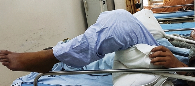
Maintaining the knee flexion over pillows, after the arthroscopic release
Discussion
The management of knee stiffness offers a significant challenge to the treating surgeon and is a significant cause of pain and disability to the patients. The aims of treatment in these individuals are (a) to control pain, (b) to resolve inflammation, (c) to regain an early functional arc of motion (3). The management of knee stiffness is guided by several factors like the type of initial injury, amount of loss of ROM, time since injury, and the status of articular cartilage (9). Initially, a conservative approach is applied in the form of supervised physiotherapy, dynamic splinting, and the use of continuous passive motion (CPM). Manipulation under Anesthesia (MUA) is reserved for more resistant cases and should be carefully by an experienced surgeon. A joint affected by arthrofibrosis is less tolerant of the high forces generated during MUA, compared to normal joints (12). Hence, a forceful manipulation can lead to the development of extreme and excessive contact forces in the already jeopardized joint structures, leading to complications like peri-articular fractures, chondral damage, tear of ligaments and muscles, etc. Sassoon et al. [13] reported from their 40 patients undergoing MUA for post-traumatic knee arthrofibrosis, a mean arc of movement improvement from 59° to 110○. The timing of MUA is crucial for its success. Haller et al. [14] reported success when the MUA was performed within 3 months of fracture management. However, if the knee stiffness motion is more severe or chronic (> 3 months), arthroscopic surgery is preferred.
Nowadays, open surgical procedures, like excision of infrapatellar and prepatellar adhesions and quadricepsplasty, are not done commonly as these are associated with additional surgical morbidity and may be less favorable for management of flexion contracture. Hence, arthroscopic surgical procedures have paved into replace open procedures in the management of stiff knees. The pros and cons of arthroscopic surgery have been listed in Table 4. The procedure is minimally invasive (arthroscopic) with small incisions and is cosmetically viable. Arthroscopic surgical release offers several advantages to an open technique being a minimally invasive surgery. It carries lower morbidity, a lower chance of wound complications, and a decreased overall surgical risk. There is also a lesser chance of causing an iatrogenic fracture (compared with MUA alone) in a stiff knee and osteoporotic bone [9]. Furthermore, the patients can immediately start aggressive physiotherapy and, thus, decrease the chance of relapse of knee stiffness. The arthroscopy also offers the opportunity to address any concomitant lesions inside the knee.
Table 4.
Pros and cons of arthroscopic surgery for the stiff knees
| Arthroscopic procedure | PROS | CONS |
|---|---|---|
| Intra-articular release | Minimally invasive procedure | Cannot address the extra-articular cause of stiffness |
| Posterior release | Associated with lesser surgical morbidity | Neuro-vascular structures are at risk |
| Effectiveness | Best achieved in the earlier phase of stiffness | May not be so effective in long-standing cases |
| Deformity correction | Best for lesser deformities | Severe deformities may not be corrected fully |
| Surgical ease | Can be done by a trained arthroscopic surgeon, with conventional instruments | A prior understanding and knowledge of the pathology encountered is needed |
It is crucial to identify the likely cause and site of adhesions before the arthroscopic procedure. The intra-articular causes of loss of flexion include capsular adhesions in the supra-patellar region, medial recess, lateral recess, and inter-condylar region [9, 15]. The extension loss may be due to a ‘cyclops lesion’ in the intercondylar notch or due to adhesions in the posterior capsule. The ability of arthroscopic surgery to access both anterior and posterior compartments in a minimally invasive manner makes it a preferred mode of treatment for knee stiffness. In addition to arthroscopic lysis of the adhesions, pie-crusting of the tight quadriceps muscle is beneficial to gain greater ROM. Shang et al. [16] reported from the experience of five post-traumatic stiff knees that a mean maximum flexion was increased from 35° preoperatively to 80°, after arthroscopic adhesiolysis and further increased to 120° after pie-crusting. At the mean follow-up of 6 months, the maximum flexion achieved was 110°. Bansal et al. [12] have described an interesting technique of using saline-soaked ribbon gauze packing in the patella-femoral joint to prevent direct friction between chondral surfaces and, thus, avoiding inevitable complications such as cartilage damage, subchondral fractures. They also felt that it breaks the remaining adhesions involving the quadriceps expansion in the lateral and medial recesses, the supra-patellar bursa, and the muscle adhesions to the distal femur, thus achieving further improvement in the range of flexion.
The definitive management of knee stiffness should be done as early as possible, as good results are achievable in the early phase by physiotherapy, MUA, and arthroscopic release. Delay or neglect of knee stiffness may lead to the development of extra-articular contractures and adhesions, which may not be amenable to arthroscopic surgery. The best time for considering arthroscopic release is between 3 and 6 months after definitive fracture management [3]. It is, however, debatable as to after which duration and the severity of stiffness, arthroscopic surgery should not be done. Vaishya and Sharma [4], in 1999, reported promising results of it on a series of 17 patients, where the range of motion (ROM) improved from an average of 23°–100°, and their patients had arthroscopic surgery 4 months to 6 years (average 2.3 years). They also noted some loss of movement postoperatively, as compared to intra-operatively. Gittings et al. [17] reported their experience of arthroscopic release in the post-traumatic stiff knees, which were treated by open reduction and internal fixation (ORIF). They found an intra-operative improvement in the ROM from an average of 72° (preoperatively)–128° (intra-operatively), with a sustained clinical improvement to a mean ROM of 101°, at the final follow-up. Kim and Joo [18] also reported good results of arthroscopic lysis of adhesions in post-traumatic cases (43 of 68 total patients). An increase in ROM was seen from an average of about 70° preoperatively to 118° postoperatively, at the mean final follow-up of 17.8 months. Klein et al. [19] retrospectively studied 58 patients with postoperative joint stiffness, mainly after arthrotomy (87%), who underwent arthroscopic fibro-arthrolysis during 1987–1991. The average time between the previous surgical procedure and the following arthroscopic arthrolysis was 22.8 months (range 4–105). They found excellent to good results in 75.7% cases and concluded that arthroscopic surgery is beneficial to patients with knee joint stiffness even after a prolonged period.
For the flexion contractures of the knee, an open or arthroscopic posteromedial release of the capsule is required. Arthroscopic posterior release needs the use of a posteromedial portal. LaPrade et al. [20] reported good results in their first 15 patients in 2008 and concluded that the arthroscopic posterior release is as effective as an open procedure.
Shelbourne et al. [21] have proposed a classification of knee stiffness following ACL reconstruction and have classified the arthrofibrosis into four types. They have suggested anterior scar resection up to the proximal tibia, in types 2, 3, and 4 arthrofibrosis. Notchplasty was required some time and the medial and lateral capsular releases, with the knee manipulation in type 3 or 4 arthrofibrosis. Paulos et al. [22] described an uncommon cause of knee stiffness and named it ‘infrapatellar contracture syndrome.’ According to them, it may happen after a knee injury or surgery and may be quite disabling for the patient than the actual primary surgery like ACL reconstruction. Patella baja is an uncommon but devastating complication after knee surgery and may cause loss of knee extension. It can happen after prolonged immobilization, surgery around the tibial tubercle, and patella tendon surgery. It causes not only knee stiffness but also pain and functional limitations. Excision of the scar with arthroscopic surgery is recommended when the conservative treatments have failed. In resistant cases, an open patellar tendon tenotomy is required [23]. Acquired patella baja is the result of the shortening of patellar tendon secondary to the scarring of the patellar tendon to the upper tibia or shrinkage of the fibers of the tendon. Drexler et al. [24] treated patella baja with proximalization of the tibial tuberosity and have reported satisfactory outcomes.
Stiffness after TKA is a major problem. Ipach et al. [25] found its prevalence after primary TKA to be 4.54%, and for revision TKA to be 5.11%, from the study of 1344 patients operated between 2004 and 2009. The causes of stiffness after TKA could be due to inadequate pain control, reflex sympathetic dystrophy, infection, abnormal component positioning or sizing, insufficient soft-tissue balancing, and aseptic loosening [22]. Several options are available for the management of stiffness after TKA. These include non-operative treatment in the form of using adequate pain-relieving measures, intense physiotherapy, and dynamic knee bracing. The operative options include MUA, arthroscopic or open arthrolysis, and revision of the abnormally placed components. Ipach et al. [25] found MUA to be a successful and effective procedure in improving the ROM in these cases. The MUA was done at a mean of 95.17 days (range 9–130 days), after the primary surgery. Flexion was improved significantly after the MUA in both the groups, with a mean gain of 35.13° in primary TKA and of 41.31° in the revision TKA group. Arbuthnot and Brink [26] treated 22 cases of knee stiffness following TKA, by arthroscopic arthrolysis. They recorded a mean ROM improvement from 8°–69° to 3°–105° on the table, but it declined slightly to 4°–93° (P: 0.05) at follow-up. They recommended arthroscopic arthrolysis to be a useful measure, which compares well with other methods of treatment for stiffness. Knee arthroscopy is considered a safe procedure in the diagnosis and or treatment of complications after TKA, associated with a low rate of adverse effects [28, 29]. The most common indications for arthroscopy after TKA could be a soft-tissue impingement, arthrofibrosis, peri-prosthetic infection, and the removal of free bodies or cement fragments. The approximate percentages of the effectiveness of arthroscopic surgery after TKA vary according to the indication: 85% in soft-tissue impingement; 90% in arthrofibrosis; and 55% in peri-prosthetic infection [27]. Complication rates after the arthroscopic procedure are reported to be around 0.5%, in a systematic review [28]. The likely complications could be the procedure-related infection, abrasion of the artificial joint components, and technical difficulties in orientation and visualization due to the reflection of light against the metal component, causing a ‘mirror phenomenon’ [28]. Rarely, an instrument breakage is also possible.
In a systematic review of various procedures used for stiffness after TKA, Ghani et al. [29] observed that no significant differences were found in the KSS of the four treatment modalities. They observed that MUA provided a mean increase in ROM of 38.4°, the arthroscopic release had a mean increase of 36.2°, the open surgical release had a mean increase of 43.4°, and revision TKA had a mean increase of 24.7°. Therefore, the selection of treatment should be tailored according to the patient’s problem and the experience of the surgeon.
Although a successful and patient-friendly procedure, the arthroscopic release of the knee is a technically challenging surgery, which requires significant experience of the surgeon in arthroscopy. It has some caveat and shortcoming also like it can release all the scarred tissue intra-articularly; the extra-articular causes of stiffness cannot be addressed by it. There may be a chance of injury to the adjacent ligaments, articular cartilage, and neurovascular structures. Moreover, the patient co-operation, compliance, motivation, and enthusiasm are vital in the postoperative period during physical therapy for achieving success.
Conclusion
A stiff knee due to any cause is a troublesome proposition to both the patients and treating surgeons. It is quite common and a significant cause of pain and disability after a knee injury, infection, and surgery. The commonest cause of stiffness is after a peri-articular injury to the knee. Various methods of management have been described to deal with knee stiffness. Amongst the operative treatment, MUA and arthroscopic surgery were found to be the most effective. Arthroscopic surgery offers a good option of releasing the stiff knees in the majority of cases of post-traumatic, post-ORIF, and post-TKA cases. However, there are some caveats and limitations attached to this procedure. Arthroscopic surgery is most valuable and useful if done earlier in the course of the stiffness (preferably between 3 and 6 months). However, its success in long-standing cases has also been documented. Open surgery, nowadays, is going out of favor and is only done in selected cases.
Electronic Supplementary Material
Below is the link to the electronic supplementary material.
Supplementary video 1: Arthroscopic release of a stiff knee after prior arthrotomy procedure. (MPG 31396 kb)
Compliance with Ethical Standards
Conflict of Interest
The authors declare that they have no conflict of interest.
Ethical Standard Statement
This article does not contain any studies with human or animal subjects performed by any of the authors.
Informed Consent
For this type of study an informed consent is not required.
Footnotes
Publisher's Note
Springer Nature remains neutral with regard to jurisdictional claims in published maps and institutional affiliations
References
- 1.Lindenfeld TN, Wojtvs EM, Husain A. Surgical treatment of arthrofibrosis of the knee. Instructional Course Lectures. 2020;49:211–221. [PubMed] [Google Scholar]
- 2.Haller JM, Holt DC, McFadden ML, Higgins TF, Kubiak EN. Arthrofibrosis of the knee following a fracture of the tibial plateau. The Bone & Joint Journal. 2015;97-B:109–114. doi: 10.1302/0301-620X.97B1.34195. [DOI] [PubMed] [Google Scholar]
- 3.Steifel EC, McIntyre L. Arthroscopic lysis of adhesions for treatment of post-traumatic arthrofibrosis of the knee joint. Arthroscopy Techniques. 2017;6(4):e939–e944. doi: 10.1016/j.eats.2017.03.001. [DOI] [PMC free article] [PubMed] [Google Scholar]
- 4.Vaishya R, Sharma P. Arthroscopic management of stiff knees. Indian Journal of Orthopaedics. 1999;33(4):274–275. [Google Scholar]
- 5.Pujol N, Boisrenoult B, Beaufils P. Post-traumatic knee stiffness: Surgical techniques. Orthopaedics & Traumatology: Surgery & Research. 2015;101:S179–S186. doi: 10.1016/j.otsr.2014.06.026. [DOI] [PubMed] [Google Scholar]
- 6.Nicoll EA. Quadricepsplasty. The Journal of Bone and Joint Surgery. 1963;45B:483–490. doi: 10.1302/0301-620X.45B3.483. [DOI] [PubMed] [Google Scholar]
- 7.Jackson DW, Schaefer RK. Cyclops syndrome: Loss of extension following intra-articular anterior cruciate ligament reconstruction. Arthroscopy. 1990;6:171–178. doi: 10.1016/0749-8063(90)90072-L. [DOI] [PubMed] [Google Scholar]
- 8.McNamara IR, Smith TO, Shepherd KL, Clark AB, Nielsen DM, Donell S, Hing CB, et al. Surgical fixation methods for tibial plateau fractures. Cochrane Database of Systematic Reviews. 2015 doi: 10.1002/14651858.CD009679.pub2. [DOI] [PMC free article] [PubMed] [Google Scholar]
- 9.Sprague NF, O’Connor RL, Fox JM. Arthroscopic treatment of postoperative knee fibroarthrosis. Clinical Orthopaedics and Related Research. 1982;166:165–172. [PubMed] [Google Scholar]
- 10.KimD H, Gill TJ, Millett PJ. Arthroscopic treatment of the arthrofibrotic knee. Arthroscopy. 2004;20(suppl2):187–194. doi: 10.1016/j.arthro.2004.04.036. [DOI] [PubMed] [Google Scholar]
- 11.Kukreja M, Kang J, Curry EJ, Li X. Arthroscopic lysis of adhesions and anterior interval release with manipulation under anesthesia for severe post-traumatic knee stiffness: A simple and reproducible step-by-step guide. Arthroscopy Techniques. 2019;8(5):e429–e435. doi: 10.1016/j.eats.2019.01.005. [DOI] [PMC free article] [PubMed] [Google Scholar]
- 12.Bansal H, Nag HL, Mehta N, Sain A. A new method of arthroscopic assisted management of stiff knee: A technical note. Arthroscopy and Orthopedic Sports Medicine. 2018;5(2):53–56. doi: 10.14517/aosm17019. [DOI] [Google Scholar]
- 13.Sassoon AA, Adigweme OO, Langford J, KovalK J, HaidukewychG J. Manipulation under anesthesia: A safe and effective treatment for posttraumatic arthrofibrosis of the knee. Journal of Orthopaedic Trauma. 2015;29:e464–e468. doi: 10.1097/BOT.0000000000000395. [DOI] [PubMed] [Google Scholar]
- 14.Haller JM, Holt DC, McFadden ML, Higgins TF, Kubiak EN. Arthrofibrosis of the knee following a fracture of the tibial plateau. The Bone & Joint Journal. 2015;97:109–114. doi: 10.1302/0301-620x.97b1.34195. [DOI] [PubMed] [Google Scholar]
- 15.Paulos LE, Rosenberg TD, Drawbert J, Manning J, Abbott P. Infrapatellar contracture syndrome. An unrecognized cause of knee stiffness with patella entrapment and patella infera. American Journal of Sports Medicine. 1987;15:331–341. doi: 10.1177/036354658701500407. [DOI] [PubMed] [Google Scholar]
- 16.Shang P, Liu H, Zhang Y, Xue E, Pan X. A mini-invasive technique for severe arthrofibrosis of the knee: A technical note. Injury. 2016;47(8):1867–1870. doi: 10.1016/j.injury.2016.06.015. [DOI] [PubMed] [Google Scholar]
- 17.Gittings D, Hesketh P, Dattilo J, Zgonis M, Kelly J, Mehta S. Arthroscopic lysis of adhesions improves knee range of motion after fixation of intra-articular fractures about the knee. Archives of Orthopaedic and Trauma Surgery. 2016;136:1631–1635. doi: 10.1007/s00402-016-2561-2. [DOI] [PubMed] [Google Scholar]
- 18.Kim YM, Joo YB. Prognostic factors of arthroscopic adhesiolysis for arthrofibrosis of the knee. Knee Surgery & Related Research. 2013;13(25):202–206. doi: 10.5792/ksrr.2013.25.4.202. [DOI] [PMC free article] [PubMed] [Google Scholar]
- 19.Klein W, Shah N, Gassen A. Arthroscopic management of postoperative arthrofibrosis of the knee joint: Indication, technique, and results. Arthroscopy: The Journal of Arthroscopic and Related Surgery. 1994;10(6):591–597. doi: 10.1016/S0749-8063(05)80053-2. [DOI] [PubMed] [Google Scholar]
- 20.LaPrade RF, Pedtke AC, Roethle ST. Arthroscopic posteromedial capsular release for knee flexion contractures. Knee Surgery, Sports Traumatology, Arthroscopy. 2008;16:469–475. doi: 10.1007/s00167-008-0496-z. [DOI] [PubMed] [Google Scholar]
- 21.Shelbourne KD, Patel DV, Martini DJ. Classification and management of arthrofibrosis of the knee after anterior cruciate ligament reconstruction. American Journal of Sports Medicine. 1996;24(6):857–862. doi: 10.1177/036354659602400625. [DOI] [PubMed] [Google Scholar]
- 22.Paulos LE, Wnorowski DC, Greewald AE. Infrapatellar contracture syndrome. Diagnosis, treatment, and long-term followup. The American Journal of Sports Medicine. 1994;22(4):440–449. doi: 10.1177/036354659402200402. [DOI] [PubMed] [Google Scholar]
- 23.Kennedy MI, Zach Aman BS, DePhillipo NN, LaPrade RF. Patellar tendon tenotomy for treatment of patella baja and extension deficiency. Arthroscopy Techniques. 2019;8(3):e317–e320. doi: 10.1016/j.eats.2018.11.010. [DOI] [PMC free article] [PubMed] [Google Scholar]
- 24.Drexler M, Dwyer T, Marmor M, Sternheim A, Cameron HU, Cameron JC. The treatment of acquired patella baja with proximalize the tibial tuberosity. Knee Surgery, Sports Traumatology, Arthroscopy. 2013;13(21):2578–2583. doi: 10.1007/s00167-012-2278-x. [DOI] [PubMed] [Google Scholar]
- 25.Ipach I, Schäfer R, Lahrmann J, Kluba T. Stiffness after knee arthrotomy: Evaluation of prevalence and results after manipulation under anaesthesia. Orthopaedics & Traumatology: Surgery & Research. 2011;97:292–296. doi: 10.1016/j.otsr.2011.01.006. [DOI] [PubMed] [Google Scholar]
- 26.Arbuthnot JE, Brink RB. Arthroscopic arthrolysis for the treatment of stiffness after total knee replacement gives moderate improvements in range of motion and functional knee scores. Knee Surgery, Sports Traumatology, Arthroscopy. 2010;18:346–351. doi: 10.1007/s00167-009-0878-x. [DOI] [PubMed] [Google Scholar]
- 27.Encinas-Ullán CA, Rodríguez-Merchán EC. Arthroscopic treatment of total knee arthroplasty complications. EFORT Open Reviews. 2019 doi: 10.1302/2058-5241.4.180035. [DOI] [PMC free article] [PubMed] [Google Scholar]
- 28.Hegazy AM, Elsoufy MA. Arthroscopic arthrolysis for arthrofibrosis of the knee after total knee replacement. HSSJ. 2011;7:130–133. doi: 10.1007/s11420-011-9202-7. [DOI] [PMC free article] [PubMed] [Google Scholar]
- 29.Ghani H, Maffulli N, Khanduja V. Management of stiffness following total knee arthroplasty: A systematic review. Knee. 2012;19(6):751–759. doi: 10.1016/j.knee.2012.02.010. [DOI] [PubMed] [Google Scholar]
Associated Data
This section collects any data citations, data availability statements, or supplementary materials included in this article.
Supplementary Materials
Supplementary video 1: Arthroscopic release of a stiff knee after prior arthrotomy procedure. (MPG 31396 kb)