Introduction
Antimicrobial peptides (AMPs) represent an ancient mechanism for antagonizing microbial opponents, being generated by eukaryotes, eubacteria, and archaea alike [1,2]. Given the dearth in new antibiotics, there has been increasing interest in AMPs. As our understanding has grown, a tantalizing possibility has taken shape: Might these agents of competition be at the heart of the cooperative success story that gave rise to mitochondria and chloroplasts? Striking similarities suggest that the system of protein import into these endosymbiotic organelles may derive from an interplay of AMP attack and defense [3].
AMPs can be detoxified through import
While the term “AMP” applies to any antimicrobial based on peptide bonds, this article focuses on genetically encoded, ribosomally produced AMPs (RAMPs). Many RAMPs act by destabilizing membranes, although several also have intracellular targets [2]. The peptide nature of RAMPs allows for a general detoxification mechanism via proteolytic degradation in the bacterial cytoplasm, rendering import a viable defense strategy. First proposed in 1993 based on a screen for mutants sensitive to antimicrobial peptides (sap) in Salmonella [4], RAMP internalization was finally confirmed via immunogold labeling for sap homologues in non-typeable Haemophilus influenzae [5]. Such defense by import plays a role in extant endosymbiosis: In Sinorhizobium meliloti, the BacA transporter promotes defensive uptake of nodule-specific cysteine-rich (NCR) RAMPs expressed by the host plant [2,6]. These detoxification mechanisms have striking analogies to protein targeting to eukaryotic organelles [3], in which nuclear encoded proteins addressed to mitochondria or chloroplasts harbor highly divergent N-terminal presequences termed as “targeting peptides (TPs)” [7,8]. Translocation machineries import the targeted preprotein across 2 sets of membranes, and then the TP is cleaved by dedicated peptidases [9,10].
HA-RAMPs and TPs are similar in structure and function
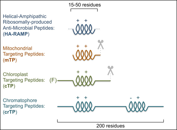
Mitochondrial TPs (mTPs) are characterized by a cationic, amphipathic α-helix [11] (Fig 1). Chloroplast TPs (cTPs) are less structured, yet examples studied via NMR were shown to contain stretches that fold into amphipathic helices in a membrane-mimetic environment [12–14], and signatures of such stretches can be found in the majority of cTPs [15]. The main difference between mTPs and cTPs therefore lies in the presence of additional, unstructured domains (Fig 1), notably an uncharged N-terminus [7,16].
Fig 1. Similarities between TPs and HA-RAMPs suggest organelle import evolved from AMP detoxification.
HA-RAMPs and TPs bear cationic (++), amphipathic α-helices. Further domains include cleavage sites (mTP and cTP, indicated by scissors), an uncharged N-terminus (cTP) with a leading Phenylalanine (F) in basal algal lineages, or a long carboxyl terminus (crTP) with a second predicted amphipathic helix carrying both positive and negative charges (–+).
The amoeba Paulinella provides a further example of TPs, having acquired a photosynthetic organelle called chromatophore in a much more recent cyanobacterial endosymbiosis (ca. 60 to 100 Mya) than Archaeplastida (ca. 1 Gya) [17,18]. Many imported proteins bear putative chromatophore TPs (crTPs). These crTPs are highly similar in sequence, indicating a single common evolutionary origin [19]; crTPs are also much longer than cTPs or mTPs and may not be cleaved, all pointing to chromatophore import being very distinct [19]. Yet crTPs are imported into plant chloroplasts and contain a positively charged amphipathic helix toward the N-terminus (Fig 1).
TPs thus resemble RAMPs of helical-amphipathic structure (HA-RAMPs) [3]. A recent analysis of physicochemical properties showed these similarities to be far from superficial [15]. In a quantitative description of peptide properties, the strength of the resemblance between TPs and HA-RAMPs equals or exceeds that between different types of signal peptides known to share a common origin [15]. It may thus be of interest to revise AMP classification based on physicochemical properties, as some of the existing AMP families are defined according to criteria, such as the source organism, that offer limited information on the nature of the peptides [15].
Not only do TPs share chemical properties with HA-RAMPs, but they also interact with membranes [7] and can even have destabilizing effects [10]. Indeed, chemically synthesized TPs exhibit antimicrobial activity [15,20]. Inversely, HA-RAMPs were recently shown to target a fluorescent reporter to either mitochondria or the chloroplast in the model alga Chlamydomonas reinhardtii [15].
From antagonism to endosymbiosis: Evolving inside out
The above examples of cross-functionality, rooted in a shared set of physicochemical properties, support an evolutionary link between TPs and HA-RAMPs [15]. Host attack with HA-RAMPs may have selected the bacterial organelle ancestor for defense via import. This interaction could then have been co-opted for protein import when gene transfer or genome rearrangements generated HA-RAMP–protein fusions. Having a system of protein import was crucial to enable the large-scale endosymbiotic gene transfer from the emerging organelles to the host nucleus. It was this gene transfer that generated the highly genetically integrated eukaryotic cells we know today.
Although extant import machineries of both organelles are evolutionary mosaics, containing proteins from host and bacterial ancestors alike [7,21–23], the majority of pore-forming proteins, presumably the most central and ancient factors of the import machineries, appear to have arisen within the bacterial partner during endosymbiosis. For example, the chloroplast outer envelope pore Toc75 is thought to derive from Omp85, a bacterial chaperone that aids assembly of certain outer membrane proteins recognized by carboxyl-terminal stretches ending in phenylalanine [24]. Toc75 switched polarity to accept polypeptides from the host cytoplasm rather than the bacterial periplasm [25], engendering a requirement for phenylalanine at the cTP N-terminus (Fig 1) that endures in basal algal lineages [26]. Also, the chloroplast inner envelope pore is most likely formed by Tic20/21, for which homologues have been identified in cyanobacteria [27], and the mitochondrial outer membrane pore Tom40 probably arose de novo within the proto-mitochondrion [28]. These results support the view that protein import evolved from within the endosymbiont [29], consistent with the AMP hypothesis, rather than being imposed by the host [30]. In addition, the Tim17 family, forming the inner mitochondrial pore, may also have arisen in the organelle ancestor [22,23,31].
Further support for protein import having evolved within the endosymbiont comes from proteases. After being imported, mTPs and cTPs are cleaved (Fig 1) by mitochondrial matrix processing peptidase (MPP) or chloroplast stromal processing peptidase (SPP) and further degraded by dual-targeted presequence peptidase (PreP) and organellar oligopeptidase (OOP) [9,10]. MPP, SPP, and PrepP are M16 metalloproteases, while OOP is a M3 metalloprotease. All have bacterial homologs, implying a prokaryotic origin. For example, MPP probably originated from a rickettsial putative peptidase (RPP)-like progenitor, with extant RPP being capable of cleaving mTPs [32,33]. M16 metalloproteases can even participate in protein translocation in bacteria [34].
Descent from AMP defense accounts for TP sequence degeneracy
The high sequence degeneracy of mTPs and cTPs has led to an alternative hypothesis that TPs could easily have arisen from any given sequence. This idea is based on the observation that promiscuous extant protein import machineries accept even approximately 3% to 5% of sequences selected randomly within genomes [35,36], provided these sequences fold into cationic, amphipathic helices [37]. In this scenario, TPs would have converged with HA-RAMPs, perhaps due to a selection for membrane interaction. However, this hypothesis leaves TP proteolysis and the origin of translocases unexplained.
By contrast, the AMP hypothesis offers a comprehensive scenario for the evolution of all components of the protein import process, including TPs, translocases, and proteases [3,15]. TP sequence degeneracy may be accounted for by recruitment of a promiscuous AMP defense system for protein import, given that extant AMP importers can accept divergent RAMPs [5,6]. Once protein import was established, individual TPs may well have derived from sources other than HA-RAMP genes, as long as the emerging TP bears the physicochemical properties recognized by the import machinery.
Acknowledgments
We would like to thank Francis-André Wollman for critical reading of the manuscript and Clotilde Garrido for sharing some of her data.
Funding Statement
Research by ODC and IL has been supported by annual funding from the Centre National de la Recherche Scientifique and Sorbonne University to UMR 7141, by the ChloroMitoRAMP ANR grant (ANR-19-CE13-0009) and by LabEx Dynamo (ANR-LABX-011). In addition, ODC has received support from the Rothschild Foundation. The funders had no role in study design, data collection and analysis, decision to publish, or preparation of the manuscript.
References
- 1.Besse A, Peduzzi J, Rebuffat S, Carré-Mlouka A. Antimicrobial peptides and proteins in the face of extremes: Lessons from archaeocins. Biochimie. 2015;118:344–55. 10.1016/j.biochi.2015.06.004 [DOI] [PubMed] [Google Scholar]
- 2.Mergaert P. Role of antimicrobial peptides in controlling symbiotic bacterial populations. Nat Prod Rep. 2018;35:336–56. 10.1039/c7np00056a [DOI] [PubMed] [Google Scholar]
- 3.Wollman F-A. An antimicrobial origin of transit peptides accounts for early endosymbiotic events. Traffic. 2016;17:1322–8. 10.1111/tra.12446 [DOI] [PubMed] [Google Scholar]
- 4.Parra-Lopez C, Baer MT, Groisman EA. Molecular genetic analysis of a locus required for resistance to antimicrobial peptides in Salmonella typhimurium. EMBO J. 1993;12:4053–62. Available from: http://www.ncbi.nlm.nih.gov/pubmed/8223423%5Cnhttp://www.pubmedcentral.nih.gov/articlerender.fcgi?artid=PMC413698. [DOI] [PMC free article] [PubMed] [Google Scholar]
- 5.Shelton CL, Raffel FK, Beatty WL, Johnson SM, Mason KM. Sap transporter mediated import and subsequent degradation of antimicrobial peptides in Haemophilus. PLoS Pathog. 2011;7:e1002360. 10.1371/journal.ppat.1002360 [DOI] [PMC free article] [PubMed] [Google Scholar]
- 6.Guefrachi I, Pierre O, Timchenko T, Alunni B, Barrière Q, Czernic P, et al. Bradyrhizobium BclA Is a Peptide Transporter Required for Bacterial Differentiation in Symbiosis with Aeschynomene Legumes. Mol Plant-Microbe Interact. 2015;28:1155–66. 10.1094/MPMI-04-15-0094-R [DOI] [PubMed] [Google Scholar]
- 7.Chotewutmontri P, Holbrook K, Bruce BDD. Plastid Protein Targeting: Preprotein Recognition and Translocation. Galluzzi L, editor. Int Rev Cell Mol Biol. 1st ed. 2017;330:227–294. 10.1016/bs.ircmb.2016.09.006 [DOI] [PubMed] [Google Scholar]
- 8.Wiedemann N, Pfanner N. Mitochondrial Machineries for Protein Import and Assembly. Annu Rev Biochem. 2017;86:685–714. 10.1146/annurev-biochem-060815-014352 [DOI] [PubMed] [Google Scholar]
- 9.Teixeira PF, Glaser E. Processing peptidases in mitochondria and chloroplasts. Biochim Biophys Acta. 2013;1833:360–70. 10.1016/j.bbamcr.2012.03.012 [DOI] [PubMed] [Google Scholar]
- 10.Kmiec B, Teixeira PF, Glaser E. Shredding the signal: targeting peptide degradation in mitochondria and chloroplasts. Trends Plant Sci. 2014;19:771–8. 10.1016/j.tplants.2014.09.004 [DOI] [PubMed] [Google Scholar]
- 11.Roise D, Theiler F, Horvath SJ, Tomich JM, Richards JH, Allison DS, et al. Amphiphilicity is essential for mitochondrial presequence function. EMBO J. 1988;7:649–53. 10.1002/j.1460-2075.1988.tb02859.x [DOI] [PMC free article] [PubMed] [Google Scholar]
- 12.Lancelin JM, Gans P, Bouchayer E, Bally I, Arlaud GJ. Jacquot JP. NMR structures of a mitochondrial transit peptide from the green alga Chlamydomonas reinhardtii. FEBS Lett. 1996;391:203–8. 10.1016/0014-5793(96)00734-x [DOI] [PubMed] [Google Scholar]
- 13.Krimm I, Gans P, Hernandez JF, Arlaud GJ. Lancelin JM. A coil-helix instead of a helix-coil motif can be induced in a chloroplast transit peptide from Chlamydomonas reinhardtii. Eur J Biochem. 1999;265:171–80. 10.1046/j.1432-1327.1999.00701.x [DOI] [PubMed] [Google Scholar]
- 14.Wienk HLJ, Wechselberger RW, Czisch M, De Kreuijff B. Structure, Dynamics, and Insertion of a Chloroplast Targeting Peptide in Mixed Micelles. Biochemistry. 2000:8219–27. 10.1021/bi000110i [DOI] [PubMed] [Google Scholar]
- 15.Garrido C, Caspari OD, Choquet Y, Wollman FA, Lafontaine I. Evidence Supporting an Antimicrobial Origin of Targeting Peptides to Endosymbiotic Organelles. Cell. 2020;9:1795. 10.3390/cells9081795 [DOI] [PMC free article] [PubMed] [Google Scholar]
- 16.Lee DW, Lee S, Lee J, Woo S, Razzak MA, Vitale A, et al. Molecular Mechanism of the Specificity of Protein Import into Chloroplasts and Mitochondria in Plant Cells. Mol Plant. 2019. 10.1016/j.molp.2019.03.003 [DOI] [PubMed] [Google Scholar]
- 17.Delaye L, Valadez-Cano C, Pérez-Zamorano B. How Really Ancient Is Paulinella Chromatophora? PLoS Curr. 2016;8. 10.1371/currents.tol.e68a099364bb1a1e129a17b4e06b0c6b [DOI] [PMC free article] [PubMed] [Google Scholar]
- 18.Shih PM, Matzke NJ. Primary endosymbiosis events date to the later Proterozoic with cross-calibrated phylogenetic dating of duplicated ATPase proteins. Proc Natl Acad Sci U S A. 2013;110:12355–60. 10.1073/pnas.1305813110 [DOI] [PMC free article] [PubMed] [Google Scholar]
- 19.Singer A, Poschmann G, Mühlich C, Valadez-Cano C, Hänsch S, Hüren V, et al. Massive Protein Import into the Early-Evolutionary-Stage Photosynthetic Organelle of the Amoeba Paulinella chromatophora. Curr Biol. 2017;27:2763–73. 10.1016/j.cub.2017.08.010 [DOI] [PubMed] [Google Scholar]
- 20.Hugosson M, Andreu D, Boman HG, Glaser E. Antibacterial peptides and mitochondrial presequences affect mitochondrial coupling, respiration and protein import. Eur J Biochem. 1994;223:1027–33. 10.1111/j.1432-1033.1994.tb19081.x [DOI] [PubMed] [Google Scholar]
- 21.Shi LX, Theg SM. The chloroplast protein import system: From algae to trees. Biochim Biophys Acta. 2013;1833:314–31. 10.1016/j.bbamcr.2012.10.002 [DOI] [PubMed] [Google Scholar]
- 22.Dolezal P, Likic V, Tachezy J, Lithgow T. Evolution of the molecular machines for protein import into mitochondria. Science. 2006;313:314–8. 10.1126/science.1127895 [DOI] [PubMed] [Google Scholar]
- 23.Fukasawa Y, Oda T, Tomii K, Origin IK. Evolutionary Alteration of the Mitochondrial Import System in Eukaryotic Lineages. Mol Biol Evol. 2017;34:1574–86. 10.1093/molbev/msx096 [DOI] [PMC free article] [PubMed] [Google Scholar]
- 24.Robert V, Volokhina EB, Senf F, Bos MP, Van Gelder P, Tommassen J. Assembly factor Omp85 recognizes its outer membrane protein substrates by a species-specific C-terminal motif. PLoS Biol. 2006;4:1984–1995. 10.1371/journal.pbio.0040377 [DOI] [PMC free article] [PubMed] [Google Scholar]
- 25.Sommer MS, Daum B, Gross LE, Weis BLM, Mirus O, Abram L, et al. Chloroplast Omp85 proteins change orientation during evolution. Proc Natl Acad Sci U S A. 2011;108:13841–6. 10.1073/pnas.1108626108 [DOI] [PMC free article] [PubMed] [Google Scholar]
- 26.Knopp M, Garg SG, Handrich M, Gould SB. Major Changes in Plastid Protein Import and the Origin of the Chloroplastida. iScience. 2020;23:100896. 10.1016/j.isci.2020.100896 [DOI] [PMC free article] [PubMed] [Google Scholar]
- 27.Lv HX, Guo GQ, Yang ZN. Translocons on the inner and outer envelopes of chloroplasts share similar evolutionary origin in Arabidopsis thaliana. J Evol Biol. 2009;22:1418–28. 10.1111/j.1420-9101.2009.01755.x [DOI] [PubMed] [Google Scholar]
- 28.Pereira J, Lupas AN. The origin of mitochondria-specific outer membrane β-barrels from an ancestral bacterial fragment. Genome Biol Evol. 2018;10:2759–65. 10.1093/gbe/evy216 [DOI] [PMC free article] [PubMed] [Google Scholar]
- 29.Alcock F, Clements A, Webb C, Lithgow T. Tinkering Inside the Organelle. Science. 2010;327:649–50. 10.1126/science.1182129 [DOI] [PubMed] [Google Scholar]
- 30.Gross J, Bhattacharya D. Mitochondrial and plastid evolution in eukaryotes: An outsiders’ perspective. Nat Rev Genet. 2009;10:495–505. 10.1038/nrg2610 [DOI] [PubMed] [Google Scholar]
- 31.Žárský V, Doležal P. Evolution of the Tim17 protein family. Biol Direct. 2016;11:1–13. 10.1186/s13062-015-0100-7 [DOI] [PMC free article] [PubMed] [Google Scholar]
- 32.Bolhuis A, Koetje E, price J-Y, Vehmaanper J, Venema G, Bron S, et al. Did the Mitochondrial Processing Peptidase Evolve from a Eubacterial Regulator of Gene Expression? Mol Biol Evol. 2000;17:198–201. 10.1093/oxfordjournals.molbev.a026232 [DOI] [PubMed] [Google Scholar]
- 33.Kitada S, Uchiyama T, Funatsu T, Kitada Y, Ogishima T, Ito AA. Protein from a Parasitic Microorganism, Rickettsia prowazekii, Can Cleave the Signal Sequences of Proteins Targeting Mitochondria. J Bacteriol. 2007;189:844–50. 10.1128/JB.01261-06 [DOI] [PMC free article] [PubMed] [Google Scholar]
- 34.Grinter R, Hay ID, Song J, Wang J, Teng D, Dhanesakaran V, et al. FusC, a member of the M16 protease family acquired by bacteria for iron piracy against plants. PLoS Biol. 2018;16:1–21. 10.1371/journal.pbio.2006026 [DOI] [PMC free article] [PubMed] [Google Scholar]
- 35.Baker A, Schatz G. Sequences from a prokaryotic genome or the mouse dihydrofolate reductase gene can restore the import of a truncated precursor protein into yeast mitochondria. Proc Natl Acad Sci. 1987;84:3117–21. 10.1073/pnas.84.10.3117 [DOI] [PMC free article] [PubMed] [Google Scholar]
- 36.Lucattini R, Likić VA, Lithgow T. Bacterial Proteins Predisposed for Targeting to Mitochondria. Mol Biol Evol. 2004;21:652–8. 10.1093/molbev/msh058 [DOI] [PubMed] [Google Scholar]
- 37.Lemire BD, Fankhauser C, Baker A, Schatz G. The mitochondrial targeting function of randomly generated peptide sequences correlates with predicted helical amphiphilicity. J Biol Chem. 1989;264:20206–15. [PubMed] [Google Scholar]


