Abstract
Amongst the neurologic complications of COVID-19 disease, very few reports have shown the presence of the virus in the cerebrospinal fluid (CSF). Seizure and rarely status epilepticus can be associated with COVID-19 disease. Here we present a 73-year-old male with prior history of stroke who has never experienced seizure before. He had no systemic presentation of COVID-19 disease. The presenting symptoms were two consecutive generalized tonic-clonic seizures that after initial resolution turned into a nonconvulsive status epilepticus despite antiepileptic treatment (a presentation similar to NORSE (new-onset refractory status epilepticus)). There was no new lesion in the brain magnetic resonance imaging (MRI). The CSF analysis only showed an increased protein levels and positive reverse transcription polymerase chain reaction (RT-PCR) of 2019-nCoV. Patient recovered partially after anesthetic, IVIG, steroid, and remdesivir. To our knowledge, this is the first report of a refractory status epilepticus with the presence of SARS-CoV-2 ribonucleic acid (RNA) in the CSF.
Keywords: COVID-19, Status epilepticus, CSF
Introduction
Seizure has been reported as a life-threatening neurological complication of severe acute respiratory syndrome coronavirus-2 (SARS-CoV-2) [1, 2]. Potential mechanisms have been speculated to cause seizure in the setting of COVID-19 including encephalitis, cytokine storm, hypoxia, and metabolic derangement [3]. Here we report a case of COVID-19-related refractory status epilepticus with the presence of SARS-CoV-2 ribonucleic acid (RNA) in the cerebrospinal fluid (CSF).
Case report
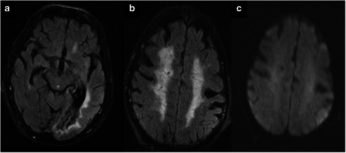
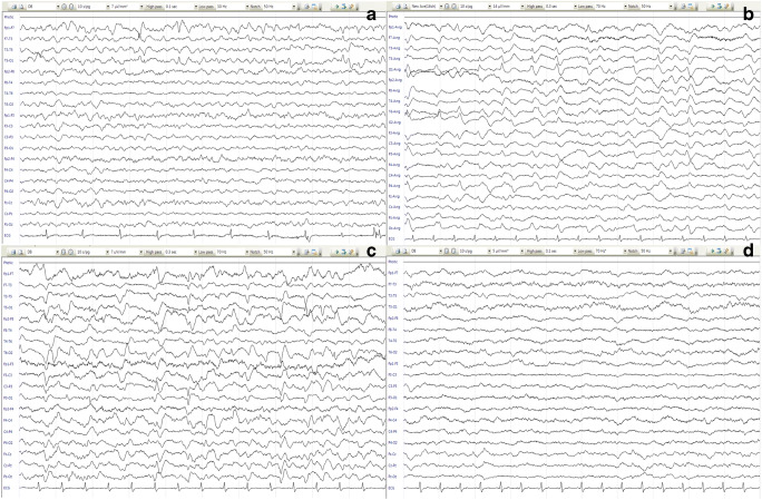
The patient is a 73-year-old man with a history of a previous left occipital lobe ischemic stroke in 2017. Although he had a mild right hemiparesis as a sequela of the stroke, he never had a seizure. He presented with two seizures on the day of admission. The seizures were focal motor (right versive seizure without loss of awareness) followed by secondary generalized tonic-clonic seizures. In the postictal phase, he was moderately confused with a gaze dominancy to the left and partial Broca-type dysphasia and increased weakness in the right arm (Todd’s paresis). There was no systemic presentation of an infection (no fever nor any respiratory symptoms) and no meningeal irritation signs like headache or neck stiffness. The patient received loading and maintenance dose of levetiracetam. The Todd’s paresis resolved, and the level of consciousness improved during the next 12 h; then he developed aphasia and fluctuations in awareness once more (aphasic seizure with loss of awareness). The brain computed tomography scan and magnetic resonance imaging (MRI) failed to show any new lesions in the brain. However, there was a slight cortical hyperintensity in diffusion-weighted (DWI) MRI without hypointensity in the apparent diffusion coefficient (ADC-map) images located in the left temporal cortex (Fig. 1). The routine lab data was normal apart from the elevation of serum leukocyte (13,000 cells/mm3). Serum and urine toxicology analyses were negative. The electroencephalography (EEG) showed a focal ictal pattern in the left temporal leads (maximum potential: F7) with a slow background rhythm consistent with a focal nonconvulsive status epilepticus (Fig. 2a). Valproate loading dose helped the patient to recover to his normal baseline cognitive and language state. Unfortunately, after 12 h, there was a recurrence of symptoms despite optimal antiepileptic serum levels. After intravenous (IV) phenytoin administration, there was no improvement in the patient’s condition, and the EEG findings aggravated and evolved into bilaterally independent lateralized periodic discharges (maximum potential: O2 andF7, respectively) and occasional EEG ictal pattern (rhythmic activity with electrical evolution, in T5) (Fig. 2b, c). CSF analysis revealed an increased protein level (98 mg/dl), with normal glucose, cell count, and opening pressure. The herpes simplex virus (HSV) polymerase chain reaction (PCR) was negative, and CSF IgG index was 0.6. The CSF microbiological assessment was negative. The chest CT scan revealed mild bilateral nodular and ground-glass opacities in the posterior and lower regions that was interpreted initially as an aspiration pneumonia. Blood oxygen saturation was normal (96%), and ceftriaxone was administered. Fever was absent. On the third day of admission, the level of consciousness decreased and fine clonic jerks were noted in the left side of the face. The patient was intubated, and a more aggressive antiepileptic treatment was applied with the impression of “refractory nonconvulsive status epilepticus in coma.” Phenobarbital and midazolam could change the EEG findings to a burst suppression pattern, and after 12 h, we gradually decreased the anesthetic doses. Immunomodulatory treatment with IVIG and dexamethasone was initiated. Meanwhile, the 2019-nCoV RT-PCR test showed the presence of the virus in both pharyngeal mucosa and the CSF. The antiviral treatment with remdesivir was also started. After 5 days of treatment with 1600 mg/day valproate, 300 mg/day phenytoin, 3000 mg/day levetiracetam, 100 mg/day phenobarbital, 25 mg/day IVIG, and 500 mg/day methylprednisolone, there was no clinical motor symptom of seizure. Patient was still comatose and showed a slight reaction (grimacing) to pain stimuli. The EEG showed generalized irregular delta slowing (Fig. 2d). During the following days, there was no significant systemic finding related to COVID-19 disease. After a week, the patient recovered partially, could open his eyes, and obeys one step command without new seizures nor focal neurological deficits.
Fig. 1.
a Previous stroke in the left PCA territory with bilateral periventricular white matter lesions. b, c Left temporal cortex flair and DWI hyperintensity
Fig. 2.
a There was very frequent rhythmic (more than 2.5 Hz) left temporal sharp compatible with EEG ictal pattern (starting from: F7> T3, T5)*. b, c There was no posterior dominant rhythm. There was continuous left temporal sharp/periodic lateralized epileptiform discharge (PLED)/EEG ictal pattern (maximum potential: T3, T5) and independent frequent right occipital sharp/PLED (maximum potentialO2)* and a continuous right temporo-occipital rhythmic and irregular delta slowing (maximum potential: O2, T6) which we assumed compatible with focal nonconvulsive status epilepticus. d After tapering the anesthetics, the background rhythm consisted of a low amplitude irregular delta, theta activity without epileptiform discharges. *F frontal, T temporal, O occipital
Discussion
Several neurological complications have been reported in connection with COVID-19 disease [4]. Few reports provide evidence to support the potential neurotropism and direct CNS invasion of COVID-19 by detecting SARS-CoV-2 RNA in CSF. So far, encephalitis, acute necrotizing encephalopathy, clinically isolated syndrome, and cerebellitis have been associated with the presence of SARS-Co-2 RNA in CSF [5–8].
In the context of COVID-19 disease, patients can be susceptible to have seizures by multiple mechanisms. Patients with established epilepsy can experience seizure breakthrough. On the other hand, new onset seizures can occur in patients who have metabolic derangement due to systemic complications of COVID-19. Seizures can be due to structural insult in the brain which can be a new lesion after COVID infection as a result of an ischemic, inflammatory, or a potential direct viral encephalitis [3].
Our patient’s MRI showed that there was no new ischemic or inflammatory lesion in the brain, and the cortical findings could be justified by the frequent epileptic activity in that region. Given that there was no hypoxia or other metabolic derangement in this patient, one can assume that the reason for the new onset seizures can be associated to a direct viral insult or secondary to inflammatory response in the CNS (compatible with an encephalitis process). Unlike previous reports of COVID-19-associated meningitis or hemorrhagic encephalitis with positive RT-PCR of viral RNA in the CSF, our patient did not have the cellular response in CSF nor the typical hemorrhagic lesions in MRI [5, 6]. Although the high CSF protein levels could be suggestive of inflammation, the normal IgG index failed to prove intrathecal IgG synthesis. We did not have access to more sophisticated laboratory tests like CSF interleukin-6, neopterin, and β2-microglubolin, and our assessment of the inflammatory response was not thorough.
Given that the old ischemic stroke was located in the left occipital region, and the current seizures come from the left temporal region, it is possible that there is a new irritated lesion due to viral invasion or inflammatory reactions that serves as the epileptogenic zone. There was no systemic condition that could explain the aggressive course of the epileptic activity, and we did not have an exact explanation to why the seizures turn into refractory status epilepticus. Status epilepticus has been reported in patients with COVID-19 [9]. In some reports, the NORSE (new-onset refractory status epilepticus) and anti-NMDA associated with COVID-19 were encountered long after the disease receded, which is expectable in a post-infectious inflammatory process [10, 11]. Nonetheless, this was not the case in our patient who experienced the status epilepticus during the acute phase of the viral infection. We assumed that similar to what occurs in NORSE, an ongoing inflammatory cascade could play a role in our patient’s condition. Therefore, we started an immunomodulatory treatment in combination with antiviral drug, which resulted in improvement in patient’s condition.
In retrospect, probably because of the delay in starting immunomodulatory and antiviral treatment, status epilepticus became refractory. This was in part due to lack of systemic and respiratory findings in this patient who had a pure neurological COVID-19 presentation. We propose that during the pandemic, all patients suspected to NORSE should be investigated for COVID-19 disease and have a PCR test. In addition, it may be beneficial to apply a strict anti-COVID-19 and immunomodulatory treatment in patients who do not have severe respiratory and systemic complications but rather rare neurologic symptoms since they may easily get aggressive and be potentially life-threatening.
Data Availability
All data not published within this article will be made available by request from any qualified investigator.
Declarations
Ethics approval
Ethical approval for this study was obtained from the Iran National Committee for ethics in biomedical research (IR.TUMS.VCR.REC.1399.520). Written informed consent for publication of this case report was obtained from his legal guardian.
Conflict of interest
The authors declare no competing interests.
Footnotes
Publisher’s note
Springer Nature remains neutral with regard to jurisdictional claims in published maps and institutional affiliations.
References
- 1.Asadi-Pooya AA, Simani L, Shahisavandi M, Barzegar Z. COVID-19, de novo seizures, and epilepsy: a systematic review. Neurol Sci. 2020;25:1–7. doi: 10.1007/s10072-020-04932-2. [DOI] [PMC free article] [PubMed] [Google Scholar]
- 2.Sohal S, Mossammat M (2020) COVID-19 presenting with seizures. IDCases:e00782 [DOI] [PMC free article] [PubMed]
- 3.Narula N, Joseph R, Katyal N, Daouk A, Acharya S, Avula A, et al. Seizure and COVID-19: association and review of potential mechanism. Neurol Psychiatry Brain Res. 2020;38:49. doi: 10.1016/j.npbr.2020.10.001. [DOI] [PMC free article] [PubMed] [Google Scholar]
- 4.Carod-Artal FJ. Neurological complications of coronavirus and COVID-19. Rev Neurol. 2020;70(9):311–322. doi: 10.33588/rn.7009.2020179. [DOI] [PubMed] [Google Scholar]
- 5.Moriguchi T, Harii N, Goto J, Harada D, Sugawara H, Takamino J, et al. A first case of meningitis/encephalitis associated with SARS-coronavirus-2. Int J Infect Dis. 2020;94:55–58. doi: 10.1016/j.ijid.2020.03.062. [DOI] [PMC free article] [PubMed] [Google Scholar]
- 6.Virhammar J, Kumlien E, Fällmar D, Frithiof R, Jackmann S, Sköld MK, et al. Acute necrotizing encephalopathy with SARS-CoV-2 RNA confirmed in cerebrospinal fluid. Neurology. 2020;95(10):445–449. doi: 10.1212/WNL.0000000000010250. [DOI] [PMC free article] [PubMed] [Google Scholar]
- 7.Domingues RB, Mendes-Correa MC, de Moura Leite FBV, Sabino EC, Salarini DZ, Claro I, et al. First case of SARS-COV-2 sequencing in cerebrospinal fluid of a patient with suspected demyelinating disease. J Neurol. 2020;267(11):3154–3156. doi: 10.1007/s00415-020-09996-w. [DOI] [PMC free article] [PubMed] [Google Scholar]
- 8.Fadakar N, Ghaemmaghami S, Masoompour SM, Shirazi Yeganeh B, Akbari A, Hooshmandi S, et al. A first case of acute cerebellitis associated with coronavirus disease (COVID-19): a case report and literature review. Cerebellum (London, England) 2020;19(6):911–914. doi: 10.1007/s12311-020-01177-9. [DOI] [PMC free article] [PubMed] [Google Scholar]
- 9.Chen W, Toprani S, Werbaneth K, Falco-Walter J. Status epilepticus and other EEG findings in patients with COVID-19: A case series. Seizure. 2020;81:198–200. doi: 10.1016/j.seizure.2020.08.022. [DOI] [PMC free article] [PubMed] [Google Scholar]
- 10.Dono F, Carrarini C, Russo M, De Angelis MV, Anzellotti F, Onofrj M et al (2020) New-onset refractory status epilepticus (NORSE) in post SARS-CoV-2 autoimmune encephalitis: a case report. Neurol Sci:1–4 [DOI] [PMC free article] [PubMed]
- 11.Monti G, Giovannini G, Marudi A, Bedin R, Melegari A, Simone AM, Santangelo M, Pignatti A, Bertellini E, Trenti T, Meletti S. Anti-NMDA receptor encephalitis presenting as new onset refractory status epilepticus in COVID-19. Seizure. 2020;81:18. doi: 10.1016/j.seizure.2020.07.006. [DOI] [PMC free article] [PubMed] [Google Scholar]
Associated Data
This section collects any data citations, data availability statements, or supplementary materials included in this article.
Data Availability Statement
All data not published within this article will be made available by request from any qualified investigator.


