PLOS ONE logoLink to PLOS ONE
. 2021 Apr 15;16(4):e0249735. doi: 10.1371/journal.pone.0249735

Mix and match: Patchwork domain evolution of the land plant-specific Ca2+-permeable mechanosensitive channel MCA

Kanae Nishii 1,2,*, Michael Möller 1, Hidetoshi Iida 3,*
Editor: Berthold Heinze4
PMCID: PMC8049495  PMID: 33857196

Abstract

Multidomain proteins can have a complex evolutionary history that may involve de novo domain evolution, recruitment and / or recombination of existing domains and domain losses. Here, the domain evolution of the plant-specific Ca2+-permeable mechanosensitive channel protein, MID1-COMPLEMENTING ACTIVITY (MCA), was investigated. MCA, a multidomain protein, possesses a Ca2+-influx-MCAfunc domain and a PLAC8 domain. Profile Hidden Markov Models (HMMs) of domains were assessed in 25 viridiplantae proteomes. While PLAC8 was detected in plants, animals, and fungi, MCAfunc was found in streptophytes but not in chlorophytes. Full MCA proteins were only found in embryophytes. We identified the MCAfunc domain in all streptophytes including charophytes where it appeared in E3 ubiquitin ligase-like proteins. Our Maximum Likelihood (ML) analyses suggested that the MCAfunc domain evolved early in the history of streptophytes. The PLAC8 domain showed similarity to Plant Cadmium Resistance (PCR) genes, and the coupling of MCAfunc and PLAC8 seemed to represent a single evolutionary event. This combination is unique in MCA, and does not exist in other plant mechanosensitive channels. Within angiosperms, gene duplications increased the number of MCAs. Considering their role in mechanosensing in roots, MCA might be instrumental for the rise of land plants. This study provides a textbook example of de novo domain emergence, recombination, duplication, and losses, leading to the convergence of function of proteins in plants.

Introduction

Proteins are essential components in any biological organism, including plants. Each protein can be assembled from smaller units, termed domains, and a protein can consist of a single or multiple domains [1]. There exist several databases for the repository of protein domains found in biological organisms [2]. Pfam, for example, currently has 19,179 entries ([3]; Pfam v.34.0, released March 2021). During organismal evolution, protein domains can combine but also evolve de novo. These de novo domains can be further combined with other de novo or existing domains to create novel proteins [1]. During plant evolution, it has been suggested that at least 500 novel protein domains unique to this evolutionary lineage have emerged [4]. A search of Arabidopsis thaliana proteomes suggested that 75% of its proteins have domains registered in Pfam [5]. This indicates that there still exist a significant amount of unknown protein domains or domain combinations even in well studied plants, let alone plants in general. The combination of domains is perhaps a cost-effective way for organisms to create novel proteins [1], and in A. thaliana, at least 25% of proteins have multiple domains [5].

Integral membrane proteins that mediate ion fluxes in response to mechanical stresses, including touch, wind, water flow, osmotic pressure, gravity, and cell division- and cell expansion-generated forces, are called mechanosensitive channels. To date, five groups of mechanosensitive channels are found in plants [6]. One of them is a group of MID1-COMPLEMENTARY ACTIVITY (MCA) proteins, which are shown to function as Ca2+-permeable mechanosensitive channels [7, 8]. The genes encoding MCAs are found exclusively in the plant kingdom [7, 9], whereas genes encoding other groups of mechanosensitive channels are found in prokaryotes and/or eukaryotes. Therefore, MCAs are unique in terms of molecular evolution and it is interesting to investigate when and where the MCA genes appeared during plant evolution.

In A. thaliana, two paralogous MCA genes, AtMCA1 and AtMCA2 have been isolated, and their functions examined in great detail. The AtMCA1 protein is involved in touch sensing at the root tip and a hypoosmotic shock-induced increase in the cytosolic free Ca2+ concentration [7]. AtMCA2 was reported to participate in Ca2+ uptake at the roots [10]. In addition, AtMCA1 and AtMCA2 respond to membrane stretch to generate cation currents when expressed in Xenopus laevis oocytes [8]. Furthermore, MCA channels appear to have common functions in plants, based on studies on Oryza sativa OsMCA1 [1113], Nicotiana tabacum NtMCA1, NtMCA2 [14], Zea mays CNR13 [15], and Streptocarpus MCA-like gene (as Saintpaulia in [16]; see [17]).

MCAs are approximately 420 amino acid (aa) residues long multidomain proteins. They retain the provisionally advocated ARPK domain (Amino-terminal domain of Rice putative Protein Kinases; 1–143 aa) [7], overlapping with the EF hand-like region at the N-terminal region (136–180 aa) (InterPro: IPR002048), and well-curated PLAC8 domain (Pfam ID: PF04749) at the C-terminal region (S1 Appendix). A coiled-coil motif is located in the middle of the proteins. An approximately 170 aa region at the N-terminus, covering the ARPK and the EF hand-like domains, has Ca2+ influx activity and is proposed to be a functional domain of MCAs [18]. In this study, we defined the N-terminal region as the MCA functional (MCAfunc) domain.

In previous work, an MCA Neighbor-Joining tree was published that included only a limited number of plants, i.e. one moss, one lycophyte, one gymnosperm, and eight angiosperms. The unrooted tree showed that MCA proteins were mostly grouped following the tree of life (e.g. tolweb.org/tree/), except for Picea sitchensis (gymnosperm) and Linum usitatissimum (angiosperm) [9]. However, information from this tree is insufficient to elucidate the evolutionary history of the protein family or their domains. To better understand the origin and evolution of MCA proteins in plants, a more comprehensive study is required. Thus, in the present study, wide-ranging phylogenetic analyses of MCA proteins were carried out on 25 viridiplantae proteomes and full MCA proteins of 55 streptophyte species. Here, for ranks, we followed the definition by Leliaert et al. [19] and NCBI Taxonomy Browser (https://www.ncbi.nlm.nih.gov/guide/taxonomy/), where viridiplantae include green algae (chlorophytes) and streptophytes, streptophytes include charophytes and embryophytes, and embryophytes (also termed as “land plants”) include bryophytes (Hornworts, Liverworts, Mosses), lycophytes, ferns, gymnosperms and angiosperms. Since MCA is a multidomain protein, we focused on the evolution, origin and fate of each domain (MCAfunc and PLAC8) as well as the full MCA protein. Comprehensive domain searches were carried out against the viridiplantae proteomes that included two chlorophytes and two charophytes. The study represents an example for the evolutionary dynamics of a multidomain protein in plants.

Materials and methods

Proteomes, genome, and transcriptomes used in this study

Twenty-five proteomes including species ranging from chlorophytes to angiosperms, were downloaded from Uniprot (https://www.uniprot.org/) and plaza (https://bioinformatics.psb.ugent.be/plaza/versions/gymno-plaza/) (S2 Appendix). Genomes / transcriptomes of 55 plant species were explored to find the full MCA genes (S3 Appendix; KEGG: [20]; Phytozome: https://phytozome.jgi.doe.gov/pz/portal.html#; OneKP: [21]; NCBI Genome: https://www.ncbi/nlm.nih.gov; Fernbase: https://www.fernbase.org; EnsemblPlants: https://plants.ensembl.org). Recently, systematic studies returned the genus Physcomitrella to the genus Physcomitrium [22], but we used the name Physcomitrella in this study for consistency with the registered names in the databases. The proteome completeness information, i.e. BUSCO completeness values (BUSCO-C) were available for most taxa on the Uniprot database. The BUSCO-C values of proteomes from plaza database (Cycas micholitzii, Taxus baccata) were newly obtained in this study using BUSCO v.4.0.6 [23], by comparisons against viridiplantae_odb10 lineage datasets.

Building profile Hidden Markov Models (HMMs)

Three profile HMMs were used in this study: for the full MCA protein the model in PANTHER (http://www.pantherdb.org/), ‘PROTEIN MID1-COMPLEMENTARY ACTIVITY 1 (MCA1): PTHR46604.SF3.pir.hmm’ (422 aa) was used. A new profile HMM was created with hmmbuild in HMMER v.3.1b2 package (http://hmmer.org/), for the 1–167 aa region of the putative MCAfunc domain (MCAfunc.hmm; 167 aa). MCAfunc.hmm was registered in Pfam v.34.0 (PF19584). The profile HMM of PLAC8, was obtained from Pfam v.33.1 (PLAC8.hmm: PF04749; 91 aa). Logos of the profile HMMs were generated with Skylign (http://www.skylign.org) (S4 and S5 Appendices).

Building domain matrices

Proteomes were interrogated for the presence of the MCAfunc and PLAC8 domains using hmmsearch (HMMER package), and the default setting (E value < 10.00). In these, proteins with the ‘full E-value’ < 0.001 and > 30 aa homologous regions were kept for further analyses. The domain sequences were aligned with MCAfunc.hmm and PLAC8.hmm, using hmmalign, respectively. The alignments were manually checked and corrected in BioEdit v.7.2.5 [24]. They were further trimmed to remove hypervariable regions with BMGE v.1.12 [25] on the Galaxy server (https://galaxy.pasteur.fr/).

The proteome of M. polymorpha subsp. ruderalis (UP000077202) did not include proteins with both domains, but the closely related M. polymorpha did. The full MCA sequence was found in the genome database of M. polymorpha subsp. ruderalis (NCBI Genome GCA_001641455.1; Mp_v4; LVLJ01003617.1:83933–90898), and was highly homologous to that in M. polymorpha (Phytozome v.12.0: Mapoly0134s0009) (S6 Appendix). Thus, the translated amino acid sequences of the genome region (LVLJ01003617.1:83933–90898) was used as “A0A176VHI1_MARPO*” (S7 and S8 Appendices).

Domain-based phylogenetic analyses

Maximum likelihood (ML) analyses were carried out with PhyML v.3.0 [26] on the ATGC server (www.atgc-montpellier.fr), with Smart Model Selection (SMS) [27]. Tree topology searches using SPR were carried out, and SH-like αLRT values obtained for branch support. ML rapid bootstrapping analyses of 2000 replicates were performed for additional clade support with RAxML v.8 [28], using models selected with ToPALi v.2 [29].

The Phyml trees were examined with Notung v.2.9 [30] for determining the root of the trees. The required species tree for this analysis (S9 Appendix) followed the Tree of Life Web Project (http://tolweb.org) and Angiosperm phylogeny website v.14 [31]. The bryophyte relationships followed [32]. For the MCAfunc, the proteins in the charophyte K. nitens were suggested as root (S10 and S11 Appendices). For the PLAC8 domain tree, no strong root position was indicated (S12 Appendix), and thus a midpoint rooted tree was shown for ease of visualization (S13 Appendix).

Partner domain HMMER searches of MCAfunc and PLAC8 domains

The retained proteins possessed either MCAfunc or PLAC8, or both domains. To determine the exact domain composition of these proteins, they were searched against the Pfam with hmmscan in HMMER (https://www.ebi.ac.uk/Tools/hmmer/). Based on their E-values they were visualized through R [33], as a colour-coded rooted phylogeny and heatmap utilizing ggplot2 [34], ggtree [35], ape v.5.0 [36], and phytools [37]. In some cases where two closely related domains were predicted for the same genome position, or domain duplications were involved, the domains with the lowest E values were selected. The data was also used for the schematic illustrations of representative domain structures visualized by R with a modified script based on Brennan (https://rforbiochemists.blogspot.com/2015/11/drawing-protein-domain-structure-using-r.html).

Phylogeny analysis of full MCA protein sequences

BLAST searches were carried out on plant genome and transcriptome databases using the AtMCA1 protein sequence. The found sequences were further evaluated using hmmsearch with MCAfunc.hmm and PLAC8.hmm. Only genes possessing both domains were included in the phylogenetic analysis. The positions of MCA sequences in genomes were examined where it was possible, and only one transcript sequence involved, e.g. in Selaginella moellendorffii, one MCA genome sequence found, whereas two identical proteins are present in the proteome (UP000001514). Thus, only one MCA from S. moellendorffii was included in the analyses. A phylogenetic tree was built with Phyml v.3.0, and subjected to Notung analyses for rooting. The bryophytes were suggested as likely root (S14 Appendix).

Because the study focussed on MCA, we specifically analysed gene duplication events for the full MCA protein tree in a Notung reconcile analysis (tree rearranged with Edge Weight Threshold = 0.6). The species tree used here (S15 Appendix) followed the Angiosperm phylogeny website v.14 [31]. The bryophyte relationship followed [32]. The relationships within angiosperm followed [38], for Brassicaceae [39], for Fabaceae [40], and for Poaceae [41].

Results

MCAfunc domain found in streptophytes, MCAfunc+PLAC8 in land plants

To determine the distribution of MCA proteins in viridiplantae, 25 proteomes (see Table 1) were interrogated for domains of the MCA protein, MCAfunc and PLAC8, with profile HMMs using HMMER. In total, 217 proteins were found possessing only the MCAfunc domain, 438 with only the PLAC8 domain, and 32 possessing both domains (Table 1; S7 and S8 Appendices). The MCAfunc domain was only present in streptophytes, whereas the PLAC8 domain was found in all proteomes examined in this study (Table 1).

Table 1. Number of proteins found in proteomes.

Result of profile HMM searches of MCAfunc and PLAC8 domains in proteomes of 25 taxa across viridiplantae. Number of proteins retaining the MCAfunc or PLAC8 domains (E value < 10−3) are listed and arranged following the Tree of Life (see S2, S7, and S8 Appendices).

Vernacular ID Taxon No of proteins with
name MCAfunc PLAC8
Chlorophytes CHLRE Chlamydomonas reinhardtii 0 12
Chlorophytes VOLCA Volvox carteri f. nagariensis 0 7
Charophytes KLENI Klebsormidium nitens 2 13
Charophytes CHABU Chara braunii 8 12
Bryophytes MARPO Marchantia polymorpha 9 13
Bryophytes MapoRu Marchantia polymorpha subsp. ruderalis 9 7
Bryophytes PHYPA Physcomitrella patens 9 23
Lycophytes SELML Selaginella moellendorffii 17 15
Gymnosperm CMI Cycas micholitzii 1 12
Gymnosperm TBA Taxus baccata 7 10
Angiosperm AMBTC Amborella trichopoda 7 9
Angiosperm MUSAM Musa acuminata subsp. malaccensis 9 26
Angiosperm ORYSJ Oryza sativa subsp. japonica 29 23
Angiosperm MAIZE Zea mays 11 25
Angiosperm SORBI Sorghum bicolor 13 19
Angiosperm AQUCA Aquilegia coerulea 10 10
Angiosperm VITVI Vitis vinifera 7 20
Angiosperm POPTR Populus trichocarpa 12 26
Angiosperm MEDTR Medicago truncatula 10 22
Angiosperm CUCSA Cucumis sativus 5 10
Angiosperm GOSRA Gossypium raimondii 14 29
Angiosperm BRAOL Brassica oleracea var. oleracea 7 29
Angiosperm ARATH Arabidopsis thaliana 5 20
Angiosperm ERYGU Erythranthe guttata 9 27
Angiosperm SOLLC Solanum lycopersicum 7 19
sum 217 438

The number of MCAfunc domain proteins within a proteome varied between species. In charophytes, Klebsormidium nitens had two proteins, but Chara braunii eight. In angiosperms, monocots possessed generally higher numbers between nine and 29, whereas dicots five to 14 per species. The number of PLAC8 domain proteins was between seven and 30 per species, the lowest in the liverwort Marchantia polymorpha subsp. ruderalis and the highest in Brassica oleracea var. oleracea. The more complete liverwort proteome of Marchantia polymorpha (BUSCO 96.7% in UniProt) had 14 PLAC8 genes. The low number in M. polymorpha subsp. ruderalis (BUSCO 91.3%) might be explained by the incompleteness of its proteome. The full MCA protein with both of MCAfunc and PLAC8 domains was not found in charophytes. In streptophytes, at least one and up to three full MCA proteins were found per species.

MCAfunc and PLAC8 domain phylogenies

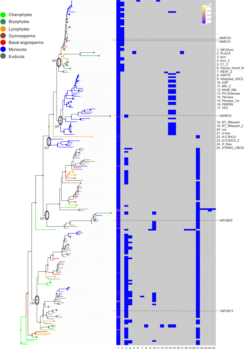
Since MCA is a multidomain protein, we studied the phylogenetic relationships of the domains MCAfunc and PLAC8 separately. In the MCAfunc domain Maximum Likelihood (ML) tree of 217 domain sequences, the samples included clustered according to the presence of partner domains (Fig 1; S11 and S16 Appendices). For example, samples of charophytes and ‘Clade a’, that included AtPUB13 (RING-type E3 ubiquitin ligase), had U-box (PF04564.15), Arm (PF00514.23) or Arm_2 (PF04826.13) as partner domains to MCAfunc (Fig 1). Arm and Arm_2 are overlapping domains. ‘Clade a’ contained two major clades each including all streptophyte lineages, suggesting a gene duplication. ‘Clade b’ (Fig 1; S11 Appendix) also contained two main clades including most streptophyte lineages, suggesting a further duplication, where most proteins in one clade had lost the Arm domain. The following clades ‘c’ and ‘d’ contained mostly monocot-specific undescribed or potential protein kinase proteins (Fig 1) (e.g. rice Q2QZY3). ‘Clade e’, is the MCA protein clade including AtMCA1 and AtMCA2, where the majority of MCAfunc domain proteins were partnered with the PLAC8 domain, which suggested that MCA as the derived proteins. A few proteins scattered across ‘Clade e’ had lost PLAC8 (Fig 1E), but there always was at least one protein with MCAfunc plus PLAC8 present in each species (Table 1). Some MCA proteins had obtained an alternative partner domain such as C1_2 (PF03107.16) and PP2 (PF14299.6) (M. polymorpha), Pkinase (PF00069.25), or Pkinase-Tyr (PF07714.17) (M. acuminata) C1_2 and Mlh1_C (PF16413.5) (Z. mays).

Fig 1. ML tree of MCAfunc domains and their partner domains predicted by HMMER website searches.

Fig 1

Left: ML tree. (a) Clade associated with E3 ubiquitin ligase AtPUB13. (b) Clade associated with AtPUB45. (c) MCAfunc only proteins. (d) Clade associated with AtARO3 and monocot U-box containing protein kinase like proteins. (e) MCA clade. Clades supported with α-LRT SH-like values > 0.8 indicated with circles at the nodes. Right: Domain individual E values (i.Evalue) resulting from HMMER website searches are shown as a heatmap. Absence of domains indicated in grey. The original tree and heatmap are shown in S11 and S16 Appendices. Numbers in legends to the right indicate the domain names (x-axis of the heatmap).

In Zea mays, A0A1D6PNG8 and A0A1D6F850 hold the protein name “MCA1” in UniProt, but they in ‘Clade d’ and also lacked PLAC8 but retained Pkinase or Pkinase_Tyr. On the other hand, CNR13 and A0AD6JP06 were found to be proper MCAs since they were in the MCA clade (‘Clade e’) and possessed PLAC8 (S11 and S16 Appendices), as previously reported [15].

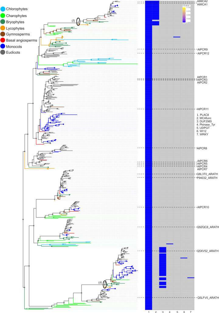
The phylogeny of the other MCA domain, PLAC8, was also examined phylogenetically. In the PLAC8 domain ML tree of 438 domain sequences, the samples also clustered according to their partner domains (Fig 2; S13 and S17 Appendices). Most of PLAC8 domain proteins appeared as single domain proteins, but the MCA clade (‘Clade I’) retained MCAfunc, while another clade (‘Clade II’) retained a DUF2985 (PF11204) domain (Fig 2; S13 and S17 Appendices) with unknown functions. In A. thaliana, PLAC8 single domain proteins are registered as “Plant Cadmium Resistance proteins (PCR)”, with the function to reduce cadmium uptake [42]. The MCA clade appeared to be closely related to a clade including AtPCR9 and AtPCR12. All proteins in Clade I’, except for two, retained MCAfunc and PLAC8. In gymnosperms, two proteins per species were found, with one having MCAfunc while the other lacked it. The ML tree topology and distribution of partner domains suggested that the coupling between MCAfunc and PLAC8 domains occurred once in the plant lineage, possibly in the common ancestor of embryophytes, and was sometimes lost after gene duplication events but was always retained in at least one copy.

Fig 2. PLAC8 domain tree and their partner domains.

Fig 2

Left: PLAC8 domain ML tree. (I) MCA ‘Clade I’. (II) ‘Clade II’ with proteins retaining DUF2985 + PLAC8. Clades supported with α-LRT SH-like support values > 0.8 indicated with circles at the node. Right: Domain combinations observed in the PLAC8 domain containing proteins associated with the ML tree (left). Domain individual E values (i.Evalue) resulting from HMMER website searches are shown as a heatmap. Absence of domains indicated in grey. The original tree and heatmap are shown in S13 and S17 Appendices. Numbers in legend to the right indicate the domain names (x-axis of the heatmap).

U-box and Arm are original partners of the MCAfunc domain

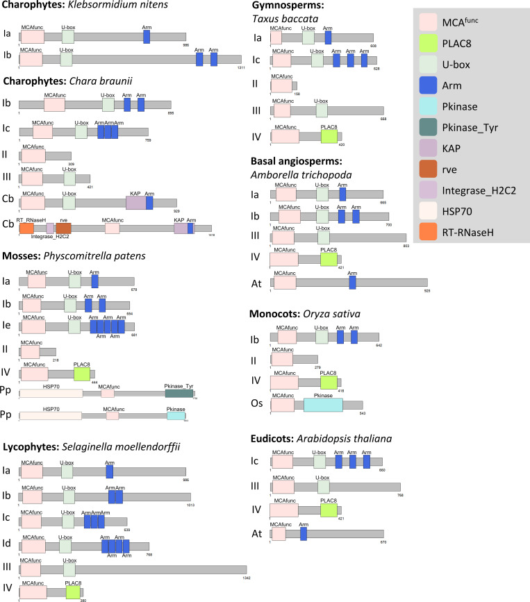
While the PLAC8 domain commonly existed within the plant, animal, and fungi kingdoms, the MCAfunc domain was only observed in streptophytes in the plant kingdom. Thus, the MCAfunc domain might be the key domain for the MCA protein, and we further assessed the coupling of the MCAfunc domain with its partner domains. The predicted domain combination for K. nitens (charophyte) was MCAfunc + U-box + Arm (type I) (Fig 3). Type I was found in all species, but the number of Arm domains varied from one to five. In C. braunii (charophyte), in addition to type I, MCAfunc only (type II) and MCAfunc + Ubox (type III) and lineage specific types (Fig 3Cb) were found. In P. patens (bryophyte-moss), type I, type II, the MCA type (MCAfunc + PLAC8: type IV), and also lineage specific types were found (Fig 3Pp). The proteome of ferns was not available, but type I to type IV, and lineage specific combinations were widely observed from lycophytes to angiosperms. The O. sativa MCAfunc + Pkinase (Os) type was widely present in angiosperm monocots, and in the moss P. patens, possessing an additional HSP70 domain (Pp). The A. thaliana MCAfunc + Arm: ARO3 (At) type was only observed in the angiosperms A. thaliana, Brassica oleracea, and Amborella trichopoda (Fig 3; Table 2).

Fig 3. Schematic illustration of protein domain combinations of the MCAfunc domain proteins in representative proteomes.

Fig 3

Type I, MCAfunc + U-box + Arm, number of Arm copies varies from 1 to 5 (Ia to Ie). Type II, MCAfunc only. Type III, MCAfunc + U-box. Type IV, MCAfunc + PLAC8. The well-studied MCA proteins in A. thaliana, AtMCA1 and AtMCA2, are type IV proteins. Lineage specific combinations are indicated by initials of the species, Cb, Pp, Os, At respectively.

Table 2. Domain partners of MCAfunc domain and their combinations found in proteomes across viridiplantae.

ID Taxon Domain combinations (Types)
I II III IV Os At sum
CHLRE Chlamydomonas reinhardtii 0 0 0 0 0 0 0
VOLCA Volvox carteri f. nagariensis 0 0 0 0 0 0 0
KLENI Klebsormidium nitens 2 0 0 0 0 0 2
CHABU Chara braunii 4 1 1 0 0 0 6
MARPO Marchantia polymorpha 4 1 1 1 0 0 7
MapoRu Marchantia polymorpha subsp. ruderalis 4 2 0 *1 0 0 7
PHYPA Physcomitrella patens 3 2 0 2 2 0 9
SELML Selaginella moellendorffii 12 0 3 2 0 0 17
CMI Cycas micholitzii 0 0 0 1 0 0 1
TBA Taxus baccata 3 2 1 1 0 0 7
AMBTC Amborella trichopoda 3 0 3 1 0 1 8
MUSAM Musa acuminata subsp. malaccensis 1 0 4 3 1 0 9
ORYSJ Oryza sativa subsp. japonica 1 14 0 1 7 0 23
MAIZE Zea mays 1 2 1 2 4 0 10
SORBI Sorghum bicolor 1 2 0 1 9 0 13
AQUCA Aquilegia coerulea 4 0 2 1 0 0 7
VITVI Vitis vinifera 3 0 3 1 0 0 7
POPTR Populus trichocarpa 6 0 2 2 0 0 10
MEDTR Medicago truncatula 4 0 2 2 0 0 8
CUCSA Cucumis sativus 2 1 0 1 0 0 4
GOSRA Gossypium raimondii 10 0 2 1 0 0 13
BRAOL Brassica oleracea var. oleracea 3 0 0 3 0 1 7
ARATH Arabidopsis thaliana 1 0 1 2 0 1 5
ERYGU Erythranthe guttata 3 1 3 2 0 0 9
SOLLC Solanum lycopersicum 4 0 1 1 0 0 6
sum 79 28 30 32 21 3

The result of HMMER searches of MCAfunc domain partners and their combinations are listed and arranged following the Tree of Life. The types of domain combinations are described as follows: Type I: MCAfunc + U-box + Arm/Arm_2, Type II: MCAfunc only, Type III: MCAfunc + U-box, Type IV: MCAfunc + PLAC8 (MCA protein type), Os (monocot type): MCAfunc + Pkinase/Pkinase_Tyr. -with HSP70, At (ARO3 type): MCAfunc + Arm

*—not found in the proteome but in the genome (see S3, S6S8 Appendices).

Full MCA protein phylogeny, duplication and diversification in land plants

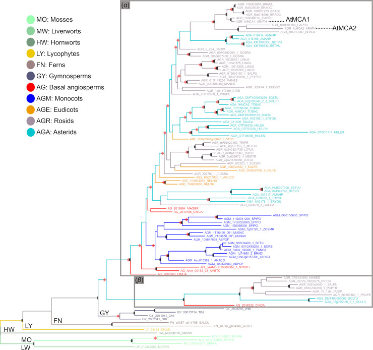
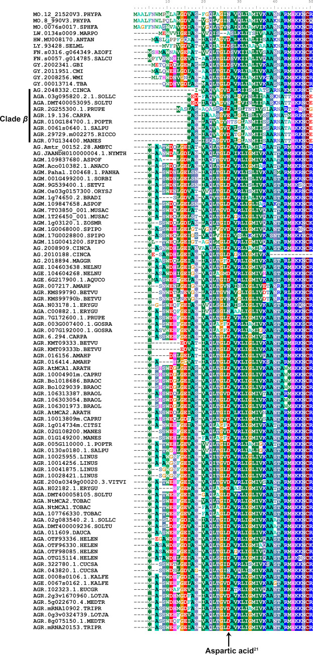
In order to unravel the history of MCA proteins in plants, a phylogeny of 106 full MCA proteins from 55 embryophyte species was reconstructed. The full MCA proteins include both MCAfunc and PLAC8 domain sequences. In this analysis, the basal grades in the ML tree, from bryophytes to gymnosperms followed the tree of life relationships (Fig 4). The angiosperm MCAs formed two clades. The majority of proteins fell in ‘Clade α’ including proteins of all angiosperm species analysed in this study. Only nine proteins formed ‘Clade β’, representing the orders Laureales (Cinnamomum camphora), Malpighiales, Rosales, Solanales and Brassicales (Carica papaya). These showed an MCA diversification and lacked approximately 10 aa in the N-terminal region (Fig 5). The predicted functional site of AtMCAs, the 21st position of aspartic acid (Asp21; Fig 5, arrow) [18], was different in mosses (asparagine) and liverworts (alanine). Hornworts, on the other hand, retained Asp21. At least one MCA per species retained Asp21 from lycophytes to angiosperms (Fig 5).

Fig 4. MCA protein ML tree, rooted on the proteins of bryophytes.

Fig 4

Proteins have diverged into two clades in angiosperms (‘α’, ‘β’). (α) Main MCA clade with all angiosperm species. (β) Diversified MCA clade showing variation at the N-terminus (see Fig 5). Black squares at the nodes indicate high values in both Phyml αLRT (> 0.7) and RaxML (> 70%) clade support analyses. Red diamonds at the nodes indicate Notung-inferred duplication events.

Fig 5. Conservation and variations in the N-terminus region of MCA proteins.

Fig 5

The gene IDs are listed in S3 Appendix. The functional site of the 21st position of Aspartic acid (Aspartic acid21, arrow) in AtMCA1 (“AGR.AtMCA1.ARATH” in the Fig 5) was well conserved within the MCA proteins, except in mosses and liverworts. Variation in the N-terminus region was observed within angiosperm species, while at least one MCA protein kept the Aspartic acid21. The angiosperm clade β MCA proteins lack approximately 10 amino acids in the N-terminal region.

A maximum of 39 duplication events were estimated across the ML tree, with two outside angiosperms (Fig 4, S18 Appendix). Three duplication events were inferred prior or at the point of diversification of angiosperms. Within the angiosperms, duplications were scattered among the lineages, but the superrosid clade stood out with an accumulation of six inferred duplications events. For several species repeated duplication events were inferred, e.g. three in Linum usitatissimum and two in Beta vulgaris and Helianthus annuus (S18 Appendix).

Discussion

The evolution of multidomain proteins can be complex, and may involve de novo domain evolution, recruitment of existing domains as partners, and recombination and domain losses [1]. In the present study where the evolution of the multidomain protein MCA was examined in detail, the results showed that it represents an example with a complex evolutionary history.

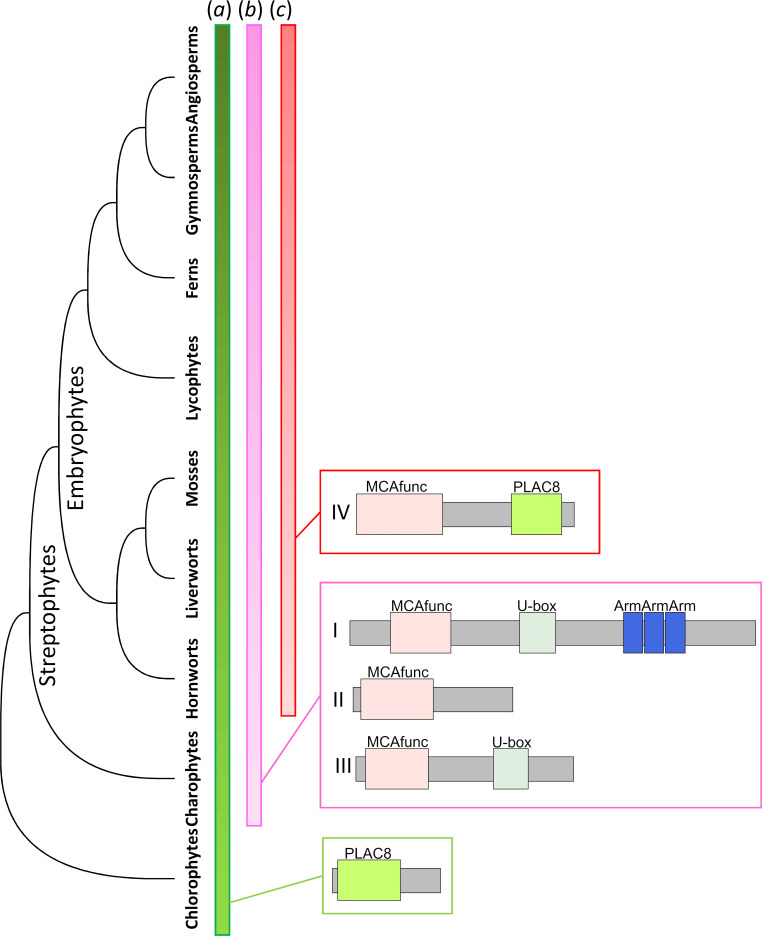
Our comprehensive proteome interrogation with profile HMMs suggested that the MCAfunc domain [18], formerly subscribed as ARPK domain plus EF hand-like [7], is a well conserved domain among plants. Accordingly, MCA can be described as a multidomain protein composed of the MCAfunc and the PLAC8 domains. PLAC8 is widely observed in eukaryotes as seen in our profile HMM searches, in which we found it in all proteomes we examined (Table 1). The MCAfunc domain, on the other hand, was streptophyte-specific and not found in chlorophytes, suggesting that the domain originated in the common ancestor of streptophytes, i.e. charophytes plus embryophytes (Fig 6) [4].

Fig 6. Schematic illustration of the MCA protein evolution.

Fig 6

PLAC8 domain genes were observed from chlorophytes to angiosperms, and many were single domain proteins (a). In the common ancestor of streptophytes, type I, type II, and type III domain combinations evolved (b). In the common ancestor of land plants, i.e. embryophytes, type IV domain combinations (MCA type, i.e. MCAfunc + PLAC8) evolved (c). In addition to the commonly observed types I-IV, lineage specific combinations were also observed.

The MCAfunc evolution further included domain recruitment, recombination and losses. The E3 ubiquitin ligase-type proteins (type I in Fig 3) found in charophytes, represent an ancestral combination (Table 1). Type I proteins were found in most streptophytes, except Cycas micholitzii possibly due to the incompleteness of its proteome. The ancestral charophyte K. nitens retained only the type I, while in the more derived C. braunii, MCAfunc obtained different partner domains or lost them all (Fig 3). Although domain-losses need to be seen with caution in some species included here due to their proteome incompleteness, single MCAfunc domain proteins were also observed in well-assembled genomes such as P. patens and O. sativa, supporting the existence of single-domain MCAfunc proteins (Fig 1 and Fig 3). Lineage-specific domain combinations were also observed in angiosperms, such as the Os- and At-types. Intriguingly, the At-type was only found in Brassicaceae and A. trichopoda, but perhaps due to unrelated parallel evolutionary events (Table 1).

A key event for the MCA evolution seemed to be the partnering of MCAfunc and PLAC8 first recruited in the common ancestor of embryophytes. MCA is seemingly streptophyte-specific and might play some basic roles, perhaps as a mechanosensor, for habit expansion to terra firma [9]. A previous study indicated that the Asp21 of MCAfunc domain is crucial for Ca2+ uptake [18]. Since Asp21 is diversified in mosses and liverworts, it could be hypothesized that their MCAs do not have Ca2+ uptake function. In angiosperms, MCA diverged into two clades and one might have changed functions from proper MCA. However, further studies would be needed to support these hypotheses.

Intriguingly, the E3 ubiquitin ligase type, with MCAfunc + U-box + Arm, seems to represent the most ancient MCAfunc protein (Fig 1). E3 ubiquitin ligase mediate substrate specificity for ubiquitylation [43] and is a large protein family. In the E3 ubiquitin ligases of A. thaliana, only plant U-Box13 (PUB13) and PUB45 retained MCAfunc domains (Fig 1; S11 and S16 Appendices). PUB13 was suggested to be involved in the abscisic acid signalling pathway, flowering time, and abiotic stress resistance [44]. The expression level of PUB45 seemed to be affected by nutrients [45]. It could be postulated that the MCAfunc domain was first utilized for these E3 ubiquitin ligases for roles for environmental adaptation, though further studies are required here. In addition, there are proteins only retaining the MCAfunc domain, but their function is not yet reported and remains unknown (S7 Appendix).

The PLAC8 domain exists in the Plant Cadmium Resistant (PCR) protein family as single domain proteins. PCRs are possibly transmembrane proteins and have roles in cadmium resistance [42] and zinc transport (PCR2; [46]). It is possible that the MCAfunc domain, initially part of E3 ubiquitin ligase, and PLAC8, an ion transporter, combined at some point in time and resulted in a novel protein, MCA, as a mechanosensor reacting to environmental calcium ions [11, 14]. The sequences between MCA and other plant mechanosensitive channels, such as MSL, are different [9], and the evolutionary history of MCA is different from that of MSL, which has originated in prokaryotes [47], and may represent an example of convergence in function.

Conclusions

In conclusion, MCA is an example of a multidomain protein, whose MCAfunc domain emerged de novo in the ancestor of streptophytes, and recruited an existing domain PLAC8 in the ancestor of embryophytes. The full MCA protein further duplicated and diversified during the evolution of land plants, involving recombination and losses of domains. However, each streptophyte species analysed had at least one complete full MCA copy, pointing to the importance of the protein. The functions of many MCA proteins are not investigated yet but they appear somewhat related to environment sensing, protein-protein interactions, and ion transport. In the basal lineage of streptophytes, i.e. charophytes, the MCAfunc domain is associated with U-box and Arm domains, supposed to play roles in the E3 ubiquitin ligase pathway. On the other hand, MCA proteins with MCAfunc and PLAC8 domains show quite different roles in ion transport. This further supports a hypothesis where domain swapping is an efficient mechanism to increase protein numbers with diversified functions during organismal evolution. Future studies will shed more light on the roles of these proteins and their interactions in relation to land plant evolution.

Supporting information

S1 Appendix. Proposed domain structure of MCA.

(a) For the MCA protein, the ARPK domain, at the N-terminus, and PLAC8, at the C-terminus, was proposed previously, and it was shown that MCA has an EF hand-like and a coiled-coil region [7]. (b) The biological function of MCA proteins was tested. As a result, the MCA functional domain at the N-terminus, which was previously described as ARPK domain and part of the EF hand-like region, was proposed [18]. This domain was analyzed in the present study and has been registered as MCAfunc domain in the Pfam database.

(PDF)

S2 Appendix. List of proteomes used in this study.

(PDF)

S3 Appendix. List of genome and transcriptome databases interrogated in this study, and the list of MCA genes included in the MCA phylogenetic analyses.

(PDF)

S4 Appendix. Profile HMM logo of the full MCA protein, PTHR46604:SF3.

(PDF)

S5 Appendix. Profile HMM logos of domains in the MCA protein.

(a) MCAfunc.hmm generated in this study, (b) PLAC8.hmm (PF04749).

(PDF)

S6 Appendix. MCA genes found in the genome of M. polymorpha subsp. ruderalis.

Top: M. polymorpha cDNA 0134s0009.1, middle: M. polymorpha genome, bottom: M. polymorpha subsp. ruderalis genome.

(PDF)

S7 Appendix. MCAfunc domain genes retrieved from proteomes.

(PDF)

S8 Appendix. PLAC8 domain genes retrieved from proteomes.

(PDF)

S9 Appendix. Species tree used for Notung rooting analyses of domain trees.

(PDF)

S10 Appendix. Result of the Notung rooting analysis of the MCAfunc domain tree.

The possible root position is marked in red (arrow).

(PDF)

S11 Appendix. MCAfunc domain ML tree.

(a) Clade associated with E3 ubiquitin ligase AtPUB13. (b) Clade associated with AtPUB45. (c) MCAfunc only proteins. (d) Clade associated with AtARO3 and monocot U-box containing protein kinase like proteins. (e) MCA clade associated with AtMCA1 and AtMCA2.

(PDF)

S12 Appendix. Result of the Notung rooting analysis of the PLAC8 domain tree.

Multiple branches show equally strong estimates as possible root positions (marked in red).

(PDF)

S13 Appendix. PLAC8 domain ML tree.

(I) MCA clade (II) Clade with proteins retaining DUF2985 + PLAC8.

(PDF)

S14 Appendix. Notung analyses of the MCA tree.

(a) Results of the Notung rooting analyses of the MCA tree. Possible root position is marked in red (arrow). (b) Results of Notung rearrangement of the MCA tree. Rearranged branches are marked in yellow. D: inferred duplication.

(PDF)

S15 Appendix. Species tree used for Notung analyses for full MCA protein sequence tree.

(PDF)

S16 Appendix. Domain partners of MCAfunc domain.

Domain partners observed in the MCAfunc domain containing proteins associated with the tree shown in Fig 1 (left). Domain individual E values (i.Evalue) resulting from HMMER website searches are shown as heatmap (right). Absence of domains indicated in grey.

(PDF)

S17 Appendix. Domain partners of PLAC8 domain.

Domain partners observed in the PLAC8 domain containing proteins associated with the ML tree (left). Domain individual E values (i.Evalue) resulting from HMMER website searches are shown as heatmap (right). Absence of domains indicated in grey.

(PDF)

S18 Appendix. MCA gene duplication events estimated by the Notung analysis.

“D” indicates inferred gene duplication events.

(PDF)

S19 Appendix. The seed alignment and profile HMM of MCAfunc domain.

(ZIP)

Acknowledgments

The authors are indebted to Dannie Durand for helpful comments and discussions pertaining to this study, particularly relating to the Notung rooting analyses. The authors also thank Daniel Barker and Frank Wright for helpful discussions. KN is grateful to the following persons for facilitating research associateships, to Pete Hollingsworth and Mark Newman at the Royal Botanic Garden Edinburgh (RBGE), UK, and to Akitoshi Iwamoto at Kanagawa University, Japan. This work was logistically supported by RBGE’s Science and ICT divisions. We also thank Duncan Reddish and Catherine Kidner for facilitating RBGE Linux server access and support, and Iain Milne for organizing access to the CropDiversity server, James Hutton Institute, Dundee, UK. We acknowledge the National Institute of Genetics, Japan, for allowing the use of their NIG-supercomputer system. We thank three anonymous reviewers and the editor for their constructive comments.

Data Availability

R codes used in this study are published in protocol.io. [dx.doi.org/10.17504/protocols.io.bkqwkvxe] Gene trees and their alignment are available from TreeBASE. [http://purl.org/phylo/treebase/phylows/study/TB2:S26880] The seed alignment of the MCAfunc domain is available in the new March 2021 release, v. 34.0, of Pfam [http://pfam.xfam.org/family/PF19584#tabview=tab0], as well as in the S19 Appendix of this study.

Funding Statement

This work was supported by the Japan Society for the Promotion of Science (JSPS) [KAKENHI Grant Number 25120708], Ministry of Education, Culture, Sports, Science & Technology of Japan, to HI. KN’s stay at RBGE is financially supported by the Edinburgh Botanic Garden Sibbald Trust [2018#18], JSPS [JSPS KAKENHI Grant Number 18K06375], and the Sumitomo Foundation [170204].

References

  • 1.Buljan M, Bateman A. The evolution of protein domain families. Biochem Soc Trans. 2009; 37: 751–755. 10.1042/BST0370751 [DOI] [PubMed] [Google Scholar]
  • 2.Bagowski CP, Bruins W, Te Velthuis AJ. The nature of protein domain evolution: shaping the interaction network. Curr Genomics. 2010; 11: 368–376. 10.2174/138920210791616725 [DOI] [PMC free article] [PubMed] [Google Scholar]
  • 3.El-Gebali S, Mistry J, Bateman A, Eddy SR, Luciani A, Potter SC, et al. The Pfam protein families database in 2019. Nucl Acids Res. 2019; 47: D427–D432. 10.1093/nar/gky995 [DOI] [PMC free article] [PubMed] [Google Scholar]
  • 4.Kersting AR, Bornberg-Bauer E, Moore AD, Grath S. Dynamics and adaptive benefits of protein domain emergence and arrangements during plant genome evolution. Genome Biol Evol. 2012; 4: 316–329. 10.1093/gbe/evs004 [DOI] [PMC free article] [PubMed] [Google Scholar]
  • 5.Zhang XC, Wang Z, Zhang X, Le MH, Sun J, Xu D, et al. Evolutionary dynamics of protein domain architecture in plants. BMC Evol Biol. 2012; 12: 6. 10.1186/1471-2148-12-6 [DOI] [PMC free article] [PubMed] [Google Scholar]
  • 6.Basu D, Haswell ES. Plant mechanosensitive ion channels: an ocean of possibilities. Curr Opin Plant Biol. 2017; 40: 43–48. 10.1016/j.pbi.2017.07.002 [DOI] [PMC free article] [PubMed] [Google Scholar]
  • 7.Nakagawa Y, Katagiri T, Shinozaki K, Qi Z, Tatsumi H, Furuichi T, et al. Arabidopsis plasma membrane protein crucial for Ca2+ influx and touch sensing in roots. Proc Natl Acad Sci USA. 2007; 104: 3639–3644. 10.1073/pnas.0607703104 [DOI] [PMC free article] [PubMed] [Google Scholar]
  • 8.Furuichi T, Iida H, Sokabe M, Tatsumi H. Expression of Arabidopsis MCA1 enhanced mechanosensitive channel activity in the Xenopus laevis oocyte plasma membrane. Plant Signal Behav. 2012; 7: 1022–1026. 10.4161/psb.20783 [DOI] [PMC free article] [PubMed] [Google Scholar]
  • 9.Kurusu T, Kuchitsu K, Nakano M, Nakayama Y, Iida H. Plant mechanosensing and Ca2+ transport. Trends Plant Sci. 2013; 18: 227–233. 10.1016/j.tplants.2012.12.002 [DOI] [PubMed] [Google Scholar]
  • 10.Yamanaka T, Nakagawa Y, Mori K, Nakano M, Imamura T, Kataoka H, et al. MCA1 and MCA2 that mediate Ca2+ uptake have distinct and overlapping roles in Arabidopsis. Plant Physiol. 2010; 152: 1284–1296. 10.1104/pp.109.147371 [DOI] [PMC free article] [PubMed] [Google Scholar]
  • 11.Kurusu T, Nishikawa D, Yamazaki Y, Gotoh M, Nakano M, Hamada H, et al. Plasma membrane protein OsMCA1 is involved in regulation of hypo-osmotic shock-induced Ca2+ influx and modulates generation of reactive oxygen species in cultured rice cells. BMC Plant Biol. 2012. a; 12: 11. 10.1186/1471-2229-12-11 [DOI] [PMC free article] [PubMed] [Google Scholar]
  • 12.Liu Z, Cheng Q, Sun Y, Dai H, Song G, Guo Z, et al. A SNP in OsMCA1 responding for a plant architecture defect by deactivation of bioactive GA in rice. Plant Mol Biol. 2015; 87: 17–30. 10.1007/s11103-014-0257-y [DOI] [PubMed] [Google Scholar]
  • 13.Liang J, He Y, Zhang Q, Wang W, Zhang Z. Plasma membrane Ca2+ permeable mechanosensitive channel OsDMT1 is involved in regulation of plant architecture and ion homeostasis in rice. Int J Mol Sci. 2020; 21: 1097. 10.3390/ijms21031097 [DOI] [PMC free article] [PubMed] [Google Scholar]
  • 14.Kurusu T, Yamanaka T, Nakano M, Takiguchi A, Ogasawara Y, Hayashi T, et al. Involvement of the putative Ca2+-permeable mechanosensitive channels, NtMCA1 and NtMCA2, in Ca2+ uptake, Ca2+-dependent cell proliferation and mechanical stress-induced gene expression in tobacco (Nicotiana tabacum) BY-2 cells. J Plant Res. 2012b; 125: 555–568. 10.1007/s10265-011-0462-6 [DOI] [PubMed] [Google Scholar]
  • 15.Rosa M, Abraham-Juárez MJ, Lewis MW, Fonseca JP, Tian W, Ramirez V, et al. 2017. The maize MID-COMPLEMENTING ACTIVITY homolog CELL NUMBER REGULATOR13/NARROW ODD DWARF coordinates organ growth and tissue patterning. Plant Cell 2017; 29: 474–490. 10.1105/tpc.16.00878 [DOI] [PMC free article] [PubMed] [Google Scholar]
  • 16.Ohnishi M, Kadohama N, Suzuki Y, Kajiyama T, Shichijo C, Ishizaki K, et al. Involvement of Ca2+ in vacuole degradation caused by a rapid temperature decrease in Saintpaulia palisade cells: a case of gene expression analysis in a specialized small tissue. Plant Cell Physiol. 2015; 56: 1297–1305. 10.1093/pcp/pcv048 [DOI] [PubMed] [Google Scholar]
  • 17.Nishii K, Hughes M, Briggs M, Haston E, Christie F, DeVilliers MJ, et al. Streptocarpus redefined to include all Afro-Malagasy Gesneriaceae: Molecular phylogenies prove congruent with geography and cytology and uncovers remarkable morphological homoplasies. Taxon 2015; 64: 1243–1274. 10.12705/646.8 [DOI] [Google Scholar]
  • 18.Nakano M, Iida K, Nyunoya H, Iida H. Determination of structural regions important for Ca2+ uptake activity in Arabidopsis MCA1 and MCA2 expressed in yeast. Plant Cell Physiol. 2011; 52: 1915–1930. 10.1093/pcp/pcr131 [DOI] [PubMed] [Google Scholar]
  • 19.Leliaert F, Smith DR, Moreau H, Herron MD, Verbruggen H, Delwiche CF, et al. Phylogeny and molecular evolution of the green algae. Critical Rev Plant Sci 2012; 31: 1–46. 10.1080/07352689.2011.615705 [DOI] [Google Scholar]
  • 20.Kanehisa M, Sato Y, Furumichi M, Morishima K, Tanabe M. New approach for understanding genome variations in KEGG. Nucl Acids Res. 2019; 47: D590–D595. 10.1093/nar/gky962 [DOI] [PMC free article] [PubMed] [Google Scholar]
  • 21.Leebens-Mack JH, Barker MS, Carpenter EJ, Deyholos MK, et al. One thousand plant transcriptomes and the phylogenomics of green plants. Nature 2019; 574: 679–685. 10.1038/s41586-019-1693-2 [DOI] [PMC free article] [PubMed] [Google Scholar]
  • 22.Medina R, Johnson MG, Liu Y, Wickett NJ, Shaw AJ, Goffinet B. Phylogenomic delineation of Physcomitrium (Bryophyta: Funariaceae) based on targeted sequencing of nuclear exons and their flanking regions rejects the retention of Physcomitrella, Physcomitridium and Aphanorrhegma. J Syst Evol. 2019; 57: 404–417. 10.1111/jse.12516 [DOI] [Google Scholar]
  • 23.Simão FA, Waterhouse RM, Ioannidis P, Kriventseva EV, Zdobnov EM. BUSCO: assessing genome assembly and annotation completeness with single-copy orthologs. Bioinformatics 2015; 31: 3210–3212. 10.1093/bioinformatics/btv351 [DOI] [PubMed] [Google Scholar]
  • 24.Hall TA. BioEdit: a user-friendly biological sequence alignment editor and analysis program for Windows 95/98/NT. Nucl Acid S. 1999; 41: 95–98. 10.1021/bk-1999-0734.ch008 [DOI] [Google Scholar]
  • 25.Criscuolo A, Gribaldo S. BMGE (Block Mapping and Gathering with Entropy): a new software for selection of phylogenetic informative regions from multiple sequence alignments. BMC Evol Biol. 2010; 10: 210. 10.1186/1471-2148-10-210 [DOI] [PMC free article] [PubMed] [Google Scholar]
  • 26.Guindon S, Dufayard JF, Lefort V, Anisimova M, Hordijk W, Gascuel O. New algorithms and methods to estimate maximum-likelihood phylogenies: assessing the performance of PhyML 3.0. Syst Biol. 2010; 59: 307–321. 10.1093/sysbio/syq010 [DOI] [PubMed] [Google Scholar]
  • 27.Lefort V, Longueville J-E, Gascuel O. SMS: Smart Model Selection in PhyML." Mol Biol Evol. 2017; 34: 2422–2424. 10.1093/molbev/msx149 [DOI] [PMC free article] [PubMed] [Google Scholar]
  • 28.Stamatakis A RAxML version 8: a tool for phylogenetic analysis and post-analysis of large phylogenies. Bioinformatics. 2014; 30: 1312–1313. 10.1093/bioinformatics/btu033 [DOI] [PMC free article] [PubMed] [Google Scholar]
  • 29.Milne I, Lindner D, Bayer M, Husmeier D, McGuire G, Marshall DF, et al. TOPALi v2: a rich graphical interface for evolutionary analyses of multiple alignments on HPC clusters and multi-core desktops. Bioinformatics. 2009; 25: 126–127. 10.1093/bioinformatics/btn575 [DOI] [PMC free article] [PubMed] [Google Scholar]
  • 30.Chen K, Durand D, Farach-Colton M. NOTUNG: a program for dating gene duplications and optimizing gene family trees. J Comput Biol. 2000; 7: 429–447. 10.1089/106652700750050871 [DOI] [PubMed] [Google Scholar]
  • 31.Stevens, P. F. 2001 onwards. Angiosperm Phylogeny Website. Version 14, July 2017 [and more or less continuously updated since] Available from: http://www.mobot.org/MOBOT/research/APweb/
  • 32.Puttick MN, Morris JL, Williams TA, Cox CJ, Edwards D, Kenrick P, et al. The interrelationships of land plants and the nature of the ancestral embryophyte. Curr Biol. 2018; 28: 733–745 e732. 10.1016/j.cub.2018.01.063 [DOI] [PubMed] [Google Scholar]
  • 33.R Core Team. R: A language and environment for statistical computing. R Foundation for Statistical Computing, Vienna, Austria. 2020. Available from: https://www.R-project.org/ [Google Scholar]
  • 34.Wickham H. ggplot2: elegant graphics for data analyses. Springer-Varlag; New York. 2016. Available from: https://ggplot2.tidyverse.org [Google Scholar]
  • 35.Yu G-C, Smith D, Zhu H, Guan Y, Tommy Lam T T-Y. ggtree: an R package for visualization and annotation of phylogenetic trees with their covariates and other associated data. Methods Ecol Evol. 2017; 8: 28–36. 10.1111/2041-210X.12628 [DOI] [Google Scholar]
  • 36.Paradis E, Schliep K. ape 5.0: an environment for modern phylogenetics and evolutionary analyses in R. Bioinformatics. 2019; 35: 526–528. 10.1093/bioinformatics/bty633 [DOI] [PubMed] [Google Scholar]
  • 37.Revell LJ. phytools: An R package for phylogenetic comparative biology (and other things). Methods Ecol Evol. 2012; 3: 217–223. 10.1111/j.2041-210X.2011.00169.x [DOI] [Google Scholar]
  • 38.Edger PP, Jocelyn C. Hall JC, Harkess A, Tang M, Coombs J, Mohammadin S, et al. Brassicales phylogeny inferred from 72 plastid genes: A reanalysis of the phylogenetic localization of two paleopolyploid events and origin of novel chemical defenses. Amer J Bot. 2017; 105: 463–469. 10.1002/ajb2.1040 [DOI] [PubMed] [Google Scholar]
  • 39.Nikolov LA, Shushkov P, Nevado B, Gan X, Al-Shehbaz IA, Filatov D, et al. Resolving the backbone of the Brassicaceae phylogeny for investigating trait diversity. New Phytol. 2019; 222: 1638–1651. 10.1111/nph.15732 [DOI] [PubMed] [Google Scholar]
  • 40.Choi IS, Ruhlman TA, Jansen RK. Comparative mitogenome analysis of the genus Trifolium reveals independent gene fission of ccmFn and intracellular gene transfers in Fabaceae. Int J Mol Sci. 2020; 21: 6. 10.3390/ijms21061959 [DOI] [PMC free article] [PubMed] [Google Scholar]
  • 41.Saarela JM, Burke SV, Wysocki WP, Barrett MD, Clark LG, Craine JM, et al. A 250 plastome phylogeny of the grass family (Poaceae): topological support under different data partitions. PeerJ. 2018; 6: e4299. 10.7717/peerj.4299 [DOI] [PMC free article] [PubMed] [Google Scholar]
  • 42.Song WY, Martinoia E, Lee J, Kim D, Kim DY, Vogt E, et al. A novel family of cys-rich membrane proteins mediates cadmium resistance in Arabidopsis. Plant Physiol. 2004; 135: 1027–1039. 10.1104/pp.103.037739 [DOI] [PMC free article] [PubMed] [Google Scholar]
  • 43.Metzger MB, Pruneda JN, Klevit RE, Weissman AM. RING-type E3 ligases: master manipulators of E2 ubiquitin-conjugating enzymes and ubiquitination. Biochim Biophys Acta. 2014; 1843: 47–60. 10.1016/j.bbamcr.2013.05.026 [DOI] [PMC free article] [PubMed] [Google Scholar]
  • 44.Liao D, Cao Y, Sun X, Espinoza C, Nguyen CT, Liang Y, et al. Arabidopsis E3 ubiquitin ligase PLANT U-BOX13 (PUB13) regulates chitin receptor LYSIN MOTIF RECEPTOR KINASE5 (LYK5) protein abundance. New Phytol. 2017; 214: 1646–1656. 10.1111/nph.14472 [DOI] [PubMed] [Google Scholar]
  • 45.Pradhan M. Understanding the regulatory basis of defense signalling in plants: The role of Argonautes in modulating defense responses in Nicotiana attenuate. PhD Dissertation Friedrich-Schiller- Universität Jena. 2019. [Google Scholar]
  • 46.Song WY, Choi KS, Kim DY, Geisler M, Park J, Vincenzetti V, et al. Arabidopsis PCR2 is a zinc exporter involved in both zinc extrusion and long-distance zinc transport. Plant Cell 2010; 22: 2237–2252. 10.1105/tpc.109.070185 [DOI] [PMC free article] [PubMed] [Google Scholar]
  • 47.Cox CD, Nakayama Y, Nomura T, Martinac B. The evolutionary ‘tinkering’ of MscS-like channels: generation of structural and functional diversity. Pflugers Arch.–Eur. J. Physiol. 2014; 467: 3–13. 10.1007/s00424-014-1522-2 [DOI] [PubMed] [Google Scholar]

Decision Letter 0

Berthold Heinze

22 Jan 2021

PONE-D-20-36297

Mix and match: patchwork domain evolution of the land plant-specific Ca2+-permeable mechanosensitive channel MCA

PLOS ONE

Dear Dr. Nishii,

Thank you for submitting your manuscript to PLOS ONE. After careful consideration, we feel that it has merit but does not fully meet PLOS ONE’s publication criteria as it currently stands. Therefore, we invite you to submit a revised version of the manuscript that addresses the points raised during the review process.

This manuscript, and the general appraoch taken, has raised interest in the review team; we encourage you to submit a revision along the lines given further below.

Please submit your revised manuscript by Mar 08 2021 11:59PM. If you will need more time than this to complete your revisions, please reply to this message or contact the journal office at plosone@plos.org. When you're ready to submit your revision, log on to https://www.editorialmanager.com/pone/ and select the 'Submissions Needing Revision' folder to locate your manuscript file.

Please include the following items when submitting your revised manuscript:

  • A rebuttal letter that responds to each point raised by the academic editor and reviewer(s). You should upload this letter as a separate file labeled 'Response to Reviewers'.

  • A marked-up copy of your manuscript that highlights changes made to the original version. You should upload this as a separate file labeled 'Revised Manuscript with Track Changes'.

  • An unmarked version of your revised paper without tracked changes. You should upload this as a separate file labeled 'Manuscript'.

If you would like to make changes to your financial disclosure, please include your updated statement in your cover letter. Guidelines for resubmitting your figure files are available below the reviewer comments at the end of this letter.

If applicable, we recommend that you deposit your laboratory protocols in protocols.io to enhance the reproducibility of your results. Protocols.io assigns your protocol its own identifier (DOI) so that it can be cited independently in the future. For instructions see: http://journals.plos.org/plosone/s/submission-guidelines#loc-laboratory-protocols

We look forward to receiving your revised manuscript.

Kind regards,

Berthold Heinze

Academic Editor

PLOS ONE

Additional Editor Comments:

The mansucript was reviewed by three independent researchers, who all indicated their (high) interest in the approach and findings. We would therefore very much like to publish an improved version, which takes into account the suggestions of the reviewers. These suggestions are all very clear and to the point, so they should be relatively easy to address (or comment). I am recommending a 'major revision' only in order for the reviewers to have a chance to look at the improved version.

Journal Requirements:

When submitting your revision, we need you to address these additional requirements.

1. Please ensure that your manuscript meets PLOS ONE's style requirements, including those for file naming. The PLOS ONE style templates can be found at

https://journals.plos.org/plosone/s/file?id=wjVg/PLOSOne_formatting_sample_main_body.pdf and

https://journals.plos.org/plosone/s/file?id=ba62/PLOSOne_formatting_sample_title_authors_affiliations.pdf

2. Thank you for stating the following in the Acknowledgments Section of your manuscript:

"RBGE is supported by the Rural and Environment Science and Analytical Services Division (RESAS) in the Scottish Government."

We note that you have provided funding information that is not currently declared in your Funding Statement. However, funding information should not appear in the Acknowledgments section or other areas of your manuscript. We will only publish funding information present in the Funding Statement section of the online submission form.

Please remove any funding-related text from the manuscript and let us know how you would like to update your Funding Statement. Currently, your Funding Statement reads as follows:

"This work was supported by the Japan Society for the Promotion of Science (JSPS) [KAKENHI Grant Number 25120708] to HI, from the Ministry of Education, Culture, Sports, Science & Technology of Japan. KN’s stay at RBGE is financially supported by the Edinburgh Botanic Garden (Sibbald) Trust and the JSPS [JSPS KAKENHI Grant Number 18K06375], and the Sumitomo Foundation."

Please include your amended statements within your cover letter; we will change the online submission form on your behalf.

3. We note that you have stated that you will provide repository information for your data at acceptance. Should your manuscript be accepted for publication, we will hold it until you provide the relevant accession numbers or DOIs necessary to access your data. If you wish to make changes to your Data Availability statement, please describe these changes in your cover letter and we will update your Data Availability statement to reflect the information you provide.

4. Please upload a new copy of S6 and S17 Figures as the detail is not clear. Please follow the link for more information: https://blogs.plos.org/plos/2019/06/looking-good-tips-for-creating-your-plos-figures-graphics/

[Note: HTML markup is below. Please do not edit.]

Reviewers' comments:

Reviewer's Responses to Questions

Comments to the Author

1. Is the manuscript technically sound, and do the data support the conclusions?

The manuscript must describe a technically sound piece of scientific research with data that supports the conclusions. Experiments must have been conducted rigorously, with appropriate controls, replication, and sample sizes. The conclusions must be drawn appropriately based on the data presented.

Reviewer #1: Partly

Reviewer #2: Yes

Reviewer #3: Yes

**********

2. Has the statistical analysis been performed appropriately and rigorously?

Reviewer #1: Yes

Reviewer #2: N/A

Reviewer #3: Yes

**********

3. Have the authors made all data underlying the findings in their manuscript fully available?

The PLOS Data policy requires authors to make all data underlying the findings described in their manuscript fully available without restriction, with rare exception (please refer to the Data Availability Statement in the manuscript PDF file). The data should be provided as part of the manuscript or its supporting information, or deposited to a public repository. For example, in addition to summary statistics, the data points behind means, medians and variance measures should be available. If there are restrictions on publicly sharing data—e.g. participant privacy or use of data from a third party—those must be specified.

Reviewer #1: Yes

Reviewer #2: Yes

Reviewer #3: Yes

**********

4. Is the manuscript presented in an intelligible fashion and written in standard English?

PLOS ONE does not copyedit accepted manuscripts, so the language in submitted articles must be clear, correct, and unambiguous. Any typographical or grammatical errors should be corrected at revision, so please note any specific errors here.

Reviewer #1: Yes

Reviewer #2: No

Reviewer #3: Yes

**********

5. Review Comments to the Author

Please use the space provided to explain your answers to the questions above. You may also include additional comments for the author, including concerns about dual publication, research ethics, or publication ethics. (Please upload your review as an attachment if it exceeds 20,000 characters)

Reviewer #1: In this project the authors explored the evolution of the MCA protein, it’s two domains MCAfunc and PLAC8, and their partner domains across all viridiplantae. They used MCA protein sequences from 55 plant species from various sources in a maximum likelihood phylogenetic analysis of protein domains identified using profile HMMs built using the HMMER program to accomplish this. The authors used their data to explain the origin of MCAfunc, the coupling of MCAfunc and PLAC8, the potential role of duplication in the evolution of the MCA, and briefly to address the role of helper domains in this process. One implication of this study is that MCA may have been fundamental to the evolution of land plants.

I was very impressed with the robustness of the methodology in this work. I cannot imagine a better strategy for building phylogenetic trees or identifying protein domains using this data. I was also impressed with the writing: the abstract is a good summary of the work, the introduction frames the problem well and introduces relevant literature, the results are direct and data-centered yet tell an interesting story, and the discussion does a great job of explaining the results in the context of relevant literature. Although I believe this work is important and well-executed there are some issues with the manuscript itself that the authors need to address, which I have detailed below as either major or minor comments:

Major comment:

1. Major: In many cases the authors refer to a protein or domain evolving in a particular modern group based on their presence in that group as well as in more derived taxa. This is not appropriate without presenting further evidence. The data suggest that the domain or protein most likely arose in the ancestor of all these taxa, which may or may not be a member of the most ancestral group. If the authors know that the common ancestor was a member of the most ancestral modern taxon that evidence must be provided in the manuscript, otherwise the claim must be made only of the common ancestor and not the most ancestral modern taxon. Examples: A) MCAfunc is said to have appeared first in charophytes rather than the ancestor of all streptophytes (lines 29, 303, 348) B) The coupling between MCAfunc and PLAC8 is said to have occurred once, “possibly in the ancestor of bryophytes” (lines 239-242, 348). This should refer to the shared ancestor of all embryophytes. C) several instances in the figure 5 legend. I recommend the authors look for this throughout the paper in case I missed something.

Minor comments:

1. Minor: Some minor grammatical issues were noticed, not enough to be distracting. The authors should edit the full text to identify and resolve as many of these as possible.

2. Minor: The groups “streptophytes” and “chlorophytes” are used extensively here. These terms can be useful, but are sometimes used to mean different things in different manuscripts. Figure 5 provides a visual explanation, but it would be much more helpful if these terms were defined in the introduction to resolve any potential confusion while readers progress through the paper.

3. Minor: Authors use incorrect language when discussing the patterns identified using HMMs referring to them as “HMMs”, when they should be referred to as either “profile HMMs” or “profiles” in brief, not “HMMs”. I double checked this with the HMMER User’s Guide. This issue was noticed on lines 25/26, and 107-114 but may also be present in other places.

4. Minor: I recommend the authors remove the statement about AtMCA1 being expressed in hamster ovary cells on lines 64-66. The contextual difference between plant roots and hamster ovaries are enormous. I therefore found it distracting and I’m not convinced it added to the story, but if the authors disagree this is not something I would insist on.

5. Minor: Authors should expand on the acronym “SMS” on line 134

6. Minor: All figures were low resolution and difficult to read, before publication the authors should replace these with higher resolution versions.

Reviewer #2: During this study, the authors examine the domain evolutionary history of the multi-domain plant-specific mechanosensitive channel, MCA. They thoroughly examined the evolution and origin of both MCA domains MCAfunc and PLAC8, via extensive phylogenetic analysis, and determined that the coupling of both domains occurred via a single event. They reveal that the MCA channel has a complex evolutionary history, however show that at least one full copy of the protein is present in streptophytes, suggesting function of the channel is highly important. The findings presented show an impressive example of how domains can combine, resulting in new protein function and opens up avenues for further studies determining the role of MCA proteins in land plant evolution.

Overall, the study is detailed covering a wide range of plant species and performing extensive analysis to track the evolutionary history of MCA domains. The introduction and discussion sections are clearly written, leading the reader through the logic of the study and are accessible to a wide audience. However, the presentation of the results text is not as clearly written or easy to follow and should be amended prior to publication. As it stands, this section is not accessible to a wider audience.

The results section text lacks suitable explanation and does not provide a narrative leading the reader through the figures. For example the results text starts “In total, 217 proteins possessing…”, without prior explanation (aside from a brief mention in the introduction) as to which species were investigated or the tools by which the search was conducted. In general, this lack of description continues throughout the results text. The authors are recommended to edit the text so that each results section starts by introducing the reasoning behind obtaining the results in the upcoming figure and the brief method by how the results were obtained. The following section should lead from the one before in the text, rather than be stand alone.

Additionally, the presentation of Table 1 as it stands is unclear as the domain contribution types are not mentioned at all until the description of Figure 3. The authors should add a description of these earlier in the text with the presentation of the table, or consider separating this information to a second table which follows Figure 3.

Finally, Appendix S18 is referred significantly within the text and is useful to look at – the authors could consider including this as a main Figure to avoid the reader having to filter through the Supplementary Information.

Additional minor edits/suggestions:

- COMPLEMENTING is spelled incorrectly on line 24 of the abstract

- There is an extremely long sentence between lines 62-68 of the Introduction which should be separated into several sentences.

- In Line 106 of the Methods the authors should define HMM

- Table 1 legend title is vague and is not stand alone- suggest the authors refine this so it is more descriptive

- Suggest adding a column with species in Table 1.

- In line 200, the authors should define ML

- Suggest that the authors add the species to the description of Fig3 in lines 253-266. For example K. nitens (charophyte).

- Appendix S18 is referred to quite a lot within the text and is useful to look at – should this be included as a Figure in its own right to avoid the reader having to filter through the Supplementary Information?

- The resolution of S6 Appendix should be improved if possible- it is difficult to see the residue numbers

- S9 S15 Appendices- only mnemonics are used. Would recommend providing the full names in the legend as well.

- S16 and S17 Appendices- would suggest the authors list the domain partners on the right of the heatmap as in Figures 1 and 2, rather than at the bottom as they are small and hard to see.

Reviewer #3: In the manuscript “Mix and match: patchwork domain evolution of the land plant-specific Ca2+-permeable mechanosensitive channel MCA” the authors present an investigation of the MCA Ca2+ permeable mechanosensitive ion channel using proteomics and bioinformatics. Their data suggests that the MCA protein is composed of many different portions. This work found that the MCA region of a MCA protein is localized to the N-terminus and is ~170 amino acids.

Comments

In lines 188-198 you discuss the Table 1 and mention the different classes of the proteomes, can you include the different classes of the genomes in the table?

You utilize a completed and partial proteome for Marchantia polymorpha as two distinct proteomes, while there is a substantial information to be gained from comparing two different experimental conditions. Are the genes identified in the partial proteome the same as the genes identified in the more complete proteome? Unless there are significant differences between the two proteomes can you eliminate the partial from the data set. If there are substantial differences that are not accounted by the completeness of the proteome then please clarify this in the manuscript.

-As multiple proteomes are incomplete, can you provide the completeness of the proteomes within the manuscript, perhaps in table 1?

-You could remove any incomplete proteomes from the analysis as it is difficult to speculate as if a protein is not present in the data set or not present in the wild type cell.

If the MCA domain is ~420 amino acids (line 71), can you speculate the role of the Type II as they appear to be smaller than the required amount.

In the conclusion can you speculate the role of these proteins and how these are similar or different than the AtMCA1 and AtMCA2 (or other known functional proteins)

In general many of the supplemental figures and figures are blurry and have made some of the interpretation difficult. Many of them required significant magnification in order to see the content. Below you can find some specific suggestions

Figure 1: the clade designations are difficult to see, can you move them to the left on the figure or highlight the regions using specific colors to make it easier to follow?

Figure 3:

As the genes are arranged by species it is unclear if all of the Type Ib are the same, should they be the same?

Is there a difference between the two genes from P. patens?

Are the known MCAs, AtMCA1 and AtMCA2, shown in this graphic?

**********

6. PLOS authors have the option to publish the peer review history of their article (what does this mean?). If published, this will include your full peer review and any attached files.

If you choose “no”, your identity will remain anonymous but your review may still be made public.

Do you want your identity to be public for this peer review? For information about this choice, including consent withdrawal, please see our Privacy Policy.

Reviewer #1: No

Reviewer #2: No

Reviewer #3: No

[NOTE: If reviewer comments were submitted as an attachment file, they will be attached to this email and accessible via the submission site. Please log into your account, locate the manuscript record, and check for the action link "View Attachments". If this link does not appear, there are no attachment files.]

While revising your submission, please upload your figure files to the Preflight Analysis and Conversion Engine (PACE) digital diagnostic tool, https://pacev2.apexcovantage.com/. PACE helps ensure that figures meet PLOS requirements. To use PACE, you must first register as a user. Registration is free. Then, login and navigate to the UPLOAD tab, where you will find detailed instructions on how to use the tool. If you encounter any issues or have any questions when using PACE, please email PLOS at figures@plos.org. Please note that Supporting Information files do not need this step.

PLoS One. 2021 Apr 15;16(4):e0249735. doi: 10.1371/journal.pone.0249735.r002

Author response to Decision Letter 0


7 Feb 2021

PONE-D-20-36297

Mix and match: patchwork domain evolution of the land plant-specific Ca2+-permeable mechanosensitive channel MCA

Authors` response to the Editor`s general comments

Thank you very much for the supportive comments from the editor. We greatly appreciate the fact that the manuscript has received such a positive interest in our approach and our findings.

We have revised the manuscript following to the editor and reviewer`s comments as detailed below.

Editor`s comments

2. Thank you for stating the following in the Acknowledgments Section of your manuscript:

"RBGE is supported by the Rural and Environment Science and Analytical Services Division (RESAS) in the Scottish Government."

We note that you have provided funding information that is not currently declared in your Funding Statement. However, funding information should not appear in the Acknowledgments section or other areas of your manuscript. We will only publish funding information present in the Funding Statement section of the online submission form.

Please remove any funding-related text from the manuscript and let us know how you would like to update your Funding Statement. Currently, your Funding Statement reads as follows:

"This work was supported by the Japan Society for the Promotion of Science (JSPS) [KAKENHI Grant Number 25120708] to HI, from the Ministry of Education, Culture, Sports, Science & Technology of Japan. KN’s stay at RBGE is financially supported by the Edinburgh Botanic Garden (Sibbald) Trust and the JSPS [JSPS KAKENHI Grant Number 18K06375], and the Sumitomo Foundation."

Please include your amended statements within your cover letter; we will change the online submission form on your behalf.

Authors` response

Financial disclosure

"RBGE is supported by the Rural and Environment Science and Analytical Services Division (RESAS) in the Scottish Government."

This sentence is removed from the Acknowledgements and inserted in the Funding Statement section.

The amended Funding Statement is also included in the cover letter.

New Funding Statement is as follows;

"This work was supported by the Japan Society for the Promotion of Science (JSPS) [KAKENHI Grant Number 25120708] to HI, from the Ministry of Education, Culture, Sports, Science & Technology of Japan. KN’s stay at RBGE is financially supported by the Edinburgh Botanic Garden (Sibbald) Trust and the JSPS [JSPS KAKENHI Grant Number 18K06375], and the Sumitomo Foundation. RBGE is supported by the Rural and Environment Science and Analytical Services Division (RESAS) in the Scottish Government."

Editor`s comment

3. We note that you have stated that you will provide repository information for your data at acceptance. Should your manuscript be accepted for publication, we will hold it until you provide the relevant accession numbers or DOIs necessary to access your data. If you wish to make changes to your Data Availability statement, please describe these changes in your cover letter and we will update your Data Availability statement to reflect the information you provide.

Authors` response

The data submitted to Treebase and protocol.io are now released and available. The Pfam release v.34.0 is under the control of “Pfam” and the “European Bioinformatics Institute” and difficult to arrange individually. Thus we also include the seed alignment of the MCAfunc domain as supplemental data.

“R codes used in this study are published on protocol.io. [dx.doi.org/10.17504/protocols.io.bkqwkvxe]

Gene trees and their alignment are available from TreeBASE. [http://purl.org/phylo/treebase/phylows/study/TB2:S26880]

The seed alignment of the MCAfunc domain will be available in the next release of the Pfam database. [Pfam release v.34.0; PF19584] as well as the S19 Appendix of this study.”

Editor`s comment

4. Please upload a new copy of S6 and S17 Figures as the detail is not clear.

Authors` Comment

We provided new versions of figures S6 and S17 with higher resolution.

Reviewer 1

Authors` response to the Reviewer 1`s general comments

We like to thank the reviewer for the very positive comments on our work.

Reviewer 1`s comment

1. Major: In many cases the authors refer to a protein or domain evolving in a particular modern group based on their presence in that group as well as in more derived taxa. This is not appropriate without presenting further evidence. The data suggest that the domain or protein most likely arose in the ancestor of all these taxa, which may or may not be a member of the most ancestral group. If the authors know that the common ancestor was a member of the most ancestral modern taxon that evidence must be provided in the manuscript, otherwise the claim must be made only of the common ancestor and not the most ancestral modern taxon. Examples: A) MCAfunc is said to have appeared first in charophytes rather than the ancestor of all streptophytes (lines 29, 303, 348) B) The coupling between MCAfunc and PLAC8 is said to have occurred once, “possibly in the ancestor of bryophytes” (lines 239-242, 348). This should refer to the shared ancestor of all embryophytes. C) several instances in the figure 5 legend. I recommend the authors look for this throughout the paper in case I missed something.

Authors` response

Thank you very much for pointing out this evolutionary issue. We agree that MCAfunc domain might have arisen in the common ancestor of streptophytes, not within charophytes. We also agree that the MCA protein might have originated in the shared ancestor of embryophytes, rather than the ancestor of bryophytes. We have checked the MS and modified all relevant sentences as listed below:

Lines 28-29 [new lines 28-30]

Original text

“Maximum likelihood (ML) analyses revealed that the MCAfunc domain first appeared in E3 ubiquitin ligases-like proteins of charophytes.”

Revised text

“Maximum likelihood (ML) analyses showed that the MCAfunc domain observed in E3 ubiquitin ligases-like proteins of charophytes, suggesting the domain arose in the common ancestor of streptophytes.”

Lines 301-303 [new Lines 332-334]

Original text

“The MCAfunc domain, on the other hand, was streptophyte-specific and not found in chlorophytes, suggesting that the domain originated in charophytes (Fig 5) [4].”

Revised text

“The MCAfunc domain, on the other hand, was streptophyte-specific and not found in chlorophytes, suggesting that the domain originated in the common ancestor of streptophytes, i.e. charophytes plus embryophytes (Fig 6) [4].”

Lines 322-323 [new Lines 354-355]

Original text

“A key event for the MCA evolution seemed to be the partnering of MCAfunc and PLAC8 first recruited in bryophytes.”

Revised text

“A key event for the MCA evolution seemed to be the partnering of MCAfunc and PLAC8 first recruited in the common ancestor of embryophytes.”

Lines 347-348 [new Lines 381-383]

Original text

“In conclusion, MCA is an example of a multidomain protein, whose MCAfunc domain emerged de novo in charophytes, and recruited an existing domain PLAC8 in bryophytes.”

Revised text

“In conclusion, MCA is an example of a multidomain protein, whose MCAfunc domain emerged de novo in the common ancestor of streptophytes, and recruited an existing domain PLAC8 in the common ancestor of embryophytes.”

Lines 239-242 [new Lines 247-250]

Original text

“The ML tree topology and distribution of partner domains suggested that the coupling between MCAfunc and PLAC8 domains occurred once in the plant lineage, possibly in the ancestor of bryophytes, and was sometimes lost after gene duplication events but was always retained in at least one copy.”

Revised text

“The ML tree topology and distribution of partner domains suggested that the coupling between MCAfunc and PLAC8 domains occurred once in the plant lineage, possibly in the common ancestor of embryophytes, and was sometimes lost after gene duplication events but was always retained in at least one copy.”

Line 348

See above

Figure 5 legend

Original text

“Fig 5. Schematic illustration of MCA evolution. PLAC8 domain genes are observed from chlorophytes to angiosperms, and many are single domain proteins (a). In charophytes, type I, type II, and type III domain combinations evolved (b). In the ancestor of land plants, from bryophytes onward, type IV domain combinations (MCA type, i.e. MCAfunc + PLAC8) evolved (c). In addition to the commonly observed types I-IV, lineage specific combinations were also observed.”

Revised text [former Fig. 5 is now Fig. 6, new lines 336-341]

“Fig 6. Schematic illustration of MCA evolution. PLAC8 domain genes were observed from chlorophytes to angiosperms, and many were single domain proteins (a). In the common ancestor of streptophytes, type I, type II, and type III domain combinations evolved (b). In the common ancestor of land plants, i.e. embryophytes, type IV domain combinations (MCA type, i.e. MCAfunc + PLAC8) evolved (c). In addition to the commonly observed types I-IV, lineage specific combinations were also observed.”

Minor comments:

Reviewer 1`s minor comments

1. Minor: Some minor grammatical issues were noticed, not enough to be distracting. The authors should edit the full text to identify and resolve as many of these as possible.

Authors` response

We revised the manuscript and made the appropriate corrections.

Reviewer 1`s minor comments

2. Minor: The groups “streptophytes” and “chlorophytes” are used extensively here. These terms can be useful, but are sometimes used to mean different things in different manuscripts. Figure 5 provides a visual explanation, but it would be much more helpful if these terms were defined in the introduction to resolve any potential confusion while readers progress through the paper.

Authors` response

We added sentences for the definitions of terminology with references.

New Lines 87-91

“Here, for ranks, we followed the definition by Leliaert et al. [19] and NCBI Taxonomy Browser (https://www.ncbi.nlm.nih.gov/guide/taxonomy/), where viridiplantae include green algae (both of chlorophyte and charophyte) and streptophytes, streptophytes include charophyte algae and embryophytes, and embryophytes (also termed as “land plants”) include bryophytes, ferns, gymnosperms and angiosperms.”

Reviewer 1`s minor comments

3. Minor: Authors use incorrect language when discussing the patterns identified using HMMs referring to them as “HMMs”, when they should be referred to as either “profile HMMs” or “profiles” in brief, not “HMMs”. I double checked this with the HMMER User’s Guide. This issue was noticed on lines 25/26, and 107-114 but may also be present in other places.

Authors` response

Thank you very much for pointing out our error. We changed HMMs to profile HMMs throughout the manuscript and legends of figure and appendix.

Reviewer 1`s minor comments

4. Minor: I recommend the authors remove the statement about AtMCA1 being expressed in hamster ovary cells on lines 64-66. The contextual difference between plant roots and hamster ovaries are enormous. I therefore found it distracting and I’m not convinced it added to the story, but if the authors disagree this is not something I would insist on.

Authors` response

The sentence is now removed from lines 64-66

Reviewer 1`s minor comments

5. Minor: Authors should expand on the acronym “SMS” on line 134

Authors` response

The explanation for the acronym SMS has been added in line 134 (new line 139)

“Maximum likelihood (ML) analyses were carried out with PhyML v.3.0 [24] on the ATGC server (www.atgc-montpellier.fr), with Smart Model Selection (SMS) [25].”

Reviewer 1`s minor comments

6. Minor: All figures were low resolution and difficult to read, before publication the authors should replace these with higher resolution versions.

Authors` response

We provided high resolution images for all figures.

General comment from reviewer 2

Reviewer #2: During this study, the authors examine the domain evolutionary history of the multi-domain plant-specific mechanosensitive channel, MCA. They thoroughly examined the evolution and origin of both MCA domains MCAfunc and PLAC8, via extensive phylogenetic analysis, and determined that the coupling of both domains occurred via a single event. They reveal that the MCA channel has a complex evolutionary history, however show that at least one full copy of the protein is present in streptophytes, suggesting function of the channel is highly important. The findings presented show an impressive example of how domains can combine, resulting in new protein function and opens up avenues for further studies determining the role of MCA proteins in land plant evolution.

Overall, the study is detailed covering a wide range of plant species and performing extensive analysis to track the evolutionary history of MCA domains. The introduction and discussion sections are clearly written, leading the reader through the logic of the study and are accessible to a wide audience. However, the presentation of the results text is not as clearly written or easy to follow and should be amended prior to publication. As it stands, this section is not accessible to a wider audience.

Authors` response

Thank you very much for the supportive comment of our work. We revised the manuscript according to the reviewers` and editor`s comments.

Reviewer 2`s comment 1

The results section text lacks suitable explanation and does not provide a narrative leading the reader through the figures. For example, the results text starts “In total, 217 proteins possessing…”, without prior explanation (aside from a brief mention in the introduction) as to which species were investigated or the tools by which the search was conducted. In general, this lack of description continues throughout the results text. The authors are recommended to edit the text so that each results section starts by introducing the reasoning behind obtaining the results in the upcoming figure and the brief method by how the results were obtained. The following section should lead from the one before in the text, rather than be stand alone.

Authors` response

We added narrative sentences at the beginning of each result section.

The species list are in Tables and appendices, and thus we cited those files in the results. Since this study included a large number of species, we like to do this in a table format in Table 1. The details of tools used in this study were stated in the Materials and Method section but we also added short descriptions as follows.

New Lines 181-182

“To determine the distribution of MCA proteins in viridiplantae, 25 proteomes (see Table 1) were interrogated for domains of the MCA protein, MCAfunc and PLAC8, with profile HMMs using HMMER.”

New line 206-207

“Since MCA is a multidomain protein, we studied the phylogenetic relationships of the domains MCAfunc and PLAC8 separately.”

Line 238

“The phylogeny of the other MCA domain, PLAC8, was also examined phylogenetically.”

New lines 261-264

“While the PLAC8 domain commonly existed within the plant, animal, and fungi kingdoms, the MCAfunc domain was only observed in streptophytes in the plant kingdom. Thus, the MCAfunc domain might be the key domain for the MCA protein, and we further assessed the coupling of the MCAfunc domain with its partner domains.”

New lines 291-293

“In order to unravel the history of MCA proteins in plants, a phylogeny of full MCA proteins from 55 embryophyte species was reconstructed. The full MCA proteins include both MCAfunc and PLAC8 domain sequences.”

Reviewer2`s comment 2

Additionally, the presentation of Table 1 as it stands is unclear as the domain contribution types are not mentioned at all until the description of Figure 3. The authors should add a description of these earlier in the text with the presentation of the table, or consider separating this information to a second table which follows Figure 3.

Authors` response

Thank you for this suggestion. The old Table 1 is now split into two, Table 1 and Table 2. Table 2 is placed after Fig.3 for the ease of reading.

Reviewer2`s comment 3

Finally, Appendix S18 is referred significantly within the text and is useful to look at – the authors could consider including this as a main Figure to avoid the reader having to filter through the Supplementary Information.

Authors` response

The S18 Appendix now appears as Fig. 5. The Figure numbers were also amended accordingly.

Reviewer2`s minor comment

Additional minor edits/suggestions:

- COMPLEMENTING is spelled incorrectly on line 24 of the abstract

Authors` response

Thank you for pointing out this error. Now it is corrected.

Reviewer2`s minor comment

- There is an extremely long sentence between lines 62-68 of the Introduction which should be separated into several sentences.

Authors` response

The sentence in lines 62-68 (new lines 63 –70) are revised as follows.

“In A. thaliana, two paralogous MCA genes, AtMCA1 and AtMCA2 have been isolated, and their functions examined in great detail. The AtMCA1 protein is involved in touch sensing at the root tip and a hypoosmotic shock-induced increase in the cytosolic free Ca2+ concentration [7]. AtMCA2 was reported to participate in Ca2+ uptake at the roots [10]. In addition, AtMCA1 and AtMCA2 respond to membrane stretch to generate cation currents when expressed in Xenopus laevis oocytes [8]. Furthermore, MCA channels appear to have common functions in plants, based on studies on Oryza sativa OsMCA1 [11,12,13], Nicotiana tabacum NtMCA1, NtMCA2 [14], Zea mays CNR13 [15], and Streptocarpus MCA-like gene (as Saintpaulia in Ohnishi et al. [16]; see Nishii et al. [17]).”

Reviewer2`s minor comment

- In Line 106 of the Methods the authors should define HMM

Authors` response

The sub-header line 106 (new line 112) has been revised according to the reviewer`s comment and “HMM” is spelled out.

Reviewer2`s minor comment

- Table 1 legend title is vague and is not stand alone- suggest the authors refine this so it is more descriptive

Authors` response

Table 1 is now split to Table 1 and Table 2 following the Reviewer 2`s comment.

The legend of Table 1 has been revised as follows.

“Table 1. Number of proteins found in proteomes. Result of profile HMM searches of MCAfunc and PLAC8 domains in proteomes of 25 taxa across viridiplantae. Number of proteins retaining the MCAfunc or PLAC8 domains (E value < 10-3) are listed and arranged following the Tree of Life (see S7, and S8 Appendix).”

Reviewer2`s minor comment

- Suggest adding a column with species in Table 1.

Authors` response

We are slightly confused by this suggestion, as Table 1 has a “Taxon” column in which the species names are presented.

Reviewer2`s minor comment

- In line 200, the authors should define ML

Authors` response

We added Maximum Likelihood in line 200 (new line 207) following to the reviewer`s comment.

Reviewer2`s minor comment

- Suggest that the authors add the species to the description of Fig3 in lines 253-266. For example K. nitens (charophyte).

Authors` response

In lines 253-266 (new lines 261-279), we added the taxonomic groups to the species.

Reviewer2`s minor comment

- Appendix S18 is referred to quite a lot within the text and is useful to look at – should this be included as a Figure in its own right to avoid the reader having to filter through the Supplementary Information?

Authors` response

Please see above response. S18 Appendix is now as Fig. 5.

Reviewer2`s minor comment

- The resolution of S6 Appendix should be improved if possible- it is difficult to see the residue numbers

Authors` response

The S6 Appendix is now revised and with higher resolution.

Reviewer2`s minor comment

- S9 S15 Appendices- only mnemonics are used. Would recommend providing the full names in the legend as well.

Authors` response

The species` full name was added in the legends of S9 and S15 Appendix.

Reviewer2`s minor comment

- S16 and S17 Appendices- would suggest the authors list the domain partners on the right of the heatmap as in Figures 1 and 2, rather than at the bottom as they are small and hard to see.

Authors` response

Authors` response

We revised S16 and S17 Appendix following to the reviewer`s comment.

Reviewer #3

Reviewer #3: In the manuscript “Mix and match: patchwork domain evolution of the land plant-specific Ca2+-permeable mechanosensitive channel MCA” the authors present an investigation of the MCA Ca2+ permeable mechanosensitive ion channel using proteomics and bioinformatics. Their data suggests that the MCA protein is composed of many different portions. This work found that the MCA region of a MCA protein is localized to the N-terminus and is ~170 amino acids.

Reviewer 3`s comment

In lines 188-198 you discuss the Table 1 and mention the different classes of the proteomes, can you include the different classes of the genomes in the table?

Authors` response

The column “Vernacular name” has been added to Table 1 for the information of taxonomic classes.

Reviewer 3`s comment

You utilize a completed and partial proteome for Marchantia polymorpha as two distinct proteomes, while there is a substantial information to be gained from comparing two different experimental conditions. Are the genes identified in the partial proteome the same as the genes identified in the more complete proteome? Unless there are significant differences between the two proteomes can you eliminate the partial from the data set. If there are substantial differences that are not accounted by the completeness of the proteome then please clarify this in the manuscript.

Authors` response

Although the proteome completeness of Marchantia polymorpha subsp. ruderalis is relatively low, adding this gene to the matrix greatly stabilized the resulting trees. Compared to the well-studied angiosperms, there are only very few proteomes available for the basal groups such as bryophyte or lycophytes, but these are important to add. Therefore, we like to keep this proteome information and sequence in this study.

Reviewer 3`s comment

-As multiple proteomes are incomplete, can you provide the completeness of the proteomes within the manuscript, perhaps in table 1?

Authors` response

The proteome completeness information is added to S2 Appendix. The numbers of proteins registered in each proteomes and their BUSCO completeness were listed. The plaza database of gymnosperm proteomes does not have BUSCO information and thus the information is obtained by the authors. It is added to the materials and methods section.

New lines 107-110

“The proteome completeness information, i.e. BUSCO completeness values (BUSCO-C) were available for most taxa on the Uniprot database. The BUSCO-C values of proteomes from plaza database (Cycas micholitzii, Taxus baccata) were newly obtained in this study using BUSCO v.4.0.6 (Simao et al. 2015), by comparisons against viridiplantae_odb10 lineage datasets.”

Reviewer 3`s comment

-You could remove any incomplete proteomes from the analysis as it is difficult to speculate as if a protein is not present in the data set or not present in the wild type cell.

Authors` response

The proteomes used here represent the reference proteome for the species. Although many proteomes are not complete (most are between 90-99.8%), they are still regarded as the current reference proteomes data for wild type lineages. The phylogenetic placement of the worst complete genome, the gymnosperm Cycas micholitzii (BUSCO-C 36.3%) is consistent with the tree of life and the MCA domain evolution patterns we found. Removal of this and other incomplete proteomes would make the analyses unnecessarily unstable, and we like to keep them included (strictly taken, no proteome is complete, even at BUSCO comparison; see Table S2 Appendix).

Reviewer 3`s comment

If the MCA domain is ~420 amino acids (line 71), can you speculate the role of the Type II as they appear to be smaller than the required amount.

Authors` response

This is a slightly imprecise comment. To be exact, the MCA protein is approximately 420 amino acids in length and retains two domains (MCAfunc and PLAC8). The MCAfunc domain defined in this study is approximately 167 aa length (New line 117).

The Type II domain combination stands for proteins only retaining the MCAfunc domain (see S7 and S16 Appendix). The function of Type II domain genes is unknown in databases such as Uniprot. It would be too speculative to describe function without functional studies, which are much beyond the scope of our study.

We added the following sentence in the discussion section.

New Lines 368-370

“In addition, there are proteins only retaining the MCAfunc domain, but their function is not yet reported and remains unknown (S7 Appendix).”

Reviewer 3`s comment

In the conclusion can you speculate the role of these proteins and how these are similar or different than the AtMCA1 and AtMCA2 (or other known functional proteins)

Authors` response

There is a sentence in the conclusion. We felt that we need to await further studies to be reported to make conclusive statements. We added the following sentences.

New lines 387-392

“In the basal lineage of streptophytes, charophytes, the MCAfunc domain is associated with U-box and Arm domains, supposed to play roles in the E3 ubiquitin ligase pathway. On the other hand, MCA proteins with MCAfunc and PLAC8 domains show quite different roles in ion transport. This further supports a hypothesis where domain swapping is an efficient mechanism to increase protein numbers with diversified functions during organismal evolution.”

Reviewer 3`s comment

In general many of the supplemental figures and figures are blurry and have made some of the interpretation difficult. Many of them required significant magnification in order to see the content.

Authors` response

We revised all main figures and most supplemental figures, and they now have higher resolution and readable text.

Below you can find some specific suggestions

Reviewer 3`s comment

Figure 1: the clade designations are difficult to see, can you move them to the left on the figure or highlight the regions using specific colors to make it easier to follow?

Authors` response

We added open ellipses to better indicate the clades.

Reviewer 3`s comment

Figure 3:

As the genes are arranged by species it is unclear if all of the Type Ib are the same, should they be the same?

Authors` response

Type Ib share the same domain composition. They sometimes vary in the length of the sequence regions between the domains.

Reviewer 3`s comment

Is there a difference between the two genes from P. patens?

Authors` response

This comment is unclear. In P. patens, three, not two, MCA proteins were recognized. These are slightly different in their amino acid sequences. There are two P. patens specific gene types that vary in their Pkinase and Pkinase_Tyr domains at the 3’end (Fig. 3).

Reviewer 3`s comment

Are the known MCAs, AtMCA1 and AtMCA2, shown in this graphic?

Authors` response

Yes, they are both type IV shown in Fig 3 for A. thaliana, having only the MCAfunc and PLAC8 domains. We added this in Fig. 3 legend.

“The well-studied MCA proteins in A. thaliana, AtMCA1 and AtMCA2, are type IV proteins.”

Attachment

Submitted filename: Response_to_Reviewers_MCAr1.docx

Decision Letter 1

Berthold Heinze

11 Mar 2021

PONE-D-20-36297R1

Mix and match: patchwork domain evolution of the land plant-specific Ca2+-permeable mechanosensitive channel MCA

PLOS ONE

Dear Dr. Nishii,

Thank you for submitting your manuscript to PLOS ONE. After careful consideration, we feel that it has merit but does not fully meet PLOS ONE’s publication criteria as it currently stands. Therefore, we invite you to submit a revised version of the manuscript that addresses the points raised during the review process.

We request to take on the two remaining issues - a re-worded statement in the Abstract, and better-quality figures (they still appear blurry).

Please submit your revised manuscript by Apr 25 2021 11:59PM. If you will need more time than this to complete your revisions, please reply to this message or contact the journal office at plosone@plos.org. When you're ready to submit your revision, log on to https://www.editorialmanager.com/pone/ and select the 'Submissions Needing Revision' folder to locate your manuscript file.

Please include the following items when submitting your revised manuscript:

  • A rebuttal letter that responds to each point raised by the academic editor and reviewer(s). You should upload this letter as a separate file labeled 'Response to Reviewers'.

  • A marked-up copy of your manuscript that highlights changes made to the original version. You should upload this as a separate file labeled 'Revised Manuscript with Track Changes'.

  • An unmarked version of your revised paper without tracked changes. You should upload this as a separate file labeled 'Manuscript'.

If you would like to make changes to your financial disclosure, please include your updated statement in your cover letter. Guidelines for resubmitting your figure files are available below the reviewer comments at the end of this letter.

If applicable, we recommend that you deposit your laboratory protocols in protocols.io to enhance the reproducibility of your results. Protocols.io assigns your protocol its own identifier (DOI) so that it can be cited independently in the future. For instructions see: http://journals.plos.org/plosone/s/submission-guidelines#loc-laboratory-protocols

We look forward to receiving your revised manuscript.

Kind regards,

Berthold Heinze

Academic Editor

PLOS ONE

Journal Requirements:

Please review your reference list to ensure that it is complete and correct. If you have cited papers that have been retracted, please include the rationale for doing so in the manuscript text, or remove these references and replace them with relevant current references. Any changes to the reference list should be mentioned in the rebuttal letter that accompanies your revised manuscript. If you need to cite a retracted article, indicate the article’s retracted status in the References list and also include a citation and full reference for the retraction notice.

Additional Editor Comments (if provided):

Many thanks for all your work in improving the manuscripts. All reviewers praise the efforts; just two minor issues remain. They can be fixed quite rapidly I hope.

[Note: HTML markup is below. Please do not edit.]

Reviewers' comments:

Reviewer's Responses to Questions

Comments to the Author

1. If the authors have adequately addressed your comments raised in a previous round of review and you feel that this manuscript is now acceptable for publication, you may indicate that here to bypass the “Comments to the Author” section, enter your conflict of interest statement in the “Confidential to Editor” section, and submit your "Accept" recommendation.

Reviewer #1: (No Response)

Reviewer #2: All comments have been addressed

Reviewer #3: All comments have been addressed

**********

2. Is the manuscript technically sound, and do the data support the conclusions?

The manuscript must describe a technically sound piece of scientific research with data that supports the conclusions. Experiments must have been conducted rigorously, with appropriate controls, replication, and sample sizes. The conclusions must be drawn appropriately based on the data presented.

Reviewer #1: Yes

Reviewer #2: Yes

Reviewer #3: (No Response)

**********

3. Has the statistical analysis been performed appropriately and rigorously?

Reviewer #1: Yes

Reviewer #2: N/A

Reviewer #3: (No Response)

**********

4. Have the authors made all data underlying the findings in their manuscript fully available?

The PLOS Data policy requires authors to make all data underlying the findings described in their manuscript fully available without restriction, with rare exception (please refer to the Data Availability Statement in the manuscript PDF file). The data should be provided as part of the manuscript or its supporting information, or deposited to a public repository. For example, in addition to summary statistics, the data points behind means, medians and variance measures should be available. If there are restrictions on publicly sharing data—e.g. participant privacy or use of data from a third party—those must be specified.

Reviewer #1: Yes

Reviewer #2: Yes

Reviewer #3: (No Response)

**********

5. Is the manuscript presented in an intelligible fashion and written in standard English?

PLOS ONE does not copyedit accepted manuscripts, so the language in submitted articles must be clear, correct, and unambiguous. Any typographical or grammatical errors should be corrected at revision, so please note any specific errors here.

Reviewer #1: Yes

Reviewer #2: Yes

Reviewer #3: (No Response)

**********

6. Review Comments to the Author

Please use the space provided to explain your answers to the questions above. You may also include additional comments for the author, including concerns about dual publication, research ethics, or publication ethics. (Please upload your review as an attachment if it exceeds 20,000 characters)

Reviewer #1: This manuscript is fundamentally an excellent paper, and it is obvious that the authors have made a strong effort to respond to my comments in the first review response. With two small exceptions I believe it is ready to be accepted for publication at PLOS One. These exceptions are both issues that I mentioned in the first review and will be described in detail below.

1) All figures are still “fuzzy” and difficult to read. I am not sure what the solution is, but I encourage the authors to seek advice and try something new in order to improve their resolution.

2) I identified an issue in my previous review where the evolution of a new domain was attributed to the current most ancestral taxon as opposed to the ancestor of that taxon as well as more derived groups. This was fixed completely in all cases but one. On lines 28-30 in the abstract a claim is still made that the MCAfunc domain first appeared in charophytes, whereas the data suggests it first appeared in the common ancestor of streptophytes. One potential solution would be to change this sentence to, “We identified The MCAfunc domain in all streptophytes including charophytes; based on our Maximum Likelihood (ML) analyses, this suggests the MCAfunc domain evolved early in the history of streptophytes”

Reviewer #2: The authors have suitably addressed all previous comments. Changes to the text and presentations of Tables make for much easier reading.

Reviewer #3: (No Response)

**********

7. PLOS authors have the option to publish the peer review history of their article (what does this mean?). If published, this will include your full peer review and any attached files.

If you choose “no”, your identity will remain anonymous but your review may still be made public.

Do you want your identity to be public for this peer review? For information about this choice, including consent withdrawal, please see our Privacy Policy.

Reviewer #1: No

Reviewer #2: No

Reviewer #3: No

[NOTE: If reviewer comments were submitted as an attachment file, they will be attached to this email and accessible via the submission site. Please log into your account, locate the manuscript record, and check for the action link "View Attachments". If this link does not appear, there are no attachment files.]

While revising your submission, please upload your figure files to the Preflight Analysis and Conversion Engine (PACE) digital diagnostic tool, https://pacev2.apexcovantage.com/. PACE helps ensure that figures meet PLOS requirements. To use PACE, you must first register as a user. Registration is free. Then, login and navigate to the UPLOAD tab, where you will find detailed instructions on how to use the tool. If you encounter any issues or have any questions when using PACE, please email PLOS at figures@plos.org. Please note that Supporting Information files do not need this step.

PLoS One. 2021 Apr 15;16(4):e0249735. doi: 10.1371/journal.pone.0249735.r004

Author response to Decision Letter 1


16 Mar 2021

PONE-D-20-36297R1

Response to Reviewers

Editor`s comment

We request to take on the two remaining issues - a re-worded statement in the Abstract, and better-quality figures (they still appear blurry).

Reviewer`s comment

Reviewer #1: This manuscript is fundamentally an excellent paper, and it is obvious that the authors have made a strong effort to respond to my comments in the first review response. With two small exceptions I believe it is ready to be accepted for publication at PLOS One. These exceptions are both issues that I mentioned in the first review and will be described in detail below.

1) All figures are still “fuzzy” and difficult to read. I am not sure what the solution is, but I encourage the authors to seek advice and try something new in order to improve their resolution.

2) I identified an issue in my previous review where the evolution of a new domain was attributed to the current most ancestral taxon as opposed to the ancestor of that taxon as well as more derived groups. This was fixed completely in all cases but one. On lines 28-30 in the abstract a claim is still made that the MCAfunc domain first appeared in charophytes, whereas the data suggests it first appeared in the common ancestor of streptophytes. One potential solution would be to change this sentence to, “We identified The MCAfunc domain in all streptophytes including charophytes; based on our Maximum Likelihood (ML) analyses, this suggests the MCAfunc domain evolved early in the history of streptophytes”

Reviewer #2: The authors have suitably addressed all previous comments. Changes to the text and presentations of Tables make for much easier reading.

Reviewer #3: (No Response)

Authors` response

We greatly appreciate the positive comments on our manuscript. Our responses to the specific comments are as follows;

Reviewer 1- Comment 1

We have contacted the PLOS ONE production team regarding the resolution of the figures and they kindly tested our figures’ quality: the figures were tested through PLOS ONE`s imaging tool (PACE) and all figures passed the review. The email from the production teams is attached below.

We also confirmed that the original TIFF figure files appeared clearly on our screen. We noticed that the generated summary PDF that included all files and figures does not show the figures at the original file quality, and these would not be their final appearance. We are wondering if the reviewer could download (by clicking the box at the top right corner of the figure pages in the pdf file) and review the original TIFF figures, rather than those in the summary PDF.

Reviewer 1- Comment 2

We have revised the sentence along the reviewer`s suggestion.

Lines 28-30

Original sentence:

Maximum likelihood (ML) analyses revealed that the MCAfunc domain first appeared in E3 ubiquitin ligase-like proteins of charophytes, suggesting the domain arose in the common ancestor of streptophytes.

Revised sentences:

We identified the MCAfunc domain in all streptophytes including charophytes where it appeared in E3 ubiquitin ligase-like proteins. Our Maximum Likelihood (ML) analyses suggested that the MCAfunc domain evolved early in the history of streptophytes.

Reviewer 2

We like to thank reviewer 2.

Attachment

Submitted filename: Response_to_Reviewers_r2.docx

Decision Letter 2

Berthold Heinze

24 Mar 2021

Mix and match: patchwork domain evolution of the land plant-specific Ca2+-permeable mechanosensitive channel MCA

PONE-D-20-36297R2

Dear Dr. Nishii,

We’re pleased to inform you that your manuscript has been judged scientifically suitable for publication and will be formally accepted for publication once it meets all outstanding technical requirements.

Within one week, you’ll receive an e-mail detailing the required amendments. When these have been addressed, you’ll receive a formal acceptance letter and your manuscript will be scheduled for publication.

An invoice for payment will follow shortly after the formal acceptance. To ensure an efficient process, please log into Editorial Manager at http://www.editorialmanager.com/pone/, click the 'Update My Information' link at the top of the page, and double check that your user information is up-to-date. If you have any billing related questions, please contact our Author Billing department directly at authorbilling@plos.org.

If your institution or institutions have a press office, please notify them about your upcoming paper to help maximize its impact. If they’ll be preparing press materials, please inform our press team as soon as possible -- no later than 48 hours after receiving the formal acceptance. Your manuscript will remain under strict press embargo until 2 pm Eastern Time on the date of publication. For more information, please contact onepress@plos.org.

Kind regards,

Berthold Heinze

Section Editor

PLOS ONE

Additional Editor Comments (optional):

My apologies from the side of the review team for the confusion with figure quality! It is a bit unfortunate that they came out blurry in the process that produced the PDF for review. The originals are acceptable, of course.

So, I am happy to accept this manuscript, which introduces an important new thought - we hope it will receive the attention it deserves.

Reviewers' comments:

Acceptance letter

Berthold Heinze

5 Apr 2021

PONE-D-20-36297R2

Mix and match: patchwork domain evolution of the land plant-specific Ca2+-permeable mechanosensitive channel MCA

Dear Dr. Nishii:

I'm pleased to inform you that your manuscript has been deemed suitable for publication in PLOS ONE. Congratulations! Your manuscript is now with our production department.

If your institution or institutions have a press office, please let them know about your upcoming paper now to help maximize its impact. If they'll be preparing press materials, please inform our press team within the next 48 hours. Your manuscript will remain under strict press embargo until 2 pm Eastern Time on the date of publication. For more information please contact onepress@plos.org.

If we can help with anything else, please email us at plosone@plos.org.

Thank you for submitting your work to PLOS ONE and supporting open access.

Kind regards,

PLOS ONE Editorial Office Staff

on behalf of

Dr. Berthold Heinze

Section Editor

PLOS ONE

Associated Data

    This section collects any data citations, data availability statements, or supplementary materials included in this article.

    Supplementary Materials

    S1 Appendix. Proposed domain structure of MCA.

    (a) For the MCA protein, the ARPK domain, at the N-terminus, and PLAC8, at the C-terminus, was proposed previously, and it was shown that MCA has an EF hand-like and a coiled-coil region [7]. (b) The biological function of MCA proteins was tested. As a result, the MCA functional domain at the N-terminus, which was previously described as ARPK domain and part of the EF hand-like region, was proposed [18]. This domain was analyzed in the present study and has been registered as MCAfunc domain in the Pfam database.

    (PDF)

    S2 Appendix. List of proteomes used in this study.

    (PDF)

    S3 Appendix. List of genome and transcriptome databases interrogated in this study, and the list of MCA genes included in the MCA phylogenetic analyses.

    (PDF)

    S4 Appendix. Profile HMM logo of the full MCA protein, PTHR46604:SF3.

    (PDF)

    S5 Appendix. Profile HMM logos of domains in the MCA protein.

    (a) MCAfunc.hmm generated in this study, (b) PLAC8.hmm (PF04749).

    (PDF)

    S6 Appendix. MCA genes found in the genome of M. polymorpha subsp. ruderalis.

    Top: M. polymorpha cDNA 0134s0009.1, middle: M. polymorpha genome, bottom: M. polymorpha subsp. ruderalis genome.

    (PDF)

    S7 Appendix. MCAfunc domain genes retrieved from proteomes.

    (PDF)

    S8 Appendix. PLAC8 domain genes retrieved from proteomes.

    (PDF)

    S9 Appendix. Species tree used for Notung rooting analyses of domain trees.

    (PDF)

    S10 Appendix. Result of the Notung rooting analysis of the MCAfunc domain tree.

    The possible root position is marked in red (arrow).

    (PDF)

    S11 Appendix. MCAfunc domain ML tree.

    (a) Clade associated with E3 ubiquitin ligase AtPUB13. (b) Clade associated with AtPUB45. (c) MCAfunc only proteins. (d) Clade associated with AtARO3 and monocot U-box containing protein kinase like proteins. (e) MCA clade associated with AtMCA1 and AtMCA2.

    (PDF)

    S12 Appendix. Result of the Notung rooting analysis of the PLAC8 domain tree.

    Multiple branches show equally strong estimates as possible root positions (marked in red).

    (PDF)

    S13 Appendix. PLAC8 domain ML tree.

    (I) MCA clade (II) Clade with proteins retaining DUF2985 + PLAC8.

    (PDF)

    S14 Appendix. Notung analyses of the MCA tree.

    (a) Results of the Notung rooting analyses of the MCA tree. Possible root position is marked in red (arrow). (b) Results of Notung rearrangement of the MCA tree. Rearranged branches are marked in yellow. D: inferred duplication.

    (PDF)

    S15 Appendix. Species tree used for Notung analyses for full MCA protein sequence tree.

    (PDF)

    S16 Appendix. Domain partners of MCAfunc domain.

    Domain partners observed in the MCAfunc domain containing proteins associated with the tree shown in Fig 1 (left). Domain individual E values (i.Evalue) resulting from HMMER website searches are shown as heatmap (right). Absence of domains indicated in grey.

    (PDF)

    S17 Appendix. Domain partners of PLAC8 domain.

    Domain partners observed in the PLAC8 domain containing proteins associated with the ML tree (left). Domain individual E values (i.Evalue) resulting from HMMER website searches are shown as heatmap (right). Absence of domains indicated in grey.

    (PDF)

    S18 Appendix. MCA gene duplication events estimated by the Notung analysis.

    “D” indicates inferred gene duplication events.

    (PDF)

    S19 Appendix. The seed alignment and profile HMM of MCAfunc domain.

    (ZIP)

    Attachment

    Submitted filename: Response_to_Reviewers_MCAr1.docx

    Attachment

    Submitted filename: Response_to_Reviewers_r2.docx

    Data Availability Statement

    R codes used in this study are published in protocol.io. [dx.doi.org/10.17504/protocols.io.bkqwkvxe] Gene trees and their alignment are available from TreeBASE. [http://purl.org/phylo/treebase/phylows/study/TB2:S26880] The seed alignment of the MCAfunc domain is available in the new March 2021 release, v. 34.0, of Pfam [http://pfam.xfam.org/family/PF19584#tabview=tab0], as well as in the S19 Appendix of this study.


    Articles from PLoS ONE are provided here courtesy of PLOS

    RESOURCES