

Abstract
Trauma is one of the most common causes of death, particularly in younger individuals. The development of specialized trauma centers, trauma-specific intensive care units, and trauma-focused medical subspecialties has led to the formation of comprehensive multidisciplinary teams and an ever-growing body of research and innovation. The field of interventional radiology provides a unique set of minimally invasive, endovascular techniques that has largely changed the way that many trauma patients are managed. This article discusses the role of interventional radiology in the care of this complex patient population, and in particular how the specialty fits into the overall team management of these patients.
Keywords: interventional radiology, trauma, minimally invasive therapy, surgery emergency medicine
Trauma is the leading cause of death in individuals younger than 45 and is the fourth leading cause of death across all age groups. 1 The ultimate goal in caring for this complex patient population is the prevention of potentially avoidable morbidity and mortality. As our understanding of the pathophysiology of the trauma patient has evolved, so too has our ability to provide care for these patients. The development of specialized trauma centers, trauma-specific intensive care units, and trauma-focused medical subspecialties has led to the formation of comprehensive multidisciplinary teams and an ever-growing body of research and innovation. The field of interventional radiology provides a unique set of minimally invasive, endovascular techniques that has largely changed the way that many trauma patients are managed. From the management of blunt solid-organ injury to ongoing hemorrhage following bony pelvic trauma, the advent of minimally invasive endovascular techniques has, in many ways, obviated the need for open surgical intervention with overall improvements in patient outcomes. As such, the importance of collaboration with interventional radiology in the management of trauma patients was reflected in the 2016 American College of Surgeons (ACS) guidelines, which mandates that all Level 1 and Level 2 trauma centers have 24-hour access to interventional radiology. 2
Upon presentation to the emergency department, a traumatically injured patient is first assessed according to standard advanced trauma life support guidelines. 3 Initial evaluation includes standardized assessment of the trauma ABCDEs (airway, breathing, circulation, disability, exposure) as part of the primary trauma survey. Patients presenting with hemodynamic instability undergo standardized interventions and resuscitation techniques including obtaining large bore intravenous access and initiation of blood transfusion. In keeping with the concepts of damage control resuscitation, resuscitation with blood products is promptly initiated, permissive hypotension is allowed, and efforts are made to determine the cause of hemodynamic instability. Bedside evaluation with ultrasonography via the focused assessment with sonography in trauma allows for expedient evaluation of the chest and abdomen for sources of hypovolemic or obstructive shock. Trauma patients with persistent hypotension, unresponsive to resuscitation, typically progress to emergent operative intervention, whereas those who respond to resuscitation may undergo further imaging evaluation, typically in the form of computed tomography (CT). In both cases, the roles for operative and endovascular intervention are constantly being assessed.
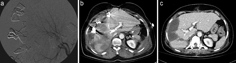
The role of interventional radiology in the care of trauma patients is clearly exemplified by the paradigm shift that has occurred in the management of solid-organ injury. The AAST Injury Scoring Scale provides a standardized guide for radiographically assessing the extent of traumatic injury to a specific organ or anatomic location, which can then help guide clinicians in further management. 4 5 In general, lower grade injuries (I–II) can often be managed with nonoperative management (NOM) and without angiographic intervention. Higher grade injuries (III–V + ) are more likely to require intervention, whether it be endovascular or operative. 6 7 The patient's hemodynamics and mechanism of injury often drive the decision-making with regard to an operative versus an endovascular approach. The current treatment of choice in the hemodynamically stable patient presenting with blunt solid organ injury is NOM with resuscitation, correction of coagulopathy and endovascular embolization as required ( Fig. 1 ). Advantages of this treatment strategy include high success rates, low complication rates, and relatively low failure rates. 8 This shift in clinical practice is reflected in both surgical and interventional radiology societal trauma guidelines and recommendations. 6 7 9
Fig. 1.

Traumatic lacerations to the spleen and left kidney. A 66-year-old man sustained a fall from 10-foot fall from ladder. ( a ) Axial and ( b ) coronal images demonstrate grade IV splenic and left kidney (black arrows) lacerations. Nonoperative management with ( c ) splenic angiography with focal distal embolization with gelatin sponge slurry and ( d ) proximal embolization with vascular plug and coils. White arrowheads denote abnormal blush. White arrow marks vascular plug and (*) coils.
Following blunt or penetrating traumatic liver injury, the first steps in patient evaluation are the assessment of hemodynamic status and the identification of additional injuries. Hemodynamic instability that is unresponsive to standard resuscitation requires immediate operative intervention with angiographic intervention being used to complement operative hemorrhage control as in a patient with grade V liver laceration in Fig. 2 . Depending on the individual patient needs and the trauma center resources, a patient may undergo concurrent operative intervention with angiographic evaluation and embolization as indicated. Oftentimes, patients presenting with radiographically identified liver injury without hemodynamic instability can be managed with NOM. AAST low-grade injuries are frequently managed successfully without the need for invasive intervention. In contrast, high-grade liver injuries have increased rates of NOM failure and may be rescued with endovascular intervention. Upon diagnosis of a AAST high-grade liver injury, the patient is routinely admitted to an ICU for serial hemodynamic monitoring and early involvement of the interventional radiology team. 6 Radiographic findings of active arterial extravasation (i.e., contrast blush on CT) warrant consideration for prompt endovascular embolization. Ultimately, the decision to pursue angiographic intervention depends on the clinical trajectory of the patient, hemodynamic monitoring, and hematologic trends.
Fig. 2.
Blunt traumatic liver injury. A 44-year-old woman pedestrian involved with motor vehicle accident was taken emergently for laparotomy where a large actively bleeding stellate laceration in the liver was noted. ( a ) Right hepatic artery angiography performed in the operating room prior to embolization with gelatin sponge slurry. CT images through the liver ( b ) 2 hours and ( c ) 7 weeks after emergent laparotomy and embolization demonstrating evolution of traumatic liver injury.
For blunt splenic injury, the rates of splenic salvage have improved with early interventional radiology consultation. 10 The management of patients presenting with blunt splenic injury depends on the initial clinical presentation, response to fluid resuscitation, the presence of peritonitis, and the constellation of concomitant injuries. Patients who are hemodynamically stable enough to obtain imaging can then be afforded a range of NOM alternatives. 7 The presence of radiographic evidence of ongoing bleeding or moderate hemoperitoneum warrants strong consideration for endovascular intervention for hemorrhage control and splenic salvage. 9 11 Asplenic patients are at increased risk of life-threatening infection due to overwhelming postsplenectomy infection, thus necessitating postoperative vaccination and lifelong vigilance. 12 Patients being treated with endovascular interventions, whether proximal or selective, retain functional splenic parenchyma, thus negating the need for vaccinations. 13 14 Angiographic interventions are not without risk, however. There are patients who may develop splenic abscess or necrosis. Providers should be aware of these potential complications and counsel patients accordingly. 15 16
The management of traumatic kidney injury follows similar patterns to spleen and liver management with some distinct differences. A multiphase radiographic evaluation with an emphasis on arterial, venous, and nephrogenic excretory phases allows for the characterization of injury, the presence of ongoing bleeding, and the presence of collecting system injury. 17 High-grade injuries in patients who are hemodynamically stable on presentation may be treated with NOM and endovascular intervention as indicated. 18 19 The presence of a concomitant collecting system injury does not preclude NOM, although delayed intervention may be required if urinary extravasation persists. 20
In addition to revolutionizing solid-organ injury, interventional radiology has significantly altered the management of traumatic pelvic hemorrhage. Blunt pelvic injury frequently can result in hemorrhagic shock from the bony pelvis or nearby arteries and veins. While arterial injury is the least common etiology of hemorrhage following blunt pelvic injury, the presence of an unstable fracture pattern significantly increases the likelihood of associated arterial injury. 21 22 23 Hemodynamically unstable patients with evidence of traumatic pelvic hemorrhage require emergent intervention given the high associated mortality. 21 These patients typically benefit from operative preperitoneal pelvic packing with or without external fixation. Oftentimes, endovascular intervention is employed as an extremely helpful adjunct in hemorrhage control to localize the area of injury and provide targeted embolization. Fig. 3 shows a patient with pelvic fractures and active hemorrhage after a fall downstairs. Alternatively, in select patients presenting to high-volume trauma centers, endovascular control of hemorrhage may be the preferred modality in patients with ongoing transfusion needs. 24 25 26 In addition to selective endovascular embolization, bilateral internal iliac artery embolization is a nonselective, endovascular embolization technique that is considered a type of damage control procedure, as it allows for expedient hemorrhage control by utilizing temporary embolic materials in a nonselective fashion. 27 This damage control technique carries the risk for pelvic and gluteal necrosis, although modern data suggest that this complication is rare. 27 More frequently, however, pelvic fractures result in hemodynamically stable injuries that can be associated with radiographic evidence of ongoing bleeding or vascular insult. These patients regularly undergo definitive management by endovascular intervention. Altogether, endovascular management of blunt pelvic injuries has high success rates with relatively low rates of failure and postprocedural complications. 23 28 As a result, many trauma centers now consider endovascular intervention the first-line therapy for patients presenting with traumatic pelvic injuries and evidence of ongoing blood loss.
Fig. 3.

Blunt pelvic injury with active arterial extravasation. An 88-year-old woman who sustained pelvic fractures with resultant active arterial hemorrhage after falling down multiple stairs. ( a ) Axial and ( b ) coronal images from CT demonstrating small foci of active extravasation (white arrow) in close proximity to right pubic rami fractures. Pelvic angiography from anterior division of the right internal iliac artery in ( c ) arterial and ( d ) delayed images demonstrating multifocal extravasation (white arrowheads). The patient was treated with embolization with gelatin sponge slurry.
Traumatic aortic injuries commonly occur following high-impact mechanisms, such as motor vehicle accidents or pedestrians struck by a moving vehicle, thoracic crush injuries, and falls. 29 30 31 There is a high associated mortality with this mechanism and injury pattern, with upward of 80% never making it to a hospital. 30 32 However, those who do survive transport to a major trauma center represent a uniquely challenging population. Due to the high-energy mechanisms that typically are associated with aortic injuries, these patients often have multiple-compartment and complex injury patterns. There are often competing priorities such as strict heart rate and blood pressure control following blunt aortic injury (impulse control) and optimizing hemodynamics for concurrent head injuries to ensure adequate cerebral perfusion. Historically, these patients underwent open operative intervention for aortic reconstruction, but these techniques were associated with a high morbidity and mortality. 30 In many cases, delayed intervention for the aortic injury is prudent as to allow the other life-threatening injuries to be definitively managed. More recently, endovascular intervention in the form of stent graft placement (thoracic endovascular aortic repair) is the preferred strategy 30 ( Fig. 4 ). These recommendations are based on data that suggest decreased mortality and morbidity, including paraplegia and renal failure, with delayed endovascular management compared with immediate and open intervention. 29 30 31 33 Higher grade aortic injuries which include aortic pseudoaneurysm (grade III) and active extravasation (grade IV) may predispose the patient to an increased rate of rupture, and for this reason delayed repair is not generally recommended. 30 33 Extensive traumatic soft-tissue injuries can result after fractures or degloving injuries and can be associated with compartment syndromes and significant blood loss. Many of these lesions are treated with the application of external compression devices and operative decompression as needed. Unfortunately, these bleeding patterns are oftentimes challenging to control with open surgery. For this reason, soft-tissue injuries, specifically those with radiographic evidence of ongoing arterial extravasation, may benefit from endovascular management. 34 If hemorrhage control is not able to be obtained with the correction of coagulopathy and the application of extrinsic pressure, angiographic intervention offers an attractive alternative to operative exploration. Early intervention may help prevent overlying tissue necrosis and secondary hematoma superinfection, which necessitate extensive surgical intervention with the associated morbidity.
Fig. 4.

Blunt traumatic aortic injury. A 39-year-old pedestrian involved with a motor vehicle accident. ( a ) Chest radiograph with finding of widened mediastinum. ( b ) Axial image from CT demonstrating traumatic aortic transection at the aortic isthmus with mediastinal hematoma (arrows). ( c ) Oblique maximum intensity image to better display extent of aortic injury (arrowheads). ( d ) Angiographic image following thoracic endovascular repair.
In conclusion, it is clear that the field for interventional radiology has had an immeasurable impact in trauma care. The field is instrumental during the initial resuscitation and management of some of the most complex patients. Additionally, interventional radiology frequently contributes to the care of trauma patients well into their hospital stay and rescues these patients from septic complications that arise during often complicated recovery periods. As the field continues to grow, it is likely that the role of the interventionalist will continue to expand. Thus, the working relationship between trauma surgeons and interventional radiologists, as well as the rest of the multidisciplinary team, is paramount for the care of the trauma patient. 35
References
- 1.WHO . Geneva World Health Organ; 2014. Injuries and Violence. The Facts 2014. [Google Scholar]
- 2.Rotondo M, Cribari C SS.Resources for optimal care of the injured patient–2014Bull Am Coll Surg2014 [PubMed]
- 3.Chicago, IL: American College of Surgeons; 2018. Advanced Trauma Life Support: Student Course Manual, 10th ed. [Google Scholar]
- 4.Moore E E, Cogbill T H, Malangoni M A, Jurkovich G J, Champion H R.Scaling system for organ specific injuriesCurr Opin Crit Care1996
- 5.Tinkoff G, Esposito T J, Reed J. American Association for the Surgery of Trauma Organ Injury Scale I: spleen, liver, and kidney, validation based on the National Trauma Data Bank. J Am Coll Surg. 2008;207(05):646–655. doi: 10.1016/j.jamcollsurg.2008.06.342. [DOI] [PubMed] [Google Scholar]
- 6.Eastern Association for the Surgery of Trauma . Stassen N A, Bhullar I, Cheng J D. Nonoperative management of blunt hepatic injury: an Eastern Association for the Surgery of Trauma practice management guideline. J Trauma Acute Care Surg. 2012;73(05) 04:S288–S293. doi: 10.1097/TA.0b013e318270160d. [DOI] [PubMed] [Google Scholar]
- 7.Eastern Association for the Surgery of Trauma . Stassen N A, Bhullar I, Cheng J D. Selective nonoperative management of blunt splenic injury: an Eastern Association for the Surgery of Trauma practice management guideline. J Trauma Acute Care Surg. 2012;73(05) 04:S294–S300. doi: 10.1097/TA.0b013e3182702afc. [DOI] [PubMed] [Google Scholar]
- 8.Green C S, Bulger E M, Kwan S W. Outcomes and complications of angioembolization for hepatic trauma: a systematic review of the literature. J Trauma Acute Care Surg. 2016;80(03):529–537. doi: 10.1097/TA.0000000000000942. [DOI] [PMC free article] [PubMed] [Google Scholar]
- 9.Padia S A, Ingraham C R, Moriarty J M. Society of Interventional Radiology Position Statement on endovascular intervention for trauma. J Vasc Interv Radiol. 2020;31(03):363–36900. doi: 10.1016/j.jvir.2019.11.012. [DOI] [PubMed] [Google Scholar]
- 10.Western Trauma Association Multi-Institutional Trials Committee . Haan J M, Biffl W, Knudson M M. Splenic embolization revisited: a multicenter review. J Trauma. 2004;56(03):542–547. doi: 10.1097/01.ta.0000114069.73054.45. [DOI] [PubMed] [Google Scholar]
- 11.Bhullar I S, Frykberg E R, Siragusa D. Selective angiographic embolization of blunt splenic traumatic injuries in adults decreases failure rate of nonoperative management. J Trauma Acute Care Surg. 2012;72(05):1127–1134. doi: 10.1097/TA.0b013e3182569849. [DOI] [PubMed] [Google Scholar]
- 12.Bonanni P, Grazzini M, Niccolai G. Recommended vaccinations for asplenic and hyposplenic adult patients. Hum Vaccines Immunother. 2017;13(02):359–368. doi: 10.1080/21645515.2017.1264797. [DOI] [PMC free article] [PubMed] [Google Scholar]
- 13.Bessoud B, Duchosal M A, Siegrist C A. Proximal splenic artery embolization for blunt splenic injury: clinical, immunologic, and ultrasound-Doppler follow-up. J Trauma. 2007;62(06):1481–1486. doi: 10.1097/TA.0b013e318047dfb8. [DOI] [PubMed] [Google Scholar]
- 14.Malhotra A K, Carter R F, Lebman D A.Preservation of splenic immunocompetence after splenic artery angioembolization for blunt splenic injury J Trauma 201069051126–1130., discussion 1130–1131 [DOI] [PubMed] [Google Scholar]
- 15.Ekeh A P, Khalaf S, Ilyas S, Kauffman S, Walusimbi M, McCarthy M C.Complications arising from splenic artery embolization: a review of an 11-year experience Am J Surg 201320503250–254., discussion 254 [DOI] [PubMed] [Google Scholar]
- 16.Cioci A C, Parreco J P, Lindenmaier L B. Readmission for infection after blunt splenic injury: a national comparison of management techniques. J Trauma Acute Care Surg. 2020;88(03):390–395. doi: 10.1097/TA.0000000000002564. [DOI] [PubMed] [Google Scholar]
- 17.in Conjunction with the Trauma and Urologic Reconstruction Network of Surgeons . Keihani S, Putbrese B E, Rogers D M. Optimal timing of delayed excretory phase computed tomography scan for diagnosis of urinary extravasation after high-grade renal trauma. J Trauma Acute Care Surg. 2019;86(02):274–281. doi: 10.1097/TA.0000000000002098. [DOI] [PubMed] [Google Scholar]
- 18.Hagiwara A, Sakaki S, Goto H. The role of interventional radiology in the management of blunt renal injury: a practical protocol. J Trauma. 2001;51(03):526–531. doi: 10.1097/00005373-200109000-00017. [DOI] [PubMed] [Google Scholar]
- 19.Altman A L, Haas C, Dinchman K H, Spirnak J P.Selective nonoperative management of blunt grade 5 renal injury J Urol 20001640127–30., discussion 30–31 [PubMed] [Google Scholar]
- 20.Erlich T, Kitrey N D. Renal trauma: the current best practice. Ther Adv Urol. 2018;10(10):295–303. doi: 10.1177/1756287218785828. [DOI] [PMC free article] [PubMed] [Google Scholar]
- 21.Dyer G SM, Vrahas M S. Review of the pathophysiology and acute management of haemorrhage in pelvic fracture. Injury. 2006;37(07):602–613. doi: 10.1016/j.injury.2005.09.007. [DOI] [PubMed] [Google Scholar]
- 22.Demetriades D, Karaiskakis M, Toutouzas K, Alo K, Velmahos G, Chan L. Pelvic fractures: epidemiology and predictors of associated abdominal injuries and outcomes. J Am Coll Surg. 2002;195(01):1–10. doi: 10.1016/s1072-7515(02)01197-3. [DOI] [PubMed] [Google Scholar]
- 23.Wijffels D J, Verbeek D O, Ponsen K J, Carel Goslings J, van Delden O M. Imaging and endovascular treatment of bleeding pelvic fractures: review article. Cardiovasc Intervent Radiol. 2019;42(01):10–18. doi: 10.1007/s00270-018-2071-4. [DOI] [PMC free article] [PubMed] [Google Scholar]
- 24.Broadwell S R, Ray C E. Transcatheter embolization in pelvic trauma. Semin Intervent Radiol. 2004;21(01):23–35. doi: 10.1055/s-2004-831402. [DOI] [PMC free article] [PubMed] [Google Scholar]
- 25.Vaidya R, Waldron J, Scott A, Nasr K. Angiography and embolization in the management of bleeding pelvic fractures. J Am Acad Orthop Surg. 2018;26(04):e68–e76. doi: 10.5435/JAAOS-D-16-00600. [DOI] [PMC free article] [PubMed] [Google Scholar]
- 26.Agolini S F, Shah K, Jaffe J, Newcomb J, Rhodes M, Reed J F., III Arterial embolization is a rapid and effective technique for controlling pelvic fracture hemorrhage. J Trauma. 1997;43(03):395–399. doi: 10.1097/00005373-199709000-00001. [DOI] [PubMed] [Google Scholar]
- 27.Bonde A, Velmahos A, Kalva S P, Mendoza A E, Kaafarani H MA, Nederpelt C J. Bilateral internal iliac artery embolization for pelvic trauma: effectiveness and safety. Am J Surg. 2020;220(02):454–458. doi: 10.1016/j.amjsurg.2019.12.013. [DOI] [PubMed] [Google Scholar]
- 28.Velmahos G C, Toutouzas K G, Vassiliu P.A prospective study on the safety and efficacy of angiographic embolization for pelvic and visceral injuries J Trauma 20025302303–308., discussion 308 [DOI] [PubMed] [Google Scholar]
- 29.Demetriades D. Blunt thoracic aortic injuries: crossing the Rubicon. J Am Coll Surg. 2012;214(03):247–259. doi: 10.1016/j.jamcollsurg.2011.11.015. [DOI] [PubMed] [Google Scholar]
- 30.Fox N, Schwartz D, Salazar J H. Evaluation and management of blunt traumatic aortic injury: a practice management guideline from the Eastern Association for the Surgery of Trauma. J Trauma Acute Care Surg. 2015;78(01):136–146. doi: 10.1097/TA.0000000000000470. [DOI] [PubMed] [Google Scholar]
- 31.American Association for the Surgery of Trauma Thoracic Aortic Injury Study Group Demetriades D, Velmahos G C, Scalea T M.Operative repair or endovascular stent graft in blunt traumatic thoracic aortic injuries: results of an American Association for the Surgery of Trauma Multicenter Study J Trauma 20086403561–570., discussion 570–571 [DOI] [PubMed] [Google Scholar]
- 32.Arthurs Z M, Starnes B W, Sohn V Y, Singh N, Martin M J, Andersen C A. Functional and survival outcomes in traumatic blunt thoracic aortic injuries: an analysis of the National Trauma Databank. J Vasc Surg. 2009;49(04):988–994. doi: 10.1016/j.jvs.2008.11.052. [DOI] [PubMed] [Google Scholar]
- 33.Demetriades D, Velmahos G C, Scalea T M. Blunt traumatic thoracic aortic injuries: early or delayed repair--results of an American Association for the Surgery of Trauma prospective study. J Trauma. 2009;66(04):967–973. doi: 10.1097/TA.0b013e31817dc483. [DOI] [PubMed] [Google Scholar]
- 34.Mirakhur A, Cormack R, Eesa M, Wong J K. Endovascular therapy for acute trauma: a pictorial review. Can Assoc Radiol J. 2014;65(02):158–167. doi: 10.1016/j.carj.2012.09.005. [DOI] [PubMed] [Google Scholar]
- 35.Goei A D, Ching B H, Meyermann M W, Nunez T, Sacks D. Tips and tricks for the trauma patient. Semin Intervent Radiol. 2010;27(01):81–98. doi: 10.1055/s-0030-1247894. [DOI] [PMC free article] [PubMed] [Google Scholar]