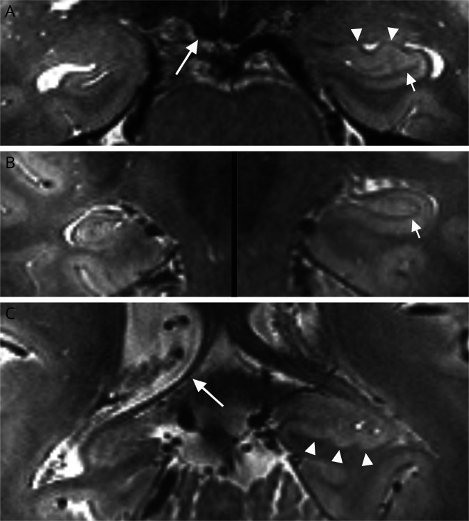
Figure 6. Hippocampal Sclerosis (HS) at 7T.
Coronal T2-weighted images at the level of the hippocampal head (A), body (B), and tail (C) show normal appearance of the left hippocampus including a continuous dark band reflecting the stratum radiatum lacunosum moleculare (arrows) and normal digitations along the head and tail (arrowheads). In contrast, the right hippocampus shows features of HS, including decreased volume, smooth outer counters, and indistinct internal architecture. Note also atrophy of the right mammillary body (long arrow in A) and fornix (long arrow in C).

