Retinoblastoma is the most common primary intraocular malignancy in children.1 Improvements in screening tactics and treatment methods have dramatically increased both patient and globe survival.1,2 Imaging methods such as fluorescein angiography have demonstrated retinal vascular abnormalities in both large- and small-caliber retinal vessels in eyes with retinoblastoma,1 and OCT can reveal or confirm otherwise subclinical lesions.2 OCT angiography (OCTA) is a noninvasive imaging method allowing for in vivo visualization of retinal and choroidal vasculature. Herein, we describe OCTA findings of a treatment-naïve, small retinoblastoma lesion in an infant using an investigational portable OCTA system (Spectralis with Flex and OCT-A module; Heidelberg Engineering, Heidelberg, Germany), under Duke University Internal Review Board protocol with parental consent. All research adhered to the tenets of the Declaration of Helsinki.
A 3-month-old male was examined under anesthesia by one of the authors (M.A.M.) after being referred for presumed bilateral retinoblastoma. Anterior segment examination was unremarkable in both eyes. Funduscopy of the right eye (Fig S1A, available at www.ophthalmologyretina.org) showed a small tumor inferotemporal to the inferior arcade measuring 4 × 4 × 1.6 mm thick by ultrasound (Fig S1A, B). The left eye demonstrated a large tumor in the macula measuring 15 × 15 × 6.27 mm with localized subretinal seeding. Fluorescein angiography of the right eye highlighted small feeder vessels and hyperfluorescence of the lesion (Fig S1C).
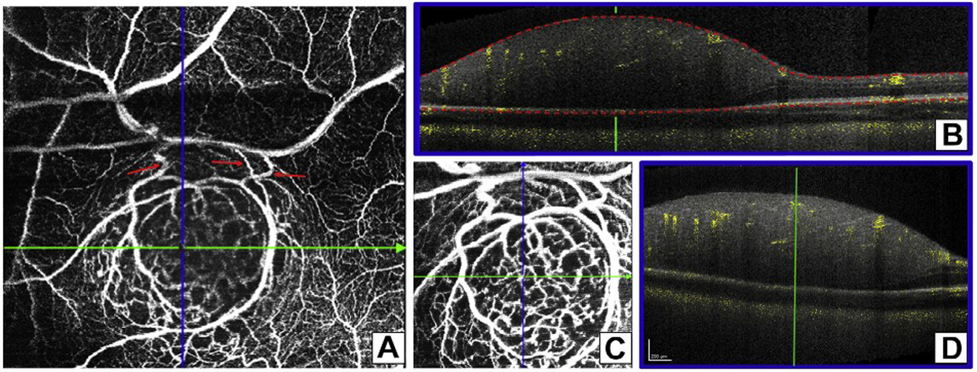
Investigational OCT of the right eye lesion showed a spherical, homogenous mass contained within the inner retina with some shadowing (Fig S1D). On the research 10° × 10° OCTA scan of the right eye tumor, acquired isotropically at 512 A-scans with an axial resolution of 1.9 mm, large superficial vessels stemmed from the inferior arcade and circumscribed the tumor, branching into finer telangiectatic vessels that led into a dense centralized vascular network (Fig 1). Fed by the encircling vessels, the intrinsic network was visible throughout the interior of the lesion at various depths on OCTA cross sections (Fig 1). Automated segmentation of the retinal layers was not possible because of obliteration of distinct retinal layers.
Figure 1.

A, 20° × 20° (5.5 × 5.5-mm) en face OCT angiography (OCTA) image of the lesion inferotemporal to the macula in the right eye with 512 A-scans/B-scan and 512 B-scans showing superficial medium-sized vessels circumscribing the tumor and feeding the intrinsic vasculature along with prominent feeder vessels (red arrows). B, Vertical line scan with OCTA flow overlay demonstrating flow at multiple levels of the tumor with some shadowing near the most inner aspects of the tumor. C, 10° × 10° (2.8 × 2.8-mm) en face OCTA image showing that the concurrent vertical line scan displayed disorganized, interwoven intrinsic, vasculature located at (D) multiple depths.
This case demonstrates the first visualization of treatment-naïve retinoblastoma tumor vasculature using OCTA in an infant, according to a PubMed search on April, 23 2018, using the keywords retinoblastoma and optical coherence tomography angiography. A prior report used OCTA-based analysis of macular vascular changes in 7- to 16-year-old patients with a history of retinoblastoma and found subtle foveal changes in both eyes 5 to 14 years after intravenous chemotherapy.3 A study of fluorescein angiography in retinoblastoma demonstrated that intrinsic tumor vessels with complex branching patterns, irregular caliber, and early termination within the tumor are distinguishable from retinal vessels.1 With OCTA, we were able to validate these distinctions with greater detail and with depth resolution that was not possible from fluorescein angiography. OCT angiography also highlighted the connection between existing retinal vasculature and the intrinsic tumor vessels through feeder vessels (Fig 1). Pathologic specimens can describe tumor vascular patterns and density, with higher vessel densities correlating with poor outcomes and metastasis risk.4 Although shadowing may obscure the deepest vessels, we demonstrated an ability to analyze tumor-intrinsic vasculature in vivo. In this case, more mature-seeming vessels led around the lesion and dove centrally into a disorganized web of smaller intrinsic vessels (possibly less mature).
OCT angiography may have future applications in the management and treatment of vascular tumors. Other imaging units (e.g., handheld OCT) have been used to characterize active retinoblastoma5 and have aided in diagnosis of lesions almost undetectable by funduscopy.2 Now that we have a novel imaging method for observing in vivo retinoblastoma tumor vasculature, further follow-up is needed to investigate OCTA measures of vessel density, feeder vessel origins, and vascular commonalities, in addition to markers of tumor regression and the microvascular response to different treatment methods and markers of tumor regression.
Supplementary Material
Acknowledgments.
The authors thank Michael P. Kelly, FOPS, for capturing the images, Maysantoine El-Dairi, MD, and Mohsin Ali, MD.
Financial Disclosure(s):
The author(s) have made the following disclosure(s): C.A.T.: Royalties, Patents — Alcon.
M.A.M.: Consultant — Castle Biosciences.
L.V.: Consultant – Alcon, Janssen Pharmaceutical, Roche, DORC, Genentech, Alimera Sciences.
Supported by the International Association of Government Officials (iGO) Fund (S.T.H., L.V.); Grant from Heidelberg Engineering, Heidelberg, Germany (all); Research to Prevent Blindness, Inc, New York, New York (all, unrestricted grant to Duke Eye Center); Knights Templar Eye Foundation (S.T.H., R.J.H., L.V.); Knights Templar Eye Foundation (S.T.H., R.J.H., L.V.); the National Institutes of Health, Bethesda, Maryland (grant no.: RO1EY025009 [C.A.T.]); and the Lions Duke Pediatric Eye Research Endowment (R.J.H.). Research equipment (Spectralis HRA + OCT with Flex module and Beta OCT-A software) provided by Heidelberg Engineering, Heidelberg, Germany.
Abbreviations and Acronyms:
- OCTA
OCT angiography
References
- 1.Kim JW, Ngai LK, Sadda S, et al. Retcam fluorescein angiography findings in eyes with advanced retinoblastoma. Br J Ophthalmol. 2014;98:1666–1671. [DOI] [PubMed] [Google Scholar]
- 2.Liu KC, Walter SD, Finn AP, Materin MA. Venous loop reveals an occult retinoblastoma tumor. Ophthalmic Surg Lasers Imaging Retina. 2017;48:768–770. [DOI] [PubMed] [Google Scholar]
- 3.Sioufi K, Say EAT, Ferenczy SC, et al. Optical coherence tomography angiography findings of deep capillary plexus microischemia after intravenous chemotherapy for retinoblastoma. Retina. 2017. [Epub ahead of print]. [DOI] [PubMed] [Google Scholar]
- 4.Rossler J, Dietrich T, Pavlakovic H, et al. Higher vessel densities in retinoblastoma with local invasive growth and metastasis. Am J Pathol. 2004;164:391–394. [DOI] [PMC free article] [PubMed] [Google Scholar]
- 5.Rootman DB, Gonzalez E, Mallipatna A, et al. Hand-held high-resolution spectral domain optical coherence tomography in retinoblastoma: clinical and morphologic considerations. Br J Ophthalmol. 2013;97:59–65. [DOI] [PubMed] [Google Scholar]
Associated Data
This section collects any data citations, data availability statements, or supplementary materials included in this article.


