

Abstract
Human umbilical cord mesenchymal stem cells (hUC-MSCs) serve as a potential cell-based therapy for degenerative disease. They provide immunomodulatory and anti-inflammatory properties, multipotent differentiation potential and are harvested with no ethical concern. It is unknown whether MSCs collected from different areas of the human umbilical cord elicit more favorable effects than others. Three MSC populations were harvested from various regions of the human umbilical cord: cord lining (CL-MSCs), perivascular region (PV-MSCs), and Wharton's jelly (WJ-MSCs). Mesenchymal markers (CD90 and CD73) were expressed by all three cell populations. Stemness marker (OCT4), endothelial cell adhesion molecular marker (CD146), and monocyte-macrophage marker (CD14) were expressed by WJ-MSCs, PV-MSCs, and CL-MSCs, respectively. Stroke presents with oxygen and glucose deprivation and leads to dysfunctional mitochondria and consequently cell death. Targeting the restoration of mitochondrial function in the stroke brain through mitochondrial transfer may be effective in treating stroke. In vitro exposure to ambient and OGD conditions resulted in CL-MSCs number decreasing the least post-OGD/R exposure, and PV-MSCs exhibiting the greatest mitochondrial activity. All three hUC-MSC populations presented similar metabolic activity and survival in normal and pathologic environments. These characteristics indicate hUC-MSCs potential as a potent therapeutic in regenerative medicine.
Keywords: Bioenergetics, ischemic diseases, mitochondria, stem cell therapy, stroke, umbilical cord mesenchymal stem cells, Wharton's Jelly
Introduction: Stroke and Mitochondria
Behind heart disease, stroke presents as the second leading cause of death and disability.[1] Stroke-induced oxygen and glucose deprivation (OGD) results in dysfunctional mitochondria further exacerbating oxidative stress, inflammation, and neuronal death while weakening oxidative metabolism.[2] Because mitochondria produce more than 90% of adenosine triphosphate for the cell,[3] energy failure, excitotoxicity, and calcium overload ensue and contribute to loss of mitochondrial membrane potential.[2] Dysfunctional mitochondria can release pro-apoptotic molecules and generate apoptosis due to increased membrane permeability.[2] Impaired mitochondrial function plays a significant role in stroke deficits.
Cell-Based Mitochondrial Transfer
Cell-based therapies replenish dead cells while facilitating the survival of injured cells. Promoting exogenous and endogenous repair mechanisms while providing trophic support, stem cells attenuate the inflammation associated with stroke.[4,5,6] Stem cells have targeted mitochondrial dysfunction restoration through transferring healthy mitochondria to afflicted cells through tunneling nanotubes, microvesicles, gap junctions, cell fusion, and direct uptake into endogenous cells.[7,8] The mechanism to initiate a mitochondrial transfer between damaged and stem cells is still unclear, however overwhelming evidence suggests the transfer of healthy mitochondria aids to restore cellular function.[9] The mitochondrial transfer has been observed in mesenchymal stem cells (MSCs), endothelial progenitor cells, neurons, and astrocytes.[10,11] Overall, these findings suggest mitochondrial transfer's potential as a potent therapeutic, however further investigation is imperative to understand the mechanism behind this phenomenon.
Human Umbilical Cord Mesenchymal Stem Cells
hUC-MSCs serve as a potential regenerative therapy due to their self-renewal abilities, multipotent differentiation, and immunomodulatory and anti-inflammatory capabilities.[5,12,13] It is unknown whether a single MSCs type derived from Wharton's jelly, perivascular region, and cord lining is more effective than others.[14] Because the human umbilical cord is made up of two arteries and a vein,[14] the cells that compose it may be therapeutically superior to other cell types because of their naturally hypoxic and glucose-lacking environment.
This review paper examines current literature that reveals mitochondrial function and the metabolic activity of various hUC-MSC populations under stroke and baseline conditions in vitro.
Effectiveness of Human Umbilical Cord Mesenchymal Stem Cells In vitro
Human umbilical cord mesenchymal stem cells (hUC-MSCs) possess anti-inflammatory, immunomodulatory, and multipotent properties. These characteristics may provide therapeutically favorable results in regenerative medicine. hUC-MSCs displaying greater efficacy compared to other stem cell types is still in question, however hUC-MSCs are easily collected with minimal ethical concern. MSCs were harvested from cord lining (CL-MSCs), perivascular region (PV-MSCs), and Wharton's jelly (WJ-MSCs) of the human umbilical cord.[15] Mesenchymal markers (CD90 and CD73) were expressed by all three cell populations. Stemness marker (OCT4), endothelial cell adhesion molecular marker (CD146), and monocyte-macrophage marker (CD14) were expressed by WJ-MSCs, PV-MSCs, and CL-MSCs, respectively.[15] OGD, apparent during stroke and reperfusion, results in dysfunctional mitochondria and consequently cell death. Evidence indicates that restoring mitochondrial function through mitochondrial transfer may be effective in treating stroke. When all three cell populations were exposed to ambient and OGD conditions in vitro, cell energy phenotype and mito stress tests were conducted to analyze the metabolic profile and mitochondrial activity. Data indicated that CL-MSCs number decreased the least post-OGD/R exposure, and PV-MSCs exhibited the greatest mitochondrial activity.[15] All three hUC-MSCs populations presented similar metabolic activity and survival in normal and hypoxic environments [Figure 1]. These beneficial characteristics bolster hUC-MSCs therapeutic potential in stroke and other ischemic diseases.
Figure 1.
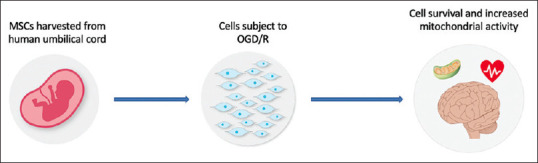
Human umbilical cord mesenchymal stem cells subject to oxygen and glucose deprivation/R treatment display increased mitochondrial activity and ability to survive in stroke conditions
Human Umbilical Cord Mesenchymal Stem Cell as a Stroke Therapeutic
Stroke-induced mitochondrial dysfunction influences neuronal death, oxidative stress, and inflammation.[2] The phenomenon of healthy mitochondrial transfer has pointed toward mitochondrial-based stem cell therapy as an effective treatment for stroke.[7,8,11] The human umbilical cord is composed of three vessels indicating that hUC-MSCS may be evolutionarily conditioned to survive in environments with decreased oxygen and glucose, further reinforcing their potential as a stroke therapy.[14] Few studies have investigated the mitochondrial function of hUC-MSCs in combination with energy metabolism.[16,17,18] It is still unclear whether PV-MSCs, WJ-MSCs, or CL-MSCs are more effective.[14,19] Furthermore, whether cells harvested from a different distance on the vessels in the umbilical cord will have differing characteristics under ischemic conditions is unknown.
Cell Survival and Energy Profile
To further understand hU-MSCs potential, PV-MSCs, WJ-MSCs, and CL-MSCs were harvested to analyze the energy metabolism profile, mitochondrial function, and survival capabilities in normal and ischemic/reperfused environments. Data indicated that all three cell populations displayed quiescent phenotypes and therefore preserved glycolytic and mitochondrial metabolism at low levels.[15] When subject to stressful conditions, hUC-MSCs augmented glycolytic metabolism to offset reduced mitochondrial activity. When the rate of decrease in oxygen concentration (OCR) was measured, only a minimal decrease in OCR was observed across OGD/R groups when compared to MSCs in normal conditions. Interestingly, while cell survival was not affected by OGD/R exposure, the cells were seen in increased concentrations. Taken together, these data indicate that hUC-MSCs retain the capacity to survive in harsh stroke environments.[15]
Reducing Cell Death
MSCs may attenuate the subacute and chronic cell death associated with stroke through their ability to increase mitochondrial function.[20,21,22,23] Targeting dysfunctional mitochondria with hUC-MSCs remains a novel stroke therapy.[24,25,26,27,28,29,30,31] Furthermore, stem-cell facilitated mitochondria transplantation may provide favorable outcomes in treating other neurological disorders not limited to stroke.[32,33,34,35,36,37,38]
Variations among Human Umbilical Cord Mesenchymal Stem Cell Types
Data indicate that PV-MSCs, WJ-MSCs, and CL-MSCs all have the capability to retain mitochondrial function in ischemic conditions.[15] While all three cell populations demonstrated comparable mitochondria and energy metabolism, PV-MSCs displayed the greatest OCR values indicating that this population has higher mitochondrial activity when compared to the other hUC-MSCs.[15] PV-MSCs also presented with a steadier decrease of OCR after OGD/R exposure which is consistent with this finding. The cells' ability to survive and function in hypoxic environments was demonstrated by CL-MSCs as they were least affected by OGD/R. Explaining the minute variations observed across the three cell populations requires further investigation on whether their origin in the human umbilical displays higher mitochondrial function in an ischemic environment.[15] Overall, hUC-MSCs demonstrate the ability to retain active and functional mitochondria in stroke conditions, further bolstering their potential as a potent mitochondria-based stem cell therapy for stroke and other ischemia-associated disorders.
Data indicate that CL-MSCs, WJ-MSCs, and PV-MSCs derived from the human umbilical cord possess a vigorous mitochondrial profile and the ability to function in ischemic environments.[15] These findings encourage hU-MSCs consideration as an effective donor cell for mitochondria-based therapy for stroke and other disorders.
Conclusion
Data indicate that CL-MSCs, WJ-MSCs, and PV-MSCs derived from human umbilical cord posses a vigorous mitochondrial profile and the ability to function in ischemic environments.[15] These findings encourage hU-MSCs consideration as an effective donor cell for mitochondria-based therapy for stroke and other disorders.
Financial support and sponsorship
Nil.
Conflicts of interest
Prof. Cesario V. Borlongan is Associate Editor of Brain Circulation.
References
- 1.Benjamin EJ, Virani SS, Callaway CW, Chamberlain AM, Chang AR, Cheng S, et al. Heart disease and stroke statistics-2018 update: A report from the American Heart Association. Circulation. 2018;137:e67–492. doi: 10.1161/CIR.0000000000000558. [DOI] [PubMed] [Google Scholar]
- 2.Yang JL, Mukda S, Chen SD. Diverse roles of mitochondria in ischemic stroke. Redox Biol. 2018;16:263–75. doi: 10.1016/j.redox.2018.03.002. [DOI] [PMC free article] [PubMed] [Google Scholar]
- 3.Watts LT, Lloyd R, Garling RJ, Duong T. Stroke neuroprotection: Targeting mitochondria. Brain Sci. 2013;3:540–60. doi: 10.3390/brainsci3020540. [DOI] [PMC free article] [PubMed] [Google Scholar]
- 4.Tajiri N, Duncan K, Antoine A, Pabon M, Acosta SA, de la Pena I, et al. Stem cell-paved biobridge facilitates neural repair in traumatic brain injury. Front Syst Neurosci. 2014;8:116. doi: 10.3389/fnsys.2014.00116. [DOI] [PMC free article] [PubMed] [Google Scholar]
- 5.Caprnda M, Kubatka P, Gazdikova K, Gasparova I, Valentova V, Stollarova N, et al. Immunomodulatory effects of stem cells: Therapeutic option for neurodegenerative disorders. Biomed Pharmacother. 2017;91:60–9. doi: 10.1016/j.biopha.2017.04.034. [DOI] [PubMed] [Google Scholar]
- 6.Chau M, Zhang J, Wei L, Yu SP. Regeneration after stroke: Stem cell transplantation and trophic factors. Brain Circ. 2016;2:86–94. doi: 10.4103/2394-8108.186279. [DOI] [PMC free article] [PubMed] [Google Scholar]
- 7.Russo E, Napoli E, Borlongan C. Healthy mitochondria for stroke cells. Brain Circ. 2018;4:95. doi: 10.4103/bc.bc_20_18. [DOI] [PMC free article] [PubMed] [Google Scholar]
- 8.Russo E, Nguyen H, Lippert T, Tuazon J, Borlongan CV, Napoli E. Mitochondrial targeting as a novel therapy for stroke. Brain Circ. 2018;4:84–94. doi: 10.4103/bc.bc_14_18. [DOI] [PMC free article] [PubMed] [Google Scholar]
- 9.Hsu YC, Wu YT, Yu TH, Wei YH. Mitochondria in mesenchymal stem cell biology and cell therapy: From cellular differentiation to mitochondrial transfer. Semin Cell Dev Biol. 2016;52:119–31. doi: 10.1016/j.semcdb.2016.02.011. [DOI] [PubMed] [Google Scholar]
- 10.Paliwal S, Chaudhuri R, Agrawal A, Mohanty S. Regenerative abilities of mesenchymal stem cells through mitochondrial transfer. J Biomed Sci. 2018;25:31. doi: 10.1186/s12929-018-0429-1. [DOI] [PMC free article] [PubMed] [Google Scholar]
- 11.Borlongan CV, Nguyen H, Lippert T, Russo E, Tuazon J, Xu K, et al. May the force be with you: Transfer of healthy mitochondria from stem cells to stroke cells. J Cereb Blood Flow Metab. 2019;39:367–70. doi: 10.1177/0271678X18811277. [DOI] [PMC free article] [PubMed] [Google Scholar]
- 12.La Rocca G, Lo Iacono M, Corsello T, Corrao S, Farina F, Anzalone R. Human Wharton's jelly mesenchymal stem cells maintain the expression of key immunomodulatory molecules when subjected to osteogenic, adipogenic and chondrogenic differentiation in vitro: New perspectives for cellular therapy. Curr Stem Cell Res Ther. 2013;8:100–13. doi: 10.2174/1574888x11308010012. [DOI] [PubMed] [Google Scholar]
- 13.Christian G. From bench to bedside. Placenta. 2016:197–215. [Google Scholar]
- 14.Davies JE, Walker JT, Keating A. Concise review: Wharton's jelly: The rich, but enigmatic, source of mesenchymal stromal cells: Wharton's jelly an enigmatic msc source. STEM CELLS Transl Med. 2017;6:1620–30. doi: 10.1002/sctm.16-0492. [DOI] [PMC free article] [PubMed] [Google Scholar]
- 15.Russo E, Lee JY, Nguyen H, Corrao S, Anzalone R, La Rocca G, et al. Energy metabolism analysis of three different mesenchymal stem cell populations of umbilical cord under normal and pathologic conditions. Stem Cell Rev Rep. 2020;16:585–95. doi: 10.1007/s12015-020-09967-8. [DOI] [PMC free article] [PubMed] [Google Scholar]
- 16.Hu C, Fan L, Cen P, Chen E, Jiang Z, Li L. Energy metabolism plays a critical role in stem cell maintenance and differentiation. Int J Mol Sci. 2016;17:253. doi: 10.3390/ijms17020253. [DOI] [PMC free article] [PubMed] [Google Scholar]
- 17.Lavrentieva A, Majore I, Kasper C, Hass R. Effects of hypoxic culture conditions on umbilical cord-derived human mesenchymal stem cells. Cell Commun Signal. 2010;8:18. doi: 10.1186/1478-811X-8-18. [DOI] [PMC free article] [PubMed] [Google Scholar]
- 18.Skiles ML, Brown KS, Tatz W, Swingle K, Brown HL. Quantitative analysis of composite umbilical cord tissue health using a standardized explant approach and an assay of metabolic activity. Cytotherapy. 2018;20:564–75. doi: 10.1016/j.jcyt.2018.01.001. [DOI] [PubMed] [Google Scholar]
- 19.Teresa Conconi M. Phenotype and differentiation potential of stromal populations obtained from various zones of human umbilical cord: An overview. TOTERMJ. 2011;4:6–20. [Google Scholar]
- 20.Stimpfel M, Jancar N, Virant-Klun I. New challenge: Mitochondrial epigenetics? Stem Cell Rev Rep. 2018;14:13–26. doi: 10.1007/s12015-017-9771-z. [DOI] [PubMed] [Google Scholar]
- 21.Kornicka K, Houston J, Marycz K. Dysfunction of mesenchymal stem cells isolated from metabolic syndrome and type 2 diabetic patients as result of oxidative stress and autophagy may limit their potential therapeutic use. Stem Cell Rev Rep. 2018;14:337–45. doi: 10.1007/s12015-018-9809-x. [DOI] [PMC free article] [PubMed] [Google Scholar]
- 22.Wu KJ, Yu SJ, Chiang CW, Lee YW, Yen BL, Hsu CS, et al. Wharton' jelly mesenchymal stromal cell therapy for ischemic brain injury. Brain Circ. 2018;4:124–7. doi: 10.4103/bc.bc_16_18. [DOI] [PMC free article] [PubMed] [Google Scholar]
- 23.Aghajani Nargesi A, Zhu XY, Hickson LJ, Conley SM, van Wijnen AJ, Lerman LO, et al. Metabolic syndrome modulates protein import into the mitochondria of porcine mesenchymal stem cells. Stem Cell Rev Rep. 2019;15:427–38. doi: 10.1007/s12015-018-9855-4. [DOI] [PMC free article] [PubMed] [Google Scholar]
- 24.Al Naem M, Bourebaba L, Kucharczyk K, Röcken M, Marycz K. Therapeutic mesenchymal stromal stem cells: Isolation, characterization and role in equine regenerative medicine and metabolic disorders. Stem Cell Rev Rep. 2020;16:301–22. doi: 10.1007/s12015-019-09932-0. [DOI] [PubMed] [Google Scholar]
- 25.Nishida F, Zappa Villar MF, Zanuzzi CN, Sisti MS, Camiña AE, Reggiani PC, et al. Intracerebroventricular delivery of human umbilical cord mesenchymal stem cells as a promising therapy for repairing the spinal cord injury induced by kainic acid. Stem Cell Rev Rep. 2020;16:167–80. doi: 10.1007/s12015-019-09934-y. [DOI] [PubMed] [Google Scholar]
- 26.Contentin R, Demoor M, Concari M, Desancé M, Audigié F, Branly T, et al. Comparison of the chondrogenic potential of mesenchymal stem cells derived from bone marrow and umbilical cord blood intended for cartilage tissue engineering. Stem Cell Rev Rep. 2020;16:126–43. doi: 10.1007/s12015-019-09914-2. [DOI] [PubMed] [Google Scholar]
- 27.Corsello T, Amico G, Corrao S, Anzalone R, Timoneri F, Lo Iacono M, et al. Wharton's jelly mesenchymal stromal cells from human umbilical cord: A close-up on immunomodulatory molecules featured in situ and in vitro. Stem Cell Rev and Rep. 2019;15:900–18. doi: 10.1007/s12015-019-09907-1. [DOI] [PubMed] [Google Scholar]
- 28.Lee JY, Tuazon JP, Corey S, Bonsack B, Acosta S, Ehrhart J, et al. A gutsy move for cell-based regenerative medicine in Parkinson's disease: Targeting the gut microbiome to sequester inflammation and neurotoxicity. Stem Cell Rev and Rep. 2019;15:690–702. doi: 10.1007/s12015-019-09906-2. [DOI] [PMC free article] [PubMed] [Google Scholar]
- 29.Lehmann M, Zappa-Villar MF, García MG, Mazzolini G, Canatelli-Mallat M, Morel GR, et al. Umbilical cord cell therapy improves spatial memory in aging rats. Stem Cell Rev Rep. 2019;15:612–7. doi: 10.1007/s12015-019-09895-2. [DOI] [PubMed] [Google Scholar]
- 30.Kong CM, Subramanian A, Biswas A, Stunkel W, Chong YS, Bongso A, et al. Changes in stemness properties, differentiation potential, oxidative stress, senescence and mitochondrial function in Wharton's jelly stem cells of umbilical cords of mothers with gestational diabetes mellitus. Stem Cell Rev Rep. 2019;15:415–26. doi: 10.1007/s12015-019-9872-y. [DOI] [PubMed] [Google Scholar]
- 31.Borlongan CV, Su TP, Wang Y. Treatment with delta opioid peptide enhances in vitro and in vivo survival of rat dopaminergic neurons. Neuroreport. 2000;11:923–6. doi: 10.1097/00001756-200004070-00005. [DOI] [PubMed] [Google Scholar]
- 32.Borlongan CV, Chopp M, Steinberg GK, Bliss TM, Li Y, Lu M, et al. Potential of stem/progenitor cells in treating stroke: The missing steps in translating cell therapy from laboratory to clinic. Regen Med. 2008;3:249–50. doi: 10.2217/17460751.3.3.249. [DOI] [PMC free article] [PubMed] [Google Scholar]
- 33.Borlongan CV, Stahl CE, Cameron DF, Saporta S, Freeman TB, Cahill DW, et al. CNS immunological modulation of neural graft rejection and survival. Neurol Res. 1996;18:297–304. doi: 10.1080/01616412.1996.11740425. [DOI] [PubMed] [Google Scholar]
- 34.Xia CF, Yin H, Borlongan CV, Chao J, Chao L. Adrenomedullin gene delivery protects against cerebral ischemic injury by promoting astrocyte migration and survival. Hum Gene Ther. 2004;15:1243–54. doi: 10.1089/hum.2004.15.1243. [DOI] [PubMed] [Google Scholar]
- 35.Borlongan CV, Sanberg PR, Freeman TB. Neural transplantation for neurodegenerative disorders. Lancet. 1999;353:S29–30. doi: 10.1016/s0140-6736(99)90229-5. [DOI] [PubMed] [Google Scholar]
- 36.Emerich DF, Thanos CG, Goddard M, Skinner SJ, Geany MS, Bell WJ, et al. Extensive neuroprotection by choroid plexus transplants in excitotoxin lesioned monkeys. Neurobiol Dis. 2006;23:471–80. doi: 10.1016/j.nbd.2006.04.014. [DOI] [PubMed] [Google Scholar]
- 37.Saporta S, Cameron DF, Borlongan CV, Sanberg PR. Survival of rat and porcine Sertoli cell transplants in the rat striatum without cyclosporine – A immunosuppression. Exp Neurol. 1997;146:299–304. doi: 10.1006/exnr.1997.6493. [DOI] [PubMed] [Google Scholar]
- 38.Acosta SA, Tajiri N, Shinozuka K, Ishikawa H, Sanberg PR, Sanchez-Ramos J, et al. Combination therapy of human umbilical cord blood cells and granulocyte colony stimulating factor reduces histopathological and motor impairments in an experimental model of chronic traumatic brain injury. PLoS One. 2014;9:e90953. doi: 10.1371/journal.pone.0090953. [DOI] [PMC free article] [PubMed] [Google Scholar]