Abstract
In this issue of Blood, Mu et al show that induced pluripotent stem cells (iPSCs) derived from patients with novel inherited bone marrow failure syndrome (IBMFS), alcohol dehydrogenase 5 (ADH5)/aldehyde dehydrogenase 2 (ALDH2) deficiency, fail to produce hematopoietic progenitors.1
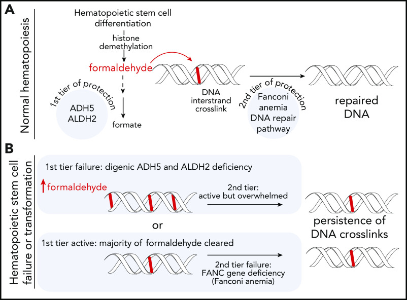
Differentiation of HSCs creates formaldehyde, which is highly reactive and forms DNA ICLs and other DNA and protein adducts. (A) Normal hematopoiesis. The majority of the formaldehyde is detoxified by the first tier of protection including ADH5, ALDH2, and possibly other enzymes active in HSCs. The leftover DNA crosslinks are repaired by the FA-repair pathway (second tier of protection) resulting in normal hematopoiesis. (B) HSC failure or transformation. Either of the tiers may fail leading to overlapping cellular and patient phenotypes including bone marrow failure, myelodysplastic syndrome, and leukemia. DNA damage in cells deficient for both ADH5 and ALDH2 (ADH5−/−ALDH2*2/+) seems to overwhelm the FA DNA-repair pathway, and patients present with a syndrome akin to FA but with a negative chromosome breakage test upon treatment with DEB or MMC. In FA, the first tier of protection works, but even the low levels of reactive aldehydes, when not cleared from the DNA, cause HSC demise.
Together with 3 recent papers,2-4 a strong case is made that the cause of this failure is DNA damage overload secondary to endogenous formaldehyde produced during hematopoietic stem cell (HSC) differentiation.
Aldehydes, such as acetaldehyde and formaldehyde, may cause DNA damage if not detoxified. Acetaldehyde is best known as a metabolite of consumed ethanol. Formaldehyde is not only an environmental carcinogen, but it is also produced in vivo during various metabolic processes, including histone demethylation, and 1-carbon metabolism.3,5
HSCs are particularly vulnerable to aldehyde-induced DNA damage, a weakness uncovered using mouse models of Fanconi anemia (FA), the most common IBMFS. FA results from a defect in the FA-repair pathway, which removes DNA interstrand crosslinks (ICLs). Concomitant loss of FANCD2 (a key protein in this pathway) and ALDH2 (which detoxifies endogenous aldehydes) resulted in loss of HSCs, induction of γ-H2AX (a marker of DNA damage), and high genomic instability.6,7 Even more striking, bone marrow failure, before the age of 2 months, was observed in mice with a combination of deficiencies in FANCD2 and ADH5, enzymes involved in formaldehyde metabolism.8 These findings demonstrated that an increased load of reactive aldehydes, caused by the deficiency in ALDH2 or ADH5 enzymes, leads to DNA damage in HSCs that requires a functional FA pathway for repair. This led to the concept of “2-tier genome protection,” in which detoxifying metabolic enzymes provide the first tier of protection by eliminating genotoxins, and the FA pathway provides the second tier of protection by repairing any DNA damage caused by such genotoxins that escaped the first line of clearance (see figure).9
The clinical relevance of these findings was validated with the identification of humans with mutant ALDH2 in the setting of FA-pathway defects. The ALDH2*2 allele, which greatly diminishes cellular ALDH2 activity, is prevalent in East Asians. When found in FA patients, it led to accelerated development of bone marrow failure, myelodysplastic syndrome, and acute leukemia, and resulted in more extensive congenital anomalies than in those who had wild-type ALDH2.10
Increased DNA damage caused by a high aldehyde load appeared to be clinically significant only in the setting of a DNA-repair disorder like FA. This was true until the discovery of a new IBMFS, ADH5/ALDH2 deficiency,2 also called AMeD syndrome (for anemia, mental retardation, and dwarfism).4 Patients with ADH5/ALDH2 deficiency have normal DNA-repair pathways; however, high levels of reactive aldehydes, likely formaldehyde, overwhelm the DNA-repair capacity in their HSCs. This results in a bone marrow dysfunction phenotype akin to FA, requiring HSC transplantation. Other shared features are skin pigmentation changes, low birth weight, and short stature with or without microcephaly. The most significant differences are the lack of chromosome fragility/breakage after treatment with ICL-inducing agents (mitomycin C [MMC] or diepoxybutane [DEB]), and the lack of radial-ray defects or thumb abnormalities. Intellectual disability, although not generally seen in FA, has been a uniform feature of ADH5/ALDH2 deficiency.2,4
In this article, Mu et al address how human cellular phenotypes and hematopoiesis are perturbed in ADH5/ALDH2 deficiency using patient-derived lymphoblasts, fibroblasts, iPSCs, and CRISPR-engineered cell lines. Patient-derived phytohemagglutinin-stimulated lymphoblasts, unlike patients’ fibroblasts, exhibit high levels of sister chromatid exchange, a marker of endogenously produced DNA damage being repaired by homologous recombination. These variations indicate that different levels of DNA damage are present in different tissues and cells, or that different types of cells have a varying degree of sensitivity to reactive aldehydes.
Experiments performed using the iPSCs were the most revealing. The authors created 2 independent cell lines from an ADH5/ALDH2-deficient patient. As expected, the patient-derived iPSCs were sensitive to exogenous treatment with formaldehyde, which induced substantial DNA damage. Importantly, these phenotypes were attenuated upon expression of ADH5. The patient iPSCs, when grown in unperturbed conditions, were able to grow well but had a significant late S/G2 delay, which is also seen in the cells of FA patients. The phenotype worsened dramatically when these cells were forced to differentiate into hematopoietic progenitors. No progenitor colonies were formed upon plating of CD34+ KDR+ cells, a defect that was reversed by expression of ADH5. Addition of an ALDH2 activator had a small positive effect on the differentiation. CD34+ KDR+ cells derived from the iPSCs activated the FA DNA-repair pathway as shown by high levels of FANCD2 present in nuclear foci. However, the repair was overloaded as γ-H2AX was also increased in these cells. Studies in the wild-type iPSCs, created to have different combinations of ADH5 and ALDH2 deficiencies, revealed that cells deficient for both enzymes stopped differentiation post CD34+ stage.
This and the other recent studies beautifully illustrate that ADH5/ALDH2 deficiency causes bone marrow failure due to increased DNA damage secondary to endogenous metabolites. The bone marrow failure is not caused by an inherited DNA-repair deficiency. This is a clear example that the DNA-repair pathways can be overwhelmed during hematopoiesis. The finding of intellectual disability in the ADH5/ALDH2 deficiency highlights that the brain cells may also experience overload of DNA-damage-repair pathways, including transcription-coupled repair and protein-crosslink repair, both of which are also expected to repair formaldehyde-induced DNA damage.
The human genome contains at least 19 ALDH and 8 ADH superfamily genes, with some overlapping substrate specificities. It remains to be seen whether other combinations of ADH/ALDH deficiencies drive DNA damage significant enough to overwhelm the second tier of genome protection and cause human disease. The fact that hematopoietic defects were improved by modulation of ADH5 or ALDH2 activity ex vivo gives hope that the levels of reactive aldehydes may be decreased leading to less DNA damage and healthier stem cells. Whether decreasing the aldehyde load by pharmacologic agents could prevent bone marrow failure or cancers in ADH5/ALDH2-deficient patients, patients with FA, or during aging needs further investigation.
Footnotes
Conflict-of-interest disclosure: Rocket Pharmaceuticals provides research funding and partial salary support to A.S. M.J. declares no competing financial interests.
REFERENCES
- 1.Mu A, Hira A, Niwa A, et al. Analysis of disease model iPSCs derived from patients with a novel Fanconi anemia–like IBMFS ADH5/ALDH2 deficiency. Blood. 2021;137(15):2021-2032. [DOI] [PubMed] [Google Scholar]
- 2.Dingler FA, Wang M, Mu A, et al. Two aldehyde clearance systems are essential to prevent lethal formaldehyde accumulation in mice and humans. Mol Cell. 2020;80(6):996-1012.e9. [DOI] [PMC free article] [PubMed] [Google Scholar]
- 3.Shen X, Wang R, Kim MJ, et al. A surge of DNA damage links transcriptional reprogramming and hematopoietic deficit in Fanconi anemia. Mol Cell. 2020;80(6):1013-1024. [DOI] [PMC free article] [PubMed] [Google Scholar]
- 4.Oka Y, Hamada M, Nakazawa Y, et al. Digenic mutations in ALDH2 and ADH5 impair formaldehyde clearance and cause a multisystem disorder, AMeD syndrome. Sci Adv. 2020;6(51):eabd7197. [DOI] [PMC free article] [PubMed] [Google Scholar]
- 5.Burgos-Barragan G, Wit N, Meiser J, et al. Mammals divert endogenous genotoxic formaldehyde into one-carbon metabolism [published correction appears in Nature. 2017;548(7669):612]. Nature. 2017;548(7669):549-554. [DOI] [PMC free article] [PubMed] [Google Scholar]
- 6.Garaycoechea JI, Crossan GP, Langevin F, et al. Alcohol and endogenous aldehydes damage chromosomes and mutate stem cells. Nature. 2018;553(7687):171-177. [DOI] [PMC free article] [PubMed] [Google Scholar]
- 7.Langevin F, Crossan GP, Rosado IV, Arends MJ, Patel KJ. Fancd2 counteracts the toxic effects of naturally produced aldehydes in mice. Nature. 2011;475(7354):53-58. [DOI] [PubMed] [Google Scholar]
- 8.Pontel LB, Rosado IV, Burgos-Barragan G, et al. Endogenous formaldehyde is a hematopoietic stem cell genotoxin and metabolic carcinogen. Mol Cell. 2015;60(1):177-188. [DOI] [PMC free article] [PubMed] [Google Scholar]
- 9.Garaycoechea JI, Patel KJ. Why does the bone marrow fail in Fanconi anemia? Blood. 2014;123(1):26-34. [DOI] [PubMed] [Google Scholar]
- 10.Hira A, Yabe H, Yoshida K, et al. Variant ALDH2 is associated with accelerated progression of bone marrow failure in Japanese Fanconi anemia patients. Blood. 2013;122(18):3206-3209. [DOI] [PMC free article] [PubMed] [Google Scholar]


