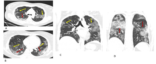
Figure 2. Typical COVID 19. Transverse CT images shows multiple ground-glass opacities (A, B yellow arrow) and mixed ground glass opacities and consolidation in both of the lower lung lobes (A, B, red arrow). Coronal reconstruction CT shows peripheral multifocal GGO (C, D yellow arrow) and consolidation (D red arrow) in both upper and lower lung lobes.

