

Abstract
Sociality has profound evolutionary roots and is observed from unicellular organisms to multicellular animals. In line with the view that social principles apply across levels of biological complexity, a growing body of data highlights the remarkable social nature of mitochondria – life-sustaining endosymbiotic organelles with their own genome that populate the cell cytoplasm. Here, we draw from organizing principles of behavior in social organisms to reveal that similar to individuals among social networks, mitochondria communicate with each other and with the cell nucleus, exhibit group formation and interdependence, synchronize their behaviors, and functionally specialize to accomplish specific functions within the organism. Mitochondria are social organelles. The extension of social principles across levels of biological complexity is a theoretical shift that emphasizes the role of communication and interdependence in cell biology, physiology, and neuroscience. With the help of emerging computational methods capable of capturing complex dynamic behavioral patterns, the implementation of social concepts in mitochondrial biology may facilitate cross-talk across disciplines towards increasingly holistic and accurate models of human health.
Keywords: Social behavior, Mitochondrial dynamics, Networks, Communication, Interdisciplinary, Psychobiology, Biomarker
1. Introduction
Social behavior refers to the interactions that take place among individuals of the same species. Social groups shape the development of their members and favor the emergence of complex behaviors that single individuals would otherwise not develop in isolation (Apicella et al., 2012). Social groups and communities also combine individuals’ abilities and limitations such that they mutually enhance and complement each other, enabling tasks and achievements that would not be possible to attain by any single individual. Social ant colonies, for example, build elaborate anthills, while social humans build institutions of higher education, multi-story buildings, and cultures. As a result, social systems and the individuals that comprise them behave in ways not readily described by the intrinsic properties of each individual. Rather, within social networks, the behavior of each individual is motivated and, thus, best explained and predicted by the combination of both biological and contextual factors, in which social forces play a prime role.
In this perspective, we discuss evidence that leads us to propose that mitochondrial behavior is similarly best understood from a social lens. This view puts a special emphasis on communication processes and the exchange of information within the mitochondrial network – both within cells and across the organism. We propose that applying social principles and social network theory to mitochondrial research will foster cross-talk among scientists from different disciplines and, as a result, help develop more comprehensive and accurate models of the biological processes that underlie human health. A social view of mitochondrial behavior can influence key aspects of the scientific process, including the kind of questions we ask (i.e., becoming more integrative and less reductionistic), the type of data we collect (i.e., richer in terms of kinetics and levels of analyses), and the analytical strategies we apply (i.e., increasingly requiring computational approaches capable of identifying complex patterns within the data). Here, we begin by comparing social principles across levels of organization from mitochondria to human cultures, then provide examples of mitochondrial behavior aligned with key social principles, and finally discuss the implications of a social mitochondria perspective for the biomedical sciences.
1.1. The route towards sociality
Throughout the living world, humans emerge as extraordinarily social (Wilson, 1974). Our capacities for cooperation, coordination, and division of labor make human social behavior particularly distinctive from other species (Boyd and Richerson, 2009). However, sociality is not a privative attribute of humans. It has deep evolutionary roots and is present among the evolutionary continuum from unicellular organisms to multicellular animals. Unlike inert molecules whose behavior and fate are determined solely by physical forces exerted upon them, living organisms actively interact with their environment and with each other in social reciprocity. Sociality is believed to have evolved for its adaptive nature: by being social, individuals would increase their fitness, defined by their probability of survival and reproductive success (Hamilton, 1964a; Silk, 2007a). Even unicellular microorganisms like bacteria – once believed to be independently behaving – rely on external sources of nutrients provided by their social counterparts of the same or other species (Zengler and Zaramela, 2018). Bacteria also routinely join forces, coordinate their gene expression and create structures (e.g., biofilm) that promote survival of the whole community (Lyon, 2007; Nadell et al., 2009). These prokaryotic unicellular life forms from which more complex animals have evolved already contained the foreshadowing traces of more complex social behavior (Damasio, 2018).
The next step in the evolution of social behavior was endosymbiosis that gave rise to mitochondria and the eukaryotic cell. A mutually beneficial interaction between two types of cells that likely created the necessary conditions for multicellularity (Lane and Martin, 2010). From this, followed small multicellular invertebrates and vertebrate organisms, including colonial insects like ants who exhibit social behaviors and establish complex cooperative social organization (Simola et al., 2016). Most other life forms that followed, including lactating mammals, have evolved in a deeply socially interdependent way where mother-infant dyads, family units, and social circles play a central role in shaping development, adult behavior, and health (Sandi and Haller, 2015).
Sociality can increase individuals’ fitness (Hamilton, 1964a; Silk, 2007b). Social interactions are essential to health outcomes and their disruption leads to pathological states (Sandi and Haller, 2015). Social support (e.g., positive interactions between adults or maternal care for babies and infants) acts as a buffering mechanism under challenging situations, protecting individuals from exaggerated physiological stress responses (Ditzen and Heinrichs, 2014; Rincon-Cortes and Sullivan, 2014). For example, social support through hand holding (physical contact) is associated with coupling of brain-to-brain activity among dyads and dampens physical pain perception (Goldstein et al., 2018). Conversely, negative social exchanges – such as peer victimization (Quinlan et al., 2018), marital conflict (Wilson et al., 2017) or loss of social rank among hierarchical animals (Larrieu and Sandi, 2018) – are strongly associated with stress-related psychopathologies, highlighting how social experiences are ingrained into our biology. Furthermore, sociality can also incur physiological costs with effects on individual health by affecting individuals’ energy balance. Individuals who live in groups can experience different degrees of intra-group competition for access to food, and – under conditions of food restriction – those with lower competitive capacities tend to have reduced access to caloric intake (Kappeler et al., 2015). There are also potentially important costs to group living, such as the higher probability of socially transmitting infections and the negative health consequences deriving from chronic stress triggered by social competition (Kappeler et al., 2015). Thus, social forces have shaped evolution of complex life and continue to shape animal behavior at multiple levels.
The idea that human societies and biological organisms present important similitudes in their principles and levels of organization has been put forward by a number of evolutionary biologists (Hallpike, 1985) and more recently as a molecular sociobiology proposal (Foster, 2011). However, the focus of these proposals remains at the organ or cellular level. These proposals rightly point out how organs within an organism, or the cells within an organ, cannot survive on their own and therefore rely on a set of reciprocal social interactions. They also emphasize the notion that elements (i.e., cells, organs) group together to acquire novel emergent functions. Here, we extend these principles to the social dimension of sub-cellular organelles.
1.2. Sociality goes sub-cellular
Considering the social dimension that takes place between cells and their organelles is essential to improve current conceptualizations of human physiology and behavior. In recent years, the prevailing biomedical model has begun to shift from purely reductionistic explanations of biology and behavior centered on a prime role of DNA and genomics (Jung and Goldman, 2018; Plomin and Crabbe, 2000) to the integration of the environment in interaction with DNA (Caspi and Moffitt, 2006). However, the influencing environment is frequently envisioned either from a narrow perspective – e.g., referring to extreme experiences, such as early life stress or trauma; or too general point of view – e.g., referring to lifestyle or sociocultural factors (Quinn et al., 2018). Such gene-environment models, focused on the individual in a way somewhat divorced from their social nature, have brought us some distance along the path towards more complete and accurate models to describe the factors that influence the development of living organisms. However, these approaches fall short of explaining critical aspects of human evolution and health states (Jacob, 1977).
Every aspect of animal life – from brain growth and maturation (Atzil et al., 2018), the development of language for communication (Hoff and Tian, 2005), specialization and division of labor within the community (Robinson and Barker, 2017), physical fitness and disease risk (Aral and Nicolaides, 2017; Datar and Nicosia, 2018; Hermes et al., 2009), and lifespan (Luo et al., 2012; Steptoe et al., 2013) are shaped not only by bottom-up intrinsic biological forces and general aspects of the environment, but also by interactions with other individuals; i.e., by social interactions. The acquisition or development of several behaviors like language, and the brain connectivity that normally subserve these behaviors, cannot proceed without social interactions – highlighting the deep integration of the social context in which individuals exist and acquire specific health-defining behaviors. Thus, to understand human health trajectories, our models must include both the biological constitution of individuals themselves as well as the social principles that describe how individuals behave and influence each other.
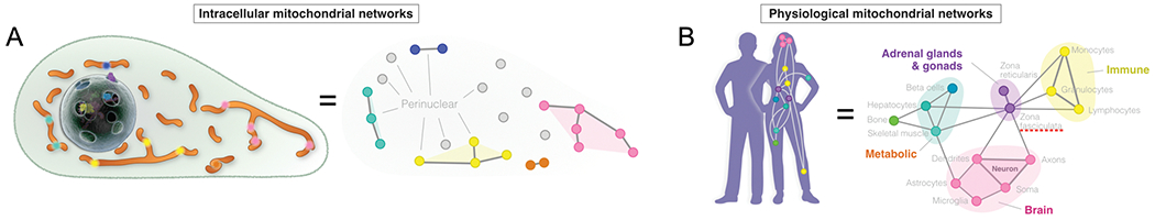
Below we draw from knowledge around known principles that best explain the behavior of organisms living in social groups, and examine how these principles apply across levels of organization, including at the cellular and sub-cellular levels (Fig. 1). Our goal is not to discuss the sociology or biology of these processes comprehensively, but rather to highlight their similarities in nature and to examine their role in sustaining health processes. We focus on mitochondria – endosymbionts with their own genome that populate the cytoplasm of most cells in the organism (Fig. 2) – as a growing body of data highlights the remarkable social nature of these organelles.
Fig. 1. Dynamic communication across multiple levels of organization.
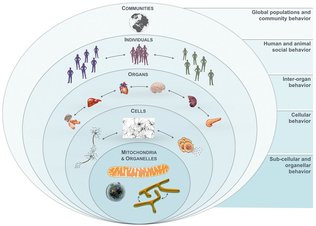
Social communities are composed of individuals, which are composed of organs, which are made of cells, which are themselves made and sustained by organelles - including mitochondria. Information flows bidirectionally, from organelle to communities, and from communities to organelles. The Community level involves organized groups of individuals bound by geographical, political, national, or institutional organizations that shape global behaviors. The Individual level includes persons with unique biological and psychological characteristics, who form groups and interact with one another. The Organ level involves genetically-identical units (organs), but with highly specialized anatomy and functions, which cooperate to sustain physiological function and allostasis. The Cellular level also involves units with the same genome but which exhibit the most developed degree of diversity and functional specialization. Cells are the simplest unitary element of life. They have the ability to perceive molecular information and to transduce it to higher levels of complexity, eventually reaching the individual’s consciousness and influencing social behavior. The Mitochondria & organellar level involves specialized sub-cellular elements that communicate, cooperate, and perform various specialized functions that collectively enable cells, organs, and organisms’ adaptation to challenges.
Fig. 2. Essentials of mitochondrial biology.
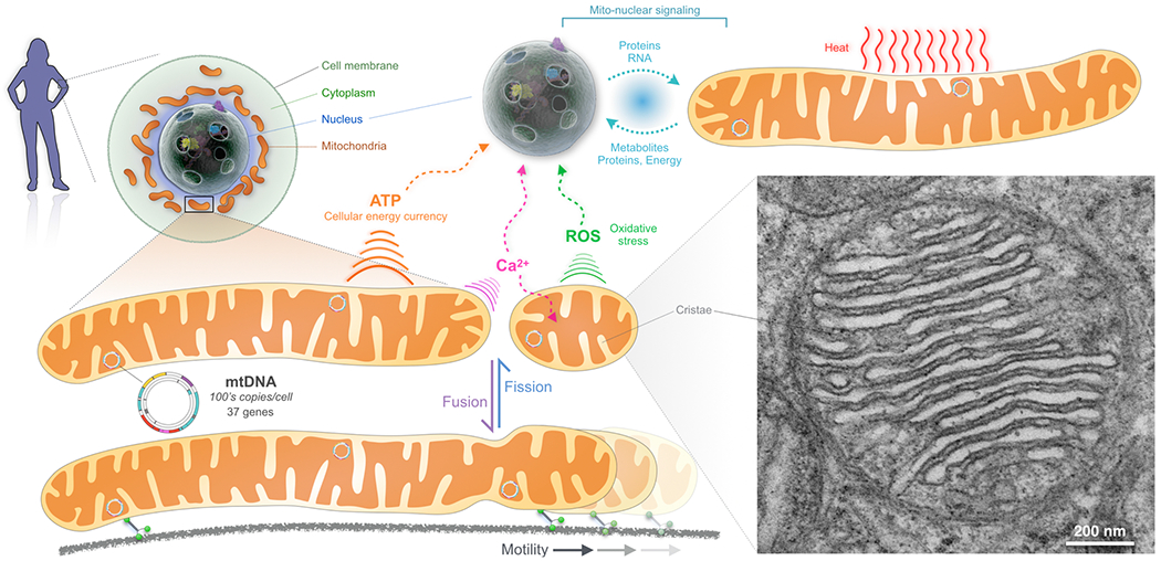
Mitochondria are small organelles that live within each cell of the human body. They are bacteria-derived, having evolved. They are bacteria-derived, having evolved over a billion years ago from aerobic bacteria incorporated into the then unicellular ancestor of the eukaryotic cell (Margulis and Bermudes, 1985). This endosymbiotic fusion made multicellular complex life possible – it gave rise to breathing, moving, thinking, feeling multicellular organisms with complex bodies like humans and other social creatures (Lane and Martin, 2010). Mitochondria share the cell cytoplasm with other organelles with whom they constantly interact (Klecker et al., 2014; Valm et al., 2017). But they are the only organelle to have their own genome – a vestige of their bacterial ancestry – the circular mitochondrial DNA (mtDNA) (Wallace, 2010). The mtDNA encodes elements of the respiratory chain located within the inner mitochondrial membrane, which consumes the oxygen we breathe and generates a membrane charge (like small batteries) from the food that we eat. This charge, called membrane potential, is used to generate adenosine triphosphate (ATP) that fuels most cellular activities, as well as other functions including ionic regulation, hormone synthesis, antioxidant detoxification, and many others (Nicholls and Fergusson, 2013). Mitochondria are also the warmest cellular component, likely maintained at >50 °C (Chretien et al., 2018). Mitochondria thus sustain life and enable stress adaptation by producing energy (Picard et al., 2018a), as well as other signals that remodel gene expression, and shape cellular and organism behavior and lifespan (Latorre-Pellicer et al., 2016; Picard et al., 2015b; Sharpley et al., 2012). Human cells and their genetic material packaged inside the nucleus depend on mitochondria for their existence, and reciprocally mitochondria depend on the nucleus for their existence – a state of co-dependence. The nuclear genome influences mtDNA stability and the evolution of the mtDNA sequence over generations (Wei et al., 2019). Reciprocally, in most cells, mitochondria are abundant near the nucleus and influence gene expression (Picard, 2015). The abundance of mitochondria (or mitochondrial mass) in a given cell can also increase following elevations in energy demand, including in response to exercise (Neufer et al., 2015; Steiner et al., 2011). Cells and their sub-cellular compartments where energy requirements are greatest have more mitochondria than less energy-demanding cells. Mitochondria are thus distributed “on demand” in different cellular compartments (e.g., synapses, cell body) and in an activity-dependent manner (Magarinos et al., 1997). Mitochondria of various sizes and shapes also move about within cells in a process driven both by mitochondria themselves and by local cellular demands. Mitochondria interact dynamically with each other in various ways including fusion, where two smaller mitochondria fuse their membranes to become a single larger one; and where a larger mitochondrion undergoes fission to generate two smaller “daughter” mitochondria (Friedman and Nunnari, 2014). Defects in dynamic mitochondrial shape changes or motility are a cause of human disease (Archer, 2013), and mitochondrial dynamics influence cellular and organismal responses to stressors of various nature (Eisner et al., 2018).
Anatomically, mitochondria have a characteristic double outer and inner membrane organization, with infoldings of the inner mitochondrial membrane where oxygen is consumed (see electron micrograph, bottom right). Although these features are ubiquitous, the morphology, set of functions, and motility of mitochondria are cell-type and tissue-specific such that mitochondria from different tissues are molecularly and functionally distinct (e.g., (Calvo et al., 2016; Picard et al., 2012)). Abbreviations: ROS, reactive oxygen species; mtDNA, mitochondrial DNA; Ca2+, calcium; ATP, adenosine triphosphate
Examining mitochondrial behavior through a social lens uncovers essential properties of mitochondria that cannot be reduced to their intrinsic molecular composition and that are best captured by the nature of their complex interactions in the context of the organism. Once implemented, this view should help to achieve a more complete understanding of the principles and mechanisms that guide human physiology and that is required to improve our predictive models of human health. Such models would explain, for example, how individuals adapt in the face of stress or infection, remain healthy or get sick, and develop as adult individuals capable of making meaningful social contributions. Eventually, this knowledge could revolutionize our capacity to intervene in personalized ways to promote adaptation, health, and human development.
2. Social principles apply across levels of biological complexity
Several contributions from evolutionary biology (Hallpike, 1985), molecular sociobiology (Foster, 2011), and cell biology (Diogo et al., 2018) have remarked a close resemblance of the organizing principles governing human societies and biological organisms. For instance, they state that, both human societies and biological organisms react to physical environments and rely on mechanisms of communication, self-maintenance, and feedback. The specialization of organ-based functions is also regarded as reminiscent of the specialization of social division of labor in societies (Smaldino, 2014) – specific organs ensure energy supply (e.g., stomach, digestive system, and adipose tissue), others ensure locomotion (e.g., skeletal system and muscles), others provide protection (e.g., bones and skin), and others ensure coordination of functions (e.g., brain, peripheral nervous system). Organs, like many social animals, can only subsist by working together.
The view that social principles apply across levels of biological complexity was greatly influenced by the consideration of sociality as one of the key drivers of the major evolutionary transitions (Szathmary and Smith, 1995). Revolutionizing the traditional evolutionary biology view of life on Earth as a linear succession of taxonomic groups, Szathmary and Maynard Smith (Szathmary and Smith, 1995) conceived evolution as the result of a few critical transitions, each of them moving from a group of independent entities to a new, more complex, living unit. After each transition, entities lose their capacity to replicate independently and cooperate together within the new unit (West et al., 2015b). Accordingly, organisms – as the essential units of adaptation – are regarded as multicellular individuals composed of groups of organs that establish high levels of cooperation and very low conflict (Queller and Strassmann, 2009). Likewise, organs represent the unitary trait that emerges from the same kind of interconnection and cooperation among cells (Smaldino, 2014). In agreement with this theory, our view considers biological organisms as social groups and goes beyond this idea by emphasizing trans-dimensional social interactions across different levels of organization and biological complexity, including the organellar level.
The fundamental determinants and characteristics of social behavior are strikingly conserved across levels of biological complexity (see Fig. 1). In order to exemplify the pervasiveness and importance across levels of organization of one of these principles, namely communication, we consider here parallels between human behavior and the brain.
In human communities and societies, communication is necessary for virtually all categories of social activities including planning, decision-making, and coordinating actions (Paarporn et al., 2018; Smith, 2010; Sueur et al., 2011). Humans employ a broad range of written, verbal, and non-verbal forms of communication and apply them to develop and disseminate norms and conventions for social organization (McAdams, 1997). Language extends the type and scope of collective actions by enabling the exchange of information about temporally and spatially distant events (Hauser et al., 2002). Valuable collective projects in human societies (e.g., constructions, sociomedical research, financial markets) could not be achieved without dedicated communication mechanisms, including face-to-face meetings, telecommunication, the internet, and other communication channels (Smith, 2010). Several of these mechanisms – e.g., email, text messaging, and telephone – are redundant, such that one or multiple systems may be used to transmit the necessary information in the most efficient manner. Communication disorders (e.g., hearing impairment or other) predict smaller social network size, fewer positive social exchanges, less integration in a social life, and higher levels of loneliness (Palmer et al., 2016). Eventually, failure to communicate causes poor interpersonal exchanges that can lead to the demise of social endeavors (Baek, 2014; Christensen and Shenk, 1991), highlighting the essentiality of communication in human social behavior.
At the level of organs and cells, communication is similarly enabled by a large number of channels – hormones and neurotransmitters, for example – many of which also have redundant actions. The sophisticated communication streams at the organ level are best exemplified by the brain, whose most fundamental feature is the communication between its cellular units, neurons and glial cells. Brain neurons display specializations for mutual interactions involving substantial diversity and complexity. Different forms of inter-neuronal communication, including the development and pruning of specialized structures (i.e., synapses), are required for normal development and function (Pereda, 2014). Beyond direct physical intercommunication through gap junctions for example, neurons can work together in groups by synchronizing their behavior in oscillatory activity (Buzsaki et al., 2013), a mechanism that, among other functions, enables higher-level behavior among individuals, including speech processing and understanding language (Meyer, 2018). And here also, communication disorders between individual units of the brain are understood to be the basis for psychopathology (e.g., depression). They also constitute the basis for pharmacological treatments that act on neurotransmitters that carry information from one cell to the next. Furthermore, brain cells and their neurotransmitters have a highly developed capacity to interact bidirectionally with other physiological systems, such as the gut (Codagnone et al., 2019), the immune system (Miller et al., 2017), or the adrenal glands (Sarrieau et al., 1986). As a result, communication also takes place across multiple organs, highlighting the essentiality of communication in the function of the organism.
At the sub-cellular level of organelles, communication occurs via the exchange of metabolites, proteins, ions, and membrane potential conveyed, as well, by multiple redundant processes. Therefore, although the substrates of communication are relatively unique to each level of complexity, the underlying principles and importance of communication for effective social behavior are shared across human, organ, cellular, and sub-cellular levels.
In summary, social behavior is not only pervasive within and across levels of organization in social animals, but social principles (such as communication) represent fundamental mechanisms in the organism that sustain animals’ existence and shape their behavior. Next, we establish the similitude of some major social principles with the life-sustaining organelles that populate the cell: mitochondria.
3. Elements of social systems and mitochondrial behavior
Mitochondria exhibit several essential characteristics of social organisms. Below, we discuss six essential principles and how they manifest in mitochondrial behavior (Fig. 3): (1) shared environment, (2) communication, (3) group formation, (4) synchronization of behavior, (5) functional interdependence, and (6) specialization and division of labor.
Fig. 3. Conserved principles of social behavior across levels of complexity.
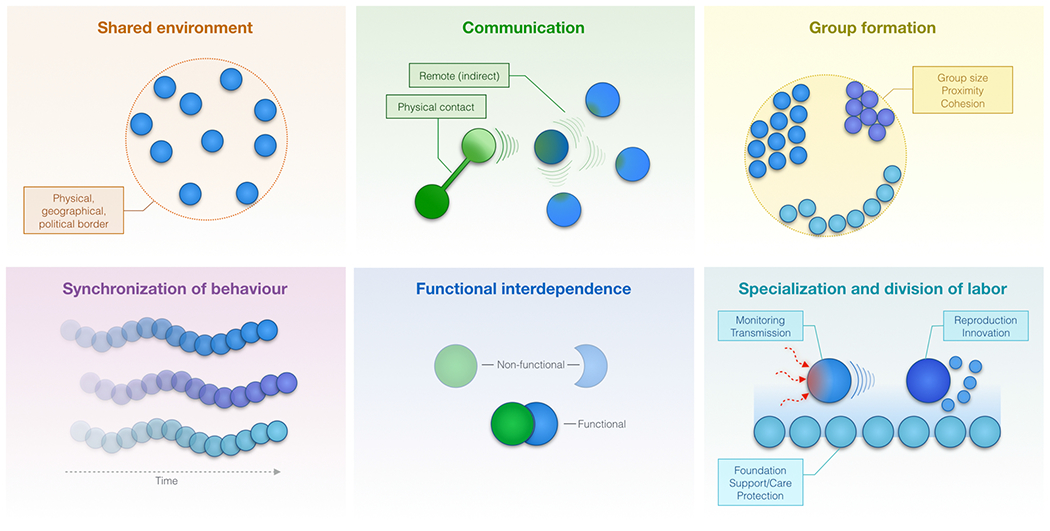
The behavior of social units is characterized by key principles, six of which are illustrated here. Social individuals generally i) share a common environment or physical space, ii) communicate with each other to share information about their own state or about the state of the environment, iii) form groups of various sizes and that acquire specific characteristics, iv) synchronize their behavior in time and space, v) are functionally interdependent, requiring other units to complement or support each other to achieve optimal function (i.e., fitness), and vi) functionally specialize and divide labor to collectively accomplish communal goals or functions. These principles apply to social networks, as well as to mitochondrial communities that populate the cell cytoplasm.
3.1. Shared environment
A shared environment is a distinctive feature of cultures and societies. In social groups living in a particular ecosystem, variation in habitat and diet are closely associated with the size and structure of the group (Clutton-Brock et al., 2009). When confined, a shared environment can largely determine individuals’ characteristics. Darwin’s finches or Galapagos tortoises, which shared distinct and confined environments in their evolutionary past, deviated in several characteristics from descendants of their common ancestors that lived in geographically more accessible areas, including the shape of their beaks and shells (Hedrick, 2019). In humans also, geographical borders produce groups of individuals that differ in their behavior and beliefs (Onnela et al., 2011), and disparities in physical and mental health among ethnic minorities can emerge in geographically-defined ways (Alarcon et al., 2016).
Similarly, large networks of mitochondria – counted in 100-1000’s when physically fragmented (Scheffler, 2008) – share the same immediate environment bound by the cell membrane. All members of the mitochondrial community within a given cell share the same cytoplasm, which includes information about the cell cycle stage, its differentiation status, activation state, and second-messengers from external stimuli as well as internal activities. Mitochondria within a given cell also share the same cell nucleus, which provides many of the gene products and proteins essential to harmonize mitochondrial behavior within the cell. Moreover, different organs are composed of specific cell types (e.g., neurons, cardiomyocytes, hepatocytes) that contain mitochondrial populations with distinct molecular compositions (Calvo et al., 2016). Thus, both social animals and mitochondria coexist in spatially-defined, shared environments that contribute to shape and preserve distinct populations of individuals, which engage in constant communication with each other.
3.2. Communication
As described above in Section 2, communication is an essential element of social organizations. It is supported by multiple redundant mechanisms, enables both close and distant exchanges among social networks, and impairment in social communication alters healthy functioning of social groups.
Similarly, several redundant mechanisms exist to facilitate communication among mitochondria. Physical membrane structures called inter-mitochondrial junctions (Picard et al., 2015a) and mitochondrial nanotunnels (Vincent et al., 2017) physically connect mitochondria and enable information exchange while retaining individual organelle boundaries. A more complete mechanism towards molecular information exchange is the fusion of two (or more) mitochondria into larger ones, leading to the more complete mixing of individual properties from the fusing mitochondria. Like human interactions, mitochondrial fusion can either be long-lasting, or partial and brief (kiss-and-run) (Liu et al., 2009). Moreover, through their life cycle, mitochondria are born from one another through the process of synonymous division (i.e., biogenesis), enabling the transmission of physical and functional properties (Scheffler, 2008) and possibly the propagation of a “trans-generational” memory. Chemical signals such as reactive oxygen species and calcium can also act as diffusible signals that transmit information without direct physical contact between mitochondria (Garcia-Perez et al., 2012).
As in human social networks and in brain neural circuits, the exchange of information among functionally networked mitochondria may enable groups of mitochondria to “perceive” incoming signals and respond as a collective unit, leading to some degree of signal integration within the mitochondrial network (Picard, 2015). Impairing communication among mitochondria by blocking mitochondrial fusion increases molecular heterogeneity among the intracellular mitochondrial community (Chen et al., 2005), causes the accumulation of damage in the mitochondrial genome (Chen et al., 2010), and reduces the energy transformation potential of the mitochondrial population (Bach et al., 2003). Moreover, these negative consequences reach beyond the malfunction of individual mitochondria and can lead to demise of the cell and of the organism during development (Chen et al., 2003). Moreover, mitochondria also communicate with other organelles through the exchange of soluble chemical signals at inter-organellar contacts, such as the endoplasmic reticulum (Csordas et al., 2018). Thus, communication is a shared principle that directly influences the behavior of both social animals and mitochondria.
3.3. Group formation
Groups are defined as a number of individuals or units that are related or located close to one another. Although not exempted from potential disadvantages (e.g., increased competition or higher rates of disease transmission) (Kappeler et al., 2015), the ubiquitous principle of aggregation (i.e., individuals living together in groups) in social animals generally provides substantial fitness advantages. Social aggregation and group living can help protect individuals from predatory threats (Krause and Ruxton, 2002), improve foraging efficiency (Pulliam and Caraco, 1984), and enhance individuals’ ability to sense and respond to environmental changes (Berdahl et al., 2013). A key driving force determining which individuals come together is the similarity in their physiological capacities (e.g., aerobic capacity and fitness) (Killen et al., 2014; Seebacher and Krause, 2017). Accordingly, the functioning as a group largely relies on the physiological underpinnings that determine how conspecifics perceive and process information from each other. Furthermore, inter-individual differences in energetic resources may affect patterns of motion and rest, determining different levels in fatigue resistance that, eventually, may contribute to passive or active sorting of social groups (Seebacher and Krause, 2017). Across evolution, social animals including humans have increased the size and complexity of groups (Dunbar and Shultz, 2007), highlighting in parallel with more complex forms of behavior the likely evolutionary advantage of group formation.
Within cells, mitochondria form groups that can differ in their fitness and topologically localize in different cellular compartments. As mentioned above, in cells with highly polarized morphology like neurons, different compartments such as the cell body (i.e., soma), dendrites, and axons have remarkably different populations of mitochondria. Somatic mitochondria vary in shape from spheroids to moderately elongated tubules; dendritic mitochondria exist as extremely long and branched stationary organellar networks; and axonal mitochondria are small motile units (Lewis et al., 2018). Mitochondria can cluster near the plasma membrane (Porat-Shliom et al., 2019), at cell-cell membrane contact sites (Gomez-Cabanas et al., 2019), around lipid droplets (Benador et al., 2018), or around the cell nucleus (Al-Mehdi et al., 2012) where they tend to aggregate with similarly behaving mitochondria and remain for the duration of their life cycle. In cases of disease where specific mitochondria undergo changes in their capacity or behavior, mutant mitochondria also aggregate, forming functionally distinct groups (Vincent et al., 2018). As another example, young and old mitochondria with different fitness selectively segregate prior to cell division, such that young and old mitochondria are differentially passed on to distinct daughter cells (which impacts downstream cell fitness) (Katajisto et al., 2015). Thus, similar to humans and other social animals, functionally and morphologically similar mitochondria exhibit group behavior.
3.4. Synchronization of behavior
Synchrony is essential to the lives of group-living organisms (Couzin, 2018) and is a vital phenomenon that manifests across all levels of biological complexity (Li and Yang, 2018). Synchronization serves multiple functions. For a group to stay cohesive, group members need to synchronize their activities, a phenomenon particularly relevant in animals during foraging and migration, where large groups migrate in unison (Seebacher and Krause, 2017). Synchronization facilitates information processing (Couzin, 2018) and allows rapid coordinated responses to threats (e.g., among a herd or swarm) (Noy et al., 2011). In humans, collective behavior is a major principle in various contexts, from collective motion (e.g., coordinated team playing in group sports) and activity synchronization (e.g., dynamic movements in crowds), to decision making (Biro et al., 2016). In fact, humans have unique capacities for coordination – the orchestration of individuals activities in an orderly manner, and coordination is essential to several facets of human societies. Mother-infant synchrony is a guiding principle for the development of positive cognitive and behavioral outcomes among children (Leclere et al., 2014). Simple and repeated synchronous interactions between individuals are also responsible for creating complex adaptive group patterns (Sumpter, 2006). Multiple group-living social animals also exhibit highly coordinated collective patterns (e.g., global movements in a school of fish) believed to correspond to a form of biological self-organization (Couzin, 2009) resulting from the local interactions among individuals located in each other’s immediate surrounding (Biro et al., 2016). The principles that support this type of self-organization include positive feedback, response thresholds, individual integrity, and variability that can lead to oscillatory behaviors (Sumpter, 2006). In the human body, coordinated processes manifesting as oscillations are also ubiquitous: a wide range of biological processes such as neuronal firing, heart beats, cell cycles, and circadian rhythms exhibit synchronized oscillatory behaviors (Li and Yang, 2018). In the brain, coordinated neuronal oscillations also allow for multiple-time-scale communication within and across neuronal networks (Buzsaki et al., 2013).
Similarly, in mitochondria, synchronized oscillations in membrane potential occur in cardiomyocytes where mitochondria are electrically coupled to one another (Aon and Cortassa, 2012; Kurz et al., 2010). In a different organ, the salivary gland, mitochondria communicate between cells through gap junctions and behave as a network of functionally coupled oscillators (Porat-Shliom et al., 2014). Spontaneous events of mitochondrial depolarization and rapid changes in superoxide levels, called “flashes”, also propagate at high speed between neighboring mitochondria – large groups of mitochondria flash synchronously (Santo-Domingo et al., 2013). Both membrane potential oscillations (Porat-Shliom et al., 2014) and flash propagation (Santo-Domingo et al., 2013) occur even when the mitochondria are not physically continuous with each other, pointing to the role of soluble signals for inter-mitochondrial communication. Moreover, the internal physical structure of mitochondrial membranes (i.e., the cristae) can also become coordinated at inter-mitochondrial junctions where two energized mitochondria are in close contact (Picard et al., 2015a), which may in part contribute to the functional coupling required for synchronization of behavior of juxtaposed mitochondria. Thus, synchronization of behavior is a shared principle extending from large groups of behaving animals to sub-cellular mitochondrial communities.
3.5. Functional interdependence
Individuals from highly social species depend on one another for various aspects of their fitness (Aktipis et al., 2018; Silk, 2007b). Interdependence among social animals account for cooperation and altruism (Roberts, 2005), including unique forms of human cooperation (Tomasello and Gonzalez-Cabrera, 2017). By helping group partners, individuals are not only investing in their partners’ fitness but also, consequently, in their own as well (Aktipis et al., 2018; Maynard Smith, 1964). Most social species limit their cooperative behaviors (i.e., acting for the common benefit of other individuals in the social group) to small groups frequently made up of relatives, and reciprocal interactions (Smaldino and Epstein, 2015). However, human societies can also reach extremely high levels of cooperative behavior, benefiting not only close relatives but also unrelated individuals. Examples of cooperative behavior in human societies range from the constructions of buildings and bridges to selfless actions such as charities and blood donations (Melis and Semmann, 2010). The latter type of actions, known as ‘altruism’ – the type of cooperative behavior directed to benefit other individuals at a cost to those that perform it – may be explained by its eventual (direct or indirect) fitness benefits for the individual that exerts the cooperative behaviors (Bourke, 2014; Hamilton, 1964a, b; West et al., 2007, 2006). Direct fitness may result when cooperation arises from the mutual dependence for survival or reproduction (termed fitness interdependence) (Aktipis et al., 2018), while indirect fitness arises when relatives are favored, increasing their reproductive success (Maynard Smith, 1964). From birth, humans are highly dependent on their caretakers. In this context, the establishment of early life attachment – a form of interdependence – between the infant and caregiver ensures optimal development and is essential for brain development, which determines long-term well-being (Sullivan et al., 2011). Globally, the spectrum of human activities embedded in human social networks progressively involves interdependent relationships, with billions of people now increasingly relying on each other for safety and the provision of various goods and skills.
At the subcellular level, fitness interdependence also accurately describes the relation between mitochondria and the cell nucleus: the nucleus requires mitochondria for energy and metabolic intermediates necessary for gene expression (Gut and Verdin, 2013), while mitochondria depend upon the nucleus to generate most of their proteins that enable their multifarious nature (Calvo et al., 2016). Thus, mitochondria and nucleus depend on each other for their fitness and survival. Mito-nuclear signaling, which consist in the exchange of proteins and chemical signals between the mitochondrial population and the nucleus, shapes how the cell as a whole responds to stress and challenges (Cardamone et al., 2018; Nargund et al., 2012; Quiros et al., 2016). In addition, mitochondrial biogenesis and the associated changes in metabolism (i.e., from glycolysis to oxidative phosphorylation) can influence cell fate decision in neural stem cells, eventually affecting neurogenesis, and thereby having an impact in cognition (Arrazola et al., 2019).
Mitochondria also exist in a cooperative relationship with each other. This interdependence is most clearly observed in mitochondrial fusion and fission processes. Upon increased cellular energy demands or reduced energy supply, mitochondria fuse to increase effectiveness (Gomes et al., 2011; Rambold et al., 2011). Exercise also increases the number of physical contacts between mitochondria in skeletal muscles (Picard et al., 2013a). Importantly, as described in the section on mitochondrial communication, perturbing mitochondrial fusion eventually leads to the accumulation of mtDNA defects and the demise of the cells (Chen et al., 2010), revealing the essential nature of fusion-type cooperative mitochondrial interactions. Conversely, new mitochondria arise from previously established ones via the reciprocal process of fission. When intracellular calcium rises during cell stimulation, mitochondria act as calcium buffer and their ability to buffer calcium is significantly enhanced by their interactions with each other in the fused state (Paltauf-Doburzynska et al., 2004). Larger populations of mitochondria also naturally have greater calcium buffering capacity (Bianchi et al., 2006), consistent with the notion that mitochondria cooperate to handle cytoplasmic environmental challenges. Genetically, mitochondria also functionally complement each other. Isolated mitochondria without a genome or with a mutant genome can be rescued by fusing with healthy conspecifics, restoring their normal function and ensuring overall mitochondrial population health (King and Attardi, 1989; Ono et al., 2001). Furthermore, as evidence that mitochondrial physical interactions influence the behavior of the cells that contain them, mitochondrial dynamics influence neurogenesis within the brain (Khacho et al., 2016, 2017). Therefore, as for social animals, normal and optimal mitochondrial function is achieved through cooperative interactions among conspecifics, such that mitochondria exist as cooperative and functionally interdependent organelles.
3.6. Specialization and division of labor
In many social species, high levels of inter-individual variation are optimal to enhance efficiency in solving the problems the group may be facing (Farine et al., 2017; Jolles et al., 2017). From social arthropods (Holldobler and Wilson, 1990) to humans (Agryris, 1973), within-group structural and functional variation enhances task efficiency and fitness (Pruitt and Riechert, 2011). A more deliberate form of functional variation is specialization: specific individuals within a population perform particular tasks, and develop tailored abilities or constitutions to optimally perform specific task(s). Division of labor is one of the hallmarks of social living. The extraordinary human capacity for division of labor is a defining characteristic of all forms of organization in our societies. It manifests as the specialization in the performance of particular social roles, professions, and division of activities, followed by the subsequent exchange of products among social members. In economics, division of labor is also considered essential for the wealth of nations (Rosenberg, 1965).
Across the organism, functionally and molecularly specialized mitochondria acquire characteristics that allow them to perform specific tasks necessary for survival. Tissues with highest energetic demands, like the heart, contain dense mitochondrial populations with proteome and internal anatomy (dense cristae) optimized for energy transformation and ATP synthesis (Johnson et al., 2007; Piquereau et al., 2010). In the adrenal glands, mitochondria are morphologically distinct and specialize not in energy production, but in steroid hormone synthesis (Midzak and Papadopoulos, 2016). Thus, across the organism, organ systems, and cell types, mitochondria assemble in groups that perform specific tasks or labor that complement each other and are essential to the functioning of the whole body (Picard et al., 2018a). For example, heart and brain mitochondria require steroid hormones for their normal maturation, but the production of these hormones is outsourced to adrenal and gonadal mitochondria that specialize in steroidogenesis.
Even within single cells, distinct groups of perinuclear, peridroplet, and synaptic mitochondria differ in composition and morphology (see Section 3.3. Group formation). For example, neuronal mitochondria located either in the cell body and in synapses differ substantially from somatic mitochondria in their protein composition (Stauch et al., 2014), marking their distinct molecular identity and functional specialization. In other cell types, such as skeletal muscle fibers, mitochondria also exist as either globular mitochondria near the nucleus, or as long tubular mitochondria optimized for energy production located among contractile myofibrils (Picard et al., 2013b; Vincent et al., 2019). Functionally, specialization and division of labor ensures that different groups of mitochondria are optimized and matched to the specific cellular environment they inhabit (Benard et al., 2006; Picard et al., 2012). Physiologically, several metabolites related to mitochondrial metabolism are produced by certain organs, and consumed by other ones, highlighting the systemic division of metabolic labor that interconnects organ-specific mitochondria as an interdependent network (Jang et al., 2019). Thus, similar to humans and other social animals who develop unique set of behaviors and outsource others based upon immediate environmental demands, different groups of mitochondria distributed across the organism functionally specialize and divide labor, enabling complex organismal functions.
3.7. An integrated social perspective of mitochondria
In this section, we have highlighted six key social principles shared between human and mitochondrial behavior. In addition, mitochondria exhibit at least three other behaviors that are not specifically “social” but that share similarities with complex social organisms. First, like other social animals, mitochondria have a life cycle: new mitochondria are born from existing ones, age, and eventually reach end of life and dye to be replaced by younger ones (Twig et al., 2008). Second, similarity to individuals of a same species, mitochondria within an organism are genetically related, sharing the same maternally inherited genome (Giles et al., 1980), except in specific cases of disease. Third, like social animals that collectively respond to environmental cues, mitochondria have the ability to “sense” and process information – to perceive, respond, and generate signals (Picard and McEwen, 2018) – such that in response to complex stressors like psychological stress, they release functional and physiologically-meaningful signals (e.g., Trumpff et al., 2019).
Mechanistically, several molecular components that account for specific aspects of the complex behaviors discussed here have been defined. For example, the motor proteins necessary for mitochondrial fusion and some of the molecular switches required for their activation have in part been defined (Friedman and Nunnari, 2014). Moreover, their regulation in response to metabolic perturbations and intracellular signaling events have also been partially elucidated (Eisner et al., 2018). Here, we propose that mitochondria are social organelles whose behavior and functions will most accurately be modeled by incorporating our growing knowledge on the molecular mechanisms enabling their behaviors, together with the social principles discussed above. The consilience of biological and sociological knowledge at the cellular and sub-cellular levels has key implications for both biomedical and behavioral sciences.
4. Biological implications of a social perspective on mitochondrial behavior
To summarize the view that we have put forward in previous sections, mitochondrial structure and function are intrinsically dependent upon their sociality. In turn, this social dimension has implications for our understanding of health and disease processes at both the organelle and organismic levels, and for guiding our scientific approaches designed to investigate biological mechanisms udnerlying cellular and sub-cellular processes (Fig. 4).
Fig. 4. Flow of information and concepts from the human social sciences, physiological sciences, and sub-cellular biology.
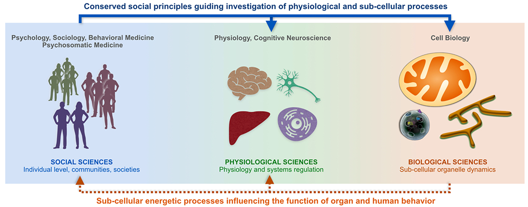
Bi-directional flow of information across disciplines can promote discovery and insight into complex behaviors. Interdisciplinary dialogue leads to insight not readily achievable by each domain in isolation (Disis and Slattery, 2010).
First, the importance of mitochondrial communication emphasized in this social perspective has direct implications for the parameters - i.e., what is being measured - that should be collected to address mechanisms of health and disease. For example, following the traditional but limited view of mitochondria as cellular powerhouses, scientific attempts to examine the role of mitochondrial involvement in disease progression naturally focus on bioenergetic parameters (i.e., measures of oxidative phosphorylation processes and ATP levels) (Chacko et al., 2014; Picard et al., 2018b). In contrast, recent work has highlighted the role of mitochondria-derived molecules, including proteins (Fuku et al., 2015; Kim et al., 2018) and DNA (Nakahira et al., 2013; Trumpff et al., 2019) as key effectors in the transduction of information in response to psychological and metabolic stressors.
In a similar way that brain disorders arise from abnormal communication between otherwise normal neurons (Gazzaniga, 2013; Jiang et al., 2019), impaired communication between otherwise normally functioning mitochondria could be a cause of dysfunction and disease. In our view, empirical approaches aiming to reveal mechanisms of health and disease need to incorporate assessments of the multiple channels whereby mitochondria communicate and interact – both locally within the cell, and systemically within the organism. If this is correct, quantifying the nature and timing of mitochondrial communication will be instrumental in generating better predictive models to explain how cells and organisms respond to challenges, including extrinsic and intrinsic stressors (Eisner et al., 2018). Developing methods to dynamically quantify the integrity of mitochondrial communication systems in health and disease could in turn lead to a new generation of biomarkers (see below).
Second, we highlight the interconnected nature of mitochondria for their normal and abnormal function and behavior. Similar to social animals for whom a sense of social connectedness is essential to well-being (Lee et al., 2001) and who form networks interconnected by social and other bonds, mitochondria are interconnected units that cannot exist as isolated organelles. In this context, it is important to mention that, in some studies, experimental approaches involving mechanical isolation of mitochondria directly alter their function in comparison to cellular permeabilization approaches that preserve mitochondrial structure (Picard et al., 2011). In humans, interconnectedness is reflected in phenomena such as ‘emotional contagion’ (Hazy and Boyatzis, 2015), forms of which also occur in other social animals (Buchanan and Preston, 2017; Carnevali et al., 2017). Infectious and non-communicable diseases (as well as health behaviors) also spread through social ties in human social networks (Hill et al., 2010). Another implication of interconnectedness is the so-called ‘bad apple’ effect in which violation of group rules by one individual (e.g., dishonest reporting, harassment, discrimination or other) may be sufficient to decrease the engagement, group identification, and performance of other members (Felps et al., 2006). Thus, behaviors can spread between individuals interconnected within a social network.
Similarly, a class of medical disorders, known as mitochondrial disease, emerge from the spread of abnormally behaving mitochondria throughout the mitochondrial network (Picard et al., 2016). In certain cases of mitochondrial diseases, a single mitochondrion can acquire a mtDNA mutation, change its behavior, and begin following rules different to those of the rest of the network. As in human social networks where the influence of bad apples propagates rapidly, and as in cancer where invasive cancerous cells proliferate to the detriment of surrounding cells, dysfunctional mitochondria with genetic anomalies develop abnormal behavior, produce aberrant signals, and highjack the cell nucleus to promote their own proliferation (Gitschlag et al., 2016; Lin et al., 2016). For reasons that remain unknown, mutant mitochondria can avoid the endogenous cellular quality control system of “mitophagy” and persist over time in human tissues, as evidenced by their accumulation with age and in disease. Thus, ‘bad apple’ mutant mitochondria can take advantage of the resources within the network to propagate as a group in social competition with normal resident mitochondria in living tissues like skeletal muscle (Vincent et al., 2018). Consequently, identification and elimination/restoration of aberrant mitochondria early in a disease could be a critical approach to prevent disease progression.
Given that mitochondria play a central role in sustaining cellular life and stress adaptation (Picard et al., 2018a), characterizing the social principles that shape mitochondrial behavior therefore expands the spectrum of the potential forces that enable life and (mal)adaptive processes generally. Physiologically, the social mitochondria perspective illustrates how health is a distributed process that depends upon multiple levels of interaction (i.e., communication, synchronization, cooperation, etc.) among organs, cells, and organelles. This emphasizes the limitation of overly reductionist models. It has been argued that the search for simple causal mechanisms (e.g., single genes) in common complex diseases has yielded relatively little insight (Moraes and Goes, 2016).
In contrast to the mainstream biomedical view, the social view of mitochondria emphasizes how dynamic molecular, organellar, cellular, and organismal processes enable the emergence of complex properties and behaviors (Gao et al., 2016; Han et al., 2017), rather than linear causal models linking single genes or proteins to disease. The obvious corollary is the substantial increase in the complexity of associated research questions that this view entails. We therefore envision that future models of both mitochondrial function and human health and behavior will benefit from the application of computational models capable of integrating variables at multiple levels. Developing testable and generalizable predictions about the behavior of mitochondria and individuals may draw inspiration from social network theory and computational social science (Fig. 5).
Fig. 5. Novel approaches to model mitochondrial behavior.
One approach to modeling human social behavior that may effectively be extended to mitochondrial behavior is graph theory. At the level of mitochondria (Left), graph theory can be applied to map the network topological distribution of mitochondria within a cell (Sukhorukov and Meyer-Hermann, 2015) and to evaluate the functional topology and clustering of individual mitochondria within cells from a particular organ (Kurz et al., 2014). Mitochondrial networks exhibit quantifiable topology based on physical interactions with one another within the cell cytoplasm. Adjacent mitochondria communicate and have the capacity to influence each other, and the networks can be dynamically remodeled within seconds to minutes via fusion/fission and motility. Mitochondria can also undergo inter-cellular transfer (Dong et al., 2017; Hayakawa et al., 2016; Tan et al., 2015) and thus migrate within and between existing networks. Across the organism (Right), mitochondria from one cell type/organ system communicate with mitochondria in other cell types (for example though the release of mitokines and steroid hormones (Durieux et al., 2011; Picard et al., 2018a)).
The central idea of social network theory is that units within a system are interconnected, such that information flows from one unit to another (Kadushin, 2012; Onnela et al., 2007). In the case of human social networks, friendship, ideas, money, disease, and behaviors are socially transmitted through existing networks (Apicella et al., 2012; Fowler and Christakis, 2010; Salali et al., 2016). In the case of organelles and mitochondrial networks, proteins, membrane potential, lipids, heat, and mtDNA are also transmitted among members of the intracellular networks (Dolgin, 2019; Ono et al., 2001).
In computational social science and other fields, networks are typically represented as graphs, developed from the branch of mathematics called graph theory (Davis, 1970; Ohtsuki et al., 2006). Among various kind of networks, information diffuses – over time – through the connections (i.e., edges) linking different units (i.e., nodes or vertices) following fundamental principles of networks and physics (Lynn and Bassett, 2019). Social networks have the ability to describe highly complex organizations and account for emergent properties, including the emergence of communities with common internal properties despite great individual diversity within a population (Han et al., 2017). Social networks can also explain the social contagion of health behaviors like smoking (Christakis and Fowler, 2008), obesity (Christakis and Fowler, 2007), and exercise (Aral and Nicolaides, 2017), as well as general properties of complex network systems such as resilience (Gao et al., 2016) that are not reducible to properties of its individual components. In this emerging systemic view, all mitochondria within the organism represent a single network of interacting organelles distributed across cells and organ systems.
Applying social network theory to build robust predictive models of mitochondrial behavior will require both technical and theoretical developments to analyze new kinds of mitochondrial datasets (e.g., time lapse, multivariate imaging or multiomic) (see next section). The resulting models should also allow explanation and prediction of not only the behavior of mitochondria themselves, but also their influence on the cells and on the organism (e.g., person) that contains them – including mapping processes that sustain human health and adaptive capacity.
5. Implications of a social mitochondria perspective for behavioral and biomedical research
When applied to behavioral and biomedical sciences, the emerging view of mitochondria as social organelles highlighted in previous sections has critical implications for the understanding of human health and disease. On the one hand, it reveals how pervasive and relevant social processes are to physiology and cell biology, opening up new ground to examine and map the physiological principles underlying human behavior. Consequently, this social view urges us to rethink the simplistic biological foundations of behavior and to consider abnormal mitochondrial social interactions among potential disease-causing mechanisms. Moreover, the social nature of mitochondria may also assist us in reconceptualizing health and disease biomarkers and offer opportunities to develop a new generation of biomarkers.
5.1. Abnormal mitochondrial communication and interconnectedness in disease
Although there has not been a systematic study of this concept, there is sufficient evidence in the literature to support the view that abnormal mitochondrial communication and interconnectedness can impact individual’s behavior and disease. Mitochondrial communication with each other relies heavily on mitochondrial dynamics, which includes fusion, fission, turnover, trafficking, and contacts with other organelles (see Fig. 2) (Gordaliza-Alaguero et al., 2019). Critical molecular players that regulate mitochondrial dynamics are dynamin-related protein 1 (DRP1) that catalyzes division, and mitofusins 1 and 2 (Mfn1/2) and optic atrophy 1 (OPA1) involved in fusion (Friedman and Nunnari, 2014). Mfn2, although not exclusively engaged in mitochondrial fusion, is a protein located in the outer mitochondrial membrane which tethers and mediates fusion between organelles, and whose expression levels can provide cues about the propensity of mitochondria to fuse and communicate. In humans, decreased Mfn2 expression in the brain is observed in neurodegeneration (Lee et al., 2012; Wang et al., 2016, 2009) and a Mfn2 single nucleotide polymorphism (SNP) was recently found to be associated with late-onset Alzheimer’s Disease (Kim et al., 2017). In animals, reduced Mfn2 expression was found following a chronic stress-induction of anxiety-like behaviors (Chakravarty et al., 2013) and in rodent models of depression (Chen et al., 2014; Liu and Zhou, 2012), suggesting that psychopathology may be associated with impaired mitochondrial social interactions. Importantly, Mfn2 is tightly regulated; whereas factors such as proinflammatory cytokines, or stress hormones (i.e., glucocorticoids) tend to block its expression, exercise tends to promote its upregulation (Zorzano et al., 2015). Specific behavioral response to nutrient challenges, feeding behavior, and obesity (Dietrich et al., 2013; Schneeberger et al., 2013), as well as the endocrine regulation of insulin secretion by pancreatic β cells (Ramirez et al., 2017) and by various stressors (Eisner et al., 2018) are also regulated by mitochondrial interactions with each other and their surroundings. Overall, animal behavior appears linked to mitochondrial social behavior.
Other signaling mechanisms involved in mitochondrial communication, such as reactive oxygen species (ROS), can also impact behavior and disease. Mitochondrial ROS production in excess of antioxidant defenses (i.e., oxidative stress) has been documented in the majority of human neurodegenerative diseases (Angelova and Abramov, 2018; Xiao et al., 2017) and psychopathologies, including high anxiety (Filiou and Sandi, 2019), depression (Siwek et al., 2013), or schizophrenia (Gonzalez-Liencres et al., 2014; Koga et al., 2016). In rodents, alterations in ROS and mitochondrial respiration in specific brain regions involved in motivation and aggression were found to be associated with high anxiety with reduced social competitiveness (Hollis et al., 2018, 2015). In addition, alterations in mitochondrial communication with the cell nucleus, an essential process for maintaining cellular homeostasis and ensuring effective responses to stress (Quiros et al., 2016), appears implicated in physiological dysfunctions typically observed in aging (Lee et al., 2013). Furthermore, increasing evidence supports the view that mitochondrial signaling through ‘mitokines’ is central to communicate mitochondrial stress from the nervous system to peripheral tissues (Picard and McManus, 2016; Zhang et al., 2018) possibly facilitating the genetic and epigenetic regulation of cellular aging and organismal lifespan (Schinzel and Dillin, 2015).
On the other hand, health-promoting interventions, such as dietary manipulations or exercise, can regulate ROS levels (Filiou and Sandi, 2019; He et al., 2016), promote mitochondrial function, brain metabolism, and expressions of fusion/fission mediators (Cherix et al., 2020; Liu and Zhou, 2012). Importantly, these health-promiting interventions can also increase the number of physical interactions among adjacent mitochondria (Picard et al., 2013a) – changes that collectively promote mitochondrial communication. Mitochondrial communication may also prevent the accumulation of mutant mitochondrial genomes over time (Picard and Turnbull, 2013). Furthermore, emerging evidence suggests a key role for mitochondrial dynamics in the regulation of exercise performance and adaptations to endurance exercise training (Moore et al., 2019), and anxiolytic (van der Kooij et al., 2018) and antidepressant (Villa et al., 2017) drugs can also regulate different aspects of brain mitochondrial function. Together, these lines of evidence suggest that the shared origin of pathogenic processes underlying common neurodegenerative and psychiatric disorders could lie in abnormal mitochondrial communication and perturbations of their social behavior.
5.2. Rethinking biomarkers to quantify mitochondrial social behavior
The extension of social principles across levels of biological complexity is a theoretical shift that leads to reconceptualize biomarkers and their use in the biomedical sciences and in neuroscience. A simplistic view of biomarkers is that they are “spill over” by-products from the diseased tissue, marking the presence of a pathological process – like smoke marks the presence of fire. However, the proposed view on the central role of signaling and communication suggests that certain biomarkers may in fact be substrates of communication whose levels rise or fall, either as part of normal adaptive processes, or to compensate for defective communication or functions. For example, in diabetes mellitus the levels of insulin rise not because of a primary pathological hypersecretory process within the pancreas, but because the communication with the target organs is impaired by insulin resistance within the target organ (Hoehn et al., 2009). This leads to a compensatory excess release of insulin in the circulation in an attempt to achieve effective signaling. Thus, impaired organ-to-organ communication in diabetes results in chronically elevated insulin. Therefore, a key question to ask in the context of this perspective is: What are the equivalent signals of impaired mitochondria-to-mitochondria communication? Given that the main purpose of communication consist in signaling and directing adaptative strategies in the face of change, we envision two main challenges and opportunities to developing novel biomarkers that reflect impaired mitochondrial communication.
First, novel biomarkers must capture dynamic processes. Because organisms are never in steady state and the flux of information changes constantly in response to internal and environmental processes, communication is naturally a dynamic process. This may be particularly true in the prodromal phase of disease, before symptoms manifest, as the organism musters multi-system neuroendocrine, immune, and metabolic processes to sustain states of health (McEwen, 2006). This process is referred to as allostasis (Schulkin and Sterling, 2019). To capture these regulatory processes from a social communication perspective, rather than relying on static and idealized measures (e.g., fasting blood draw, inflammatory marker), we predict that it will be of utmost importance to implement dynamic measures over different time scales. Some forms of communication between cells occur within seconds while other forms of communication occur over minutes to hours – both of which contain valuable information about the state of the organism.
This dynamic measures principle has been applied successfully in other fields such as cognitive neuroscience. For example, connectivity patterns extracted from time series of whole brain imaging data acquired over several minutes can yield predictive activity signatures that identify what state a person is in (e.g., pain (Kragel et al., 2018)) or can be used as a fingerprint to distinguish individuals (Finn et al., 2015). Different aspects of human behavior, such as personality and cognitive performance, are also differentially explained by functional connectivity metrics measured either dynamically at the time scale of seconds, compared to “static” measures computed from a single average over long time periods (Liegeois et al., 2019). This temporal dependence highlighting the distinct information content at different time scales is typical of complex systems. Thus, new insights into the underlying mitochondrial social behavior across cells and organs could be obtained by extending this principle to mitochondria-derived stress mediators and blood-based mitokines, including the steroid hormones that are produced in mitochondria (Bose et al., 2002; Midzak and Papadopoulos, 2016). A general framework to capture dynamic, multivariate signatures from time series mitokine data is illustrated in Fig. 6.
Fig. 6. Dynamically mapping stress-evoked systemic mitokine signatures across time scales to capture mitochondrial social behavior and regulation in health and disease states.
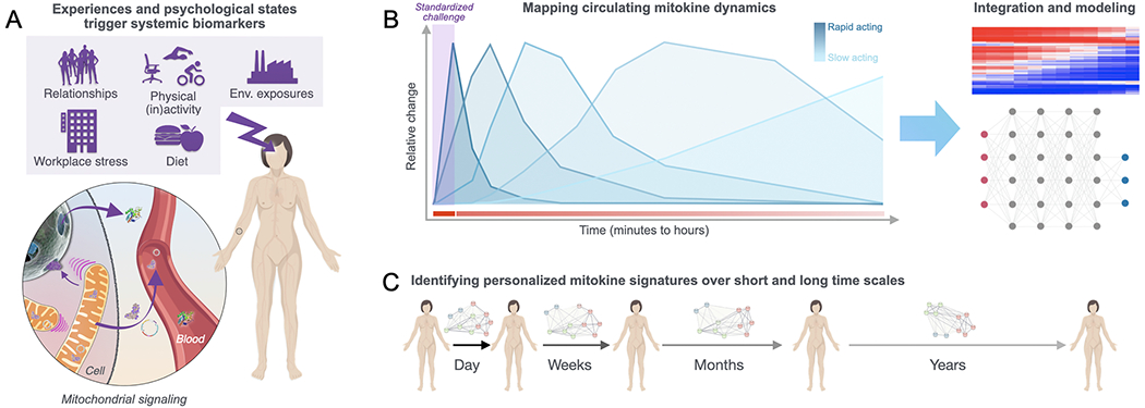
(A) Psychosocial, environmental, and behavioral factors influence the individual’s psychological states and triggers mitochondrial (and cellular) signaling and communication. Mediators of mitochondrial communication enter the circulation as mitokines. (B) Theoretical mitokine dynamics over time in response to a standardized challenge, including biomarkers with different response kinetics. Mitokines and other endocrine, metabolic, and immune parameters collected with targeted or untargeted approaches can be integrated into multivariate computational models, which are either unsupervised or constrained to different degree by prior biological knowledge. Current challenges in this direction include measuring a sizeable number of relevant markers with sufficient temporal resolution to derive (and validate) robust predictive signatures. (C) Establishing personalized mitokine signatures will require that we understand how stable/variable signatures are across time scales, and how we can leverage this information to generate insights that will guide personalized health-promoting interventions. This may help fulfill the vision of precision medicine.
Second, biomarkers of dysregulated processes are generally best revealed in response to challenge. For instance, in medicine, diabetes is diagnosed by measuring the levels of glucose and insulin following a hyperglycemic challenge, or glucose tolerance test (GTT) (Stumvoll et al., 2000). Challenge-based results offer more sensitive and reliable diagnosis than single baseline assessments (Gerstein, 2001), because at baseline a number of compensatory processes can mask underlying dysregulation. In relation to mitochondria, preclinical studies have similarly shown that complex multisystem neuroendocrine, metabolic, and transcriptional signatures of mitochondrial dysfunction, otherwise not present under baseline conditions, are revealed with a brief psychogenic stress (Picard et al., 2015b). Similarly, an emerging mitokine induced by stress is circulating cell-free mtDNA (cf-mtDNA) present in human blood, which is sensed by immune receptors and may trigger inflammatory responses in model systems (West et al., 2015a; Zhang et al., 2010). In humans, although ccf-mtDNA levels may be consistently elevated in some psychopathological states (Lindqvist et al., 2016, 2018), ccf-mtDNA selectively increases within minutes of a psychological challenge (Hummel et al., 2018; Trumpff et al., 2019) or strenuous physical activity (Stawski et al., 2017). Chronic stressors like job strain may also increase circulating levels of mitokines (Shamaei-Tousi et al., 2007), suggesting a link between psychosocial states and systemic mitochondrial signaling and communication (Picard and McEwen, 2018).
Furthermore, as indicated above, a ubiquitous life challenge that results in mitokine release is aging. Preliminary work suggests that aging-associated elevation in ccf-mtDNA may contribute to the chronic inflammatory state known as ‘inflammaging’ (Pinti et al., 2014). Recently, a protein induced in the nucleus secondary to signaling from dysfunctional mitochondria (Khan et al., 2017), GDF15, was identified as the top upregulated protein in the plasma of aged healthy individuals (Tanaka et al., 2018), indicating that signaling molecules either directly produced by mitochondria (cf-mtDNA) or indirectly induced (GDF15) represents a pervasive process not restricted to disease.
In the future, harnessing both principles of dynamic measures and response to challenge may allow us to further understand how mitochondria communicate across the organism, and how their communication (or disorders thereof) relates to states of health and disease. Outstanding questions include defining to what extent mitochondrial signaling measured at different time scales influences whole-body processes, such as vulnerability versus resilience to stress-related disorders. To what extent are mitokine signaling signatures prognostic indicators of specific disorders, or predictors of aging trajectories across the lifespan? Developing dynamic measures of mitochondrial communication mediators is an exciting prospect to tackle for interdisciplinary scientists.
6. Conclusions
Developing accurate models to predict and influence health trajectories is a shared goal across the behavioral and biological sciences. Although reductionist models have successfully been applied in some domains of biology, they are insufficient to capture and predict complex individual-level behaviors and health states. Under the impetus of increasingly large and complex datasets, major steps forward to overcome the limitations of reductionist science are taking place in other fields such as systems biology (Bzdok et al., 2019), personalized medicine (Nielsen, 2017; Yu et al., 2018), computational social science (Conte et al., 2012; Lazer et al., 2009), and computational neuroscience (Kragel et al., 2018; Lynn and Bassett, 2019). In the field of mitochondrial and cell biology, specifically, early attempts to capture the complexity of mitochondrial behavior include computational models that map the behavior of mutant mitochondria and their impact on cells and tissues (Hoitzing et al., 2019), simple multivariate indices of bioenergetic parameters (Chacko et al., 2016; Picard et al., 2018b), and other indices of mitochondrial behavior in humans (Picard et al., 2019). There remains much room for progress.
A critical drawback of existing biomedical health/disease models is that they generally do not consider the dynamic social behavior of individuals and elements across levels of organization – communities of people, organs, cells, and organelles. In our view, a social-biological perspective of mitochondria (and other biological processes) could critically advance the development of more comprehensive and accurate models to guide biomedical research. Eventually, such a perspective could also impact health care. To implement this view, it will be necessary to develop novel experimental tools that can generate high-dimensional datasets that accurately capture the dynamic social behavior of organisms, including communication among key components (i.e., nodes) in the system. Currently, it is not possible to molecularly interrogate, at scale, individual mitochondria in free living humans. However, appropriately scaled high-resolution imaging approaches in cultured cells may provide opportunities to map some aspects of complex mitochondrial behavior, in vitro. However, if our goal is to address mitochondrial behavior within the human organism, a challenge will be to generate datasets with sufficient levels of resolution and rigor necessary to test and validate complex models directly relevant to human health. Only by applying the appropriate methods and analytical framework can we establish the extent to which the social mitochondria perspective is accurate and generalizable, and the degree to which it improves our ability to predict future health states. Engaging in this interdisciplinary effort to map the social forces within organisms, as well as their underlying molecular mechanisms, is certain to contribute to the strides that appear necessary to develop predictive models of human health and behavior.
Finally, we note that a social-mitochondrial perspective emphasizes the central role of communication in biology. This leads to the inspiring possibility that social and bioenergetic principles can guide the development of communication-based biomarkers that dynamically identify and quantify both health states and global perturbations among organisms – early before disease manifests. Mapping communication and dynamic regulatory processes to predict health in a personalized way could lead to evidence-based interventions, building upon socio-bioenergetic principles, to foster health and resilience across the lifespan.
Acknowledgements
Work of the authors is supported by the Wharton Fund, NIH grants GM119793, MH119336, MH122706 (MP), the Swiss National Science Foundation (31003A-152614; 31003A-176206; and NCCR Synapsy51NF40-158776, 51NF40–185897), the EPFL-Jebsen Foundation and intramural funding from the EPFL (CS). The authors are grateful to Judyann McNamara, Alan Cohen, and members of the Mitochondrial Psychobiology laboratory for stimulating discussions.
Footnotes
Data availability statement
No datasets were generated or analysed during the current study
References
- Agryris C, 1973. Personality and Organization. Harper & Brothers, New York, NY [Google Scholar]
- Aktipis A, Cronk L, Alcock J, Ayers JD, Baciu C, Balliet D, Boddy AM, Curry OS, Krems JA, Munoz A, Sullivan D, Sznycer D, Wilkinson GS, Winfrey P, 2018. Understanding cooperation through fitness interdependence. Nat. Hum. Behav 2, 429–431. [DOI] [PubMed] [Google Scholar]
- Al-Mehdi AB, Pastukh VM, Swiger BM, Reed DJ, Patel MR, Bardwell GC, Pastukh VV, Alexeyev MF, Gillespie MN, 2012. Perinuclear mitochondrial clustering creates an oxidant-rich nuclear domain required for hypoxia-induced transcription. Sci. Signaling 5, ra47. [DOI] [PMC free article] [PubMed] [Google Scholar]
- Alarcon RD, Parekh A, Wainberg ML, Duarte CS, Araya R, Oquendo MA, 2016. Hispanic immigrants in the USA: social and mental health perspectives. Lancet Psychiat. 3, 860–870. [DOI] [PubMed] [Google Scholar]
- Angelova PR, Abramov AY, 2018. Role of mitochondrial ROS in the brain: from physiology to neurodegeneration. FEBS Lett. 592, 692–702. [DOI] [PubMed] [Google Scholar]
- Aon MA, Cortassa S, 2012. Mitochondrial network energetics in the heart. WIRES Syst. Boil. Med 4, 599–613. [DOI] [PMC free article] [PubMed] [Google Scholar]
- Apicella CL, Marlowe FW, Fowler JH, Christakis NA, 2012. Social networks and cooperation in hunter-gatherers. Nature 481, 497–501. [DOI] [PMC free article] [PubMed] [Google Scholar]
- Aral S, Nicolaides C, 2017. Exercise contagion in a global social network. Nat. Commun 8, 14753. [DOI] [PMC free article] [PubMed] [Google Scholar]
- Archer SL, 2013. Mitochondrial dynamics–mitochondrial fission and fusion in human diseases. N. Engl. J. Med 369, 2236–2251. [DOI] [PubMed] [Google Scholar]
- Arrazola MS, Andraini T, Szelechowski M, Mouledous L, Arnaune-Pelloquin L, Davezac N, Belenguer P, Rampon C, Miquel MC, 2019. Mitochondria in developmental and adult neurogenesis. Neurotoxic Res. 36, 257–267. [DOI] [PubMed] [Google Scholar]
- Atzil S, Gao W, Fradkin I, Barrett LF, 2018. Growing a social brain. Nat. Hum. Behav 2, 624–636. [DOI] [PubMed] [Google Scholar]
- Bach D, Pich S, Soriano FX, Vega N, Baumgartner B, Oriola J, Daugaard JR, Lloberas J, Camps M, Zierath JR, Rabasa-Lhoret R, Wallberg-Henriksson H, Laville M, Palacin M, Vidal H, Rivera F, Brand M, Zorzano A, 2003. Mitofusin-2 determines mitochondrial network architecture and mitochondrial metabolism. A novel regulatory mechanism altered in obesity. J. Biol. Chem 278, 17190–17197. [DOI] [PubMed] [Google Scholar]
- Baek SB, 2014. Psychopathology of social isolation. J. Exerc. Rehabil 10, 143–147. [DOI] [PMC free article] [PubMed] [Google Scholar]
- Benador IY, Veliova M, Mahdaviani K, Petcherski A, Wikstrom JD, Assali EA, Acin-Perez R, Shum M, Oliveira MF, Cinti S, Sztalryd C, Barshop WD, Wohlschlegel JA, Corkey BE, Liesa M, Shirihai OS, 2018. Mitochondria bound to lipid droplets have unique bioenergetics, composition, and dynamics that support lipid droplet expansion. Cell Metab. 27 869–885 e866. [DOI] [PMC free article] [PubMed] [Google Scholar]
- Benard G, Faustin B, Passerieux E, Galinier A, Rocher C, Bellance N, Delage JP, Casteilla L, Letellier T, Rossignol R, 2006. Physiological diversity of mitochondrial oxidative phosphorylation. Am. J. Physiol.-Cell PH 291, C1172–C1182. [DOI] [PubMed] [Google Scholar]
- Berdahl A, Torney CJ, Ioannou CC, Faria JJ, Couzin ID, 2013. Emergent sensing of complex environments by mobile animal groups. Science 339, 574–576. [DOI] [PubMed] [Google Scholar]
- Bianchi K, Vandecasteele G, Carli C, Romagnoli A, Szabadkai G, Rizzuto R, 2006. Regulation of Ca2+ signalling and Ca2+-mediated cell death by the transcriptional coactivator PGC-1alpha. Cell Death Differ. 13, 586–596. [DOI] [PubMed] [Google Scholar]
- Biro D, Sasaki T, Portugal SJ, 2016. Bringing a time-depth perspective to collective animal behaviour. Trends Ecol. Evol 31, 550–562. [DOI] [PubMed] [Google Scholar]
- Bose HS, Lingappa VR, Miller WL, 2002. Rapid regulation of steroidogenesis by mitochondrial protein import. Nature 417, 87–91. [DOI] [PubMed] [Google Scholar]
- Bourke AF, 2014. Hamilton’s rule and the causes of social evolution. Philos. Trans. R. Soc. B 369, 20130362. [DOI] [PMC free article] [PubMed] [Google Scholar]
- Boyd R, Richerson PJ, 2009. Culture and the evolution of human cooperation. Philos. Trans. R. Soc. B 364, 3281–3288. [DOI] [PMC free article] [PubMed] [Google Scholar]
- Buchanan TW, Preston SD, 2017. Commentary: social stress contagion in rats: behavioral, autonomic and neuroendocrine correlates. Front. Behav. Neurosci 11, 175. [DOI] [PMC free article] [PubMed] [Google Scholar]
- Buzsaki G, Logothetis N, Singer W, 2013. Scaling brain size, keeping timing: evolutionary preservation of brain rhythms. Neuron 80, 751–764. [DOI] [PMC free article] [PubMed] [Google Scholar]
- Bzdok D, Nichols TE, Smith SM, 2019. Towards algorithmic analytics for large-scale datasets. Nat. Mach. Intell 1, 296–306. [DOI] [PMC free article] [PubMed] [Google Scholar]
- Calvo SE, Clauser KR, Mootha VK, 2016. MitoCarta2.0: an updated inventory of mammalian mitochondrial proteins. Nucleic Acids Res. 44, D1251–1257. [DOI] [PMC free article] [PubMed] [Google Scholar]
- Carnevali L, Montano N, Statello R, Coude G, Vacondio F, Rivara S, Ferrari PF, Sgoifo A, 2017. Social stress contagion in rats: Behavioural, autonomic and neuroendocrine correlates. Psychoneuroendocrinology 82, 155–163. [DOI] [PubMed] [Google Scholar]
- Cardamone MD, Tanasa B, Cederquist CT, Huang J, Mahdaviani K, Li W, Rosenfeld MG, Liesa M, Perissi V, 2018. Mitochondrial Retrograde Signaling in Mammals Is Mediated by the Transcriptional Cofactor GPS2 via Direct Mitochondria-to-Nucleus. Translocation. Mol Cell 69 (5), 757–772 e7. [DOI] [PMC free article] [PubMed] [Google Scholar]
- Caspi A, Moffitt TE, 2006. Gene-environment interactions in psychiatry: joining forces with neuroscience. Nat. Rev. Neurosci 7, 583–590. [DOI] [PubMed] [Google Scholar]
- Chacko BK, Kramer PA, Ravi S, Benavides GA, Mitchell T, Dranka BP, Ferrick D, Singal AK, Ballinger SW, Bailey SM, Hardy RW, Zhang J, Zhi D, Darley-Usmar VM, 2014. The Bioenergetic Health Index: a new concept in mitochondrial translational research. Clin. Sci. (Lond) 127, 367–373. [DOI] [PMC free article] [PubMed] [Google Scholar]
- Chacko BK, Zhi D, Darley-Usmar VM, Mitchell T, 2016. The Bioenergetic Health Index is a sensitive measure of oxidative stress in human monocytes. Redox. Boil 8, 43–50. [DOI] [PMC free article] [PubMed] [Google Scholar]
- Chakravarty S, Reddy BR, Sudhakar SR, Saxena S, Das T, Meghah V, Brahmendra Swamy CV, Kumar A, Idris MM, 2013. Chronic unpredictable stress (CUS)-induced anxiety and related mood disorders in a zebrafish model: altered brain proteome profile implicates mitochondrial dysfunction. PLoS One 8, e63302. [DOI] [PMC free article] [PubMed] [Google Scholar]
- Chen C, Wang Y, Zhang J, Ma L, Gu J, Ho G, 2014. Contribution of neural cell death to depressive phenotypes of streptozotocin-induced diabetic mice. Dis. Model. Mech 7, 723–730. [DOI] [PMC free article] [PubMed] [Google Scholar]
- Chen H, Chomyn A, Chan DC, 2005. Disruption of fusion results in mitochondrial heterogeneity and dysfunction. J. Biol. Chem 280, 26185–26192. [DOI] [PubMed] [Google Scholar]
- Chen H, Detmer SA, Ewald AJ, Griffin EE, Fraser SE, Chan DC, 2003. Mitofusins Mfn1 and Mfn2 coordinately regulate mitochondrial fusion and are essential for embryonic development. J. Cell. Biol 160, 189–200. [DOI] [PMC free article] [PubMed] [Google Scholar]
- Chen H, Vermulst M, Wang YE, Chomyn A, Prolla TA, McCaffery JM, Chan DC, 2010. Mitochondrial fusion is required for mtDNA stability in skeletal muscle and tolerance of mtDNA mutations. Cell 141, 280–289. [DOI] [PMC free article] [PubMed] [Google Scholar]
- Cherix A, Larrieu T, Grosse J, Rodrigues J, McEwen B, Nasca C, Gruetter R, Sandi C, 2020. Metabolic signature in nucleus accumbens for anti-depressant-like effects of acetyl-L-carnitine. eLife 9, e50631. [DOI] [PMC free article] [PubMed] [Google Scholar]
- Chretien D, Benit P, Ha HH, Keipert S, El-Khoury R, Chang YT, Jastroch M, Jacobs HT, Rustin P, Rak M, 2018. Mitochondria are physiologically maintained at close to 50 degrees C. PLoS Biol 16, e2003992. [DOI] [PMC free article] [PubMed] [Google Scholar]
- Christakis NA, Fowler JH, 2007. The spread of obesity in a large social network over 32 years. N. Engl. J. Med 357, 370–379. [DOI] [PubMed] [Google Scholar]
- Christakis NA, Fowler JH, 2008. The collective dynamics of smoking in a large social network. N. Engl. J. Med 358, 2249–2258. [DOI] [PMC free article] [PubMed] [Google Scholar]
- Christensen A, Shenk JL, 1991. Communication, conflict, and psychological distance in nondistressed, clinic, and divorcing couples. J. Consult. Clin. Psychol 59, 458–463. [DOI] [PubMed] [Google Scholar]
- Clutton-Brock T, West S, Ratnieks F, Foley R, 2009. The evolution of society. Philos. Trans. R. Soc. B 364, 3127–3133. [DOI] [PMC free article] [PubMed] [Google Scholar]
- Codagnone MG, Spichak S, O’Mahony SM, O’Leary OF, Clarke G, Stanton C, Dinan TG, Cryan JF, 2019. Programming Bugs: microbiota and the developmental origins of brain health and disease. Biol. Psychiatry 85, 150–163. [DOI] [PubMed] [Google Scholar]
- Conte R, Gilbert N, Bonelli G, Cioffi-Revilla C, Deffuant G, Kertesz J, Loreto V, Moat S, Nadal J-P, Sanchez A, Nowak A, Flache A, San Miguel M, Helbing D, 2012. Manifesto of computational social science. Eur. Phys. J. Spec. Top 214, 325–346. [Google Scholar]
- Couzin ID, 2009. Collective cognition in animal groups. Trends Cogn. Sci 13, 36–43. [DOI] [PubMed] [Google Scholar]
- Couzin ID, 2018. Synchronization: the key to effective communication in animal collectives. Trends Cogn. Sci 22, 844–846. [DOI] [PubMed] [Google Scholar]
- Csordas G, Weaver D, Hajnoczky G, 2018. Endoplasmic reticulum-mitochondrial contactology: structure and signaling functions. Trends Cell Biol. 28, 523–540. [DOI] [PMC free article] [PubMed] [Google Scholar]
- Damasio A, 2018. A Strange Order of Things: Life, Feeling and the Making of Cultures. Vintage Books, New York. [Google Scholar]
- Datar A, Nicosia N, 2018. Assessing social contagion in Body Mass Index, overweight, and obesity using a natural experiment. JAMA Pediatr. 172, 239–246. [DOI] [PMC free article] [PubMed] [Google Scholar]
- Davis JA, 1970. Clustering and hierarchy in inter-personal relations: testing two graph theoretical models in 742 sociomatrices. Am. Sociol. Rev 35, 843–851. [Google Scholar]
- Dietrich MO, Liu ZW, Horvath TL, 2013. Mitochondrial dynamics controlled by mitofusins regulate Agrp neuronal activity and diet-induced obesity. Cell 155, 188–199. [DOI] [PMC free article] [PubMed] [Google Scholar]
- Diogo CV, Yambire KF, Fernandez Mosquera L, Branco FT, Raimundo N, 2018. Mitochondrial adventures at the organelle society. Biochem. Biophys. Res. Commun 500, 87–93. [DOI] [PMC free article] [PubMed] [Google Scholar]
- Disis ML, Slattery JT, 2010. The road we must take: multidisciplinary team science. Sci. Transl. Med 2, 22cm29. [DOI] [PubMed] [Google Scholar]
- Ditzen B, Heinrichs M, 2014. Psychobiology of social support: the social dimension of stress buffering. Restor. Neurol. Neurosci 32, 149–162. [DOI] [PubMed] [Google Scholar]
- Dolgin E, 2019. How secret conversations inside cells are transforming biology. Nature 567, 162–164. [DOI] [PubMed] [Google Scholar]
- Dong LF, Kovarova J, Bajzikova M, Bezawork-Geleta A, Svec D, Endaya B, Sachaphibulkij K, Coelho AR, Sebkova N, Ruzickova A, Tan AS, Kluckova K, Judasova K, Zamecnikova K, Rychtarcikova Z, Gopalan V, Andera L, Sobol M, Yan B, Pattnaik B, Bhatraju N, Truksa J, Stopka P, Hozak P, Lam AK, Sedlacek R, Oliveira PJ, Kubista M, Agrawal A, Dvorakova-Hortova K, Rohlena J, Berridge MV, Neuzil J, 2017. Horizontal transfer of whole mitochondria restores tumorigenic potential in mitochondrial DNA-deficient cancer cells. eLife 6, e22187. [DOI] [PMC free article] [PubMed] [Google Scholar]
- Dunbar RI, Shultz S, 2007. Evolution in the social brain. Science 317, 1344–1347. [DOI] [PubMed] [Google Scholar]
- Durieux J, Wolff S, Dillin A, 2011. The cell-non-autonomous nature of electron transport chain-mediated longevity. Cell 144, 79–91. [DOI] [PMC free article] [PubMed] [Google Scholar]
- Eisner V, Picard M, Hajnoczky G, 2018. Mitochondrial dynamics in adaptive and maladaptive cellular stress responses. Nat. Cell Biol 20, 755–765. [DOI] [PMC free article] [PubMed] [Google Scholar]
- Farine DR, Strandburg-Peshkin A, Couzin ID, Berger-Wolf TY, Crofoot MC, 2017. Individual variation in local interaction rules can explain emergent patterns of spatial organization in wild baboons. Proc. Biol. Sci. R. Soc 284, 20162243. [DOI] [PMC free article] [PubMed] [Google Scholar]
- Felps W, Mitchell TR, Byington E, 2006. How, when, and why bad apples spoil the barrel: negative group members and dysfunctional groups. Res. Organ. Behav 27, 175–222. [Google Scholar]
- Filiou MD, Sandi C, 2019. Anxiety and brain mitochondria: a bidirectional crosstalk. Trends Neurosci. 42, 573–588. [DOI] [PubMed] [Google Scholar]
- Finn ES, Shen X, Scheinost D, Rosenberg MD, Huang J, Chun MM, Papademetris X, Constable RT, 2015. Functional connectome fingerprinting: identifying individuals using patterns of brain connectivity. Nat. Neurosci 18, 1664–1671. [DOI] [PMC free article] [PubMed] [Google Scholar]
- Foster KR, 2011. The sociobiology of molecular systems. Nat. Rev. Genet 12, 193–203. [DOI] [PubMed] [Google Scholar]
- Fowler JH, Christakis NA, 2010. Cooperative behavior cascades in human social networks. Proc. Natl. Acad. Sci. U.S.A 107, 5334–5338. [DOI] [PMC free article] [PubMed] [Google Scholar]
- Friedman JR, Nunnari J, 2014. Mitochondrial form and function. Nature 505, 335–343. [DOI] [PMC free article] [PubMed] [Google Scholar]
- Fuku N, Pareja-Galeano H, Zempo H, Alis R, Arai Y, Lucia A, Hirose N, 2015. The mitochondrial-derived peptide MOTS-c: a player in exceptional longevity? Aging Cell 14, 921–923. [DOI] [PMC free article] [PubMed] [Google Scholar]
- Gao J, Barzel B, Barabasi AL, 2016. Universal resilience patterns in complex networks. Nature 530, 307–312. [DOI] [PubMed] [Google Scholar]
- Garcia-Perez C, Roy SS, Naghdi S, Lin X, Davies E, Hajnoczky G, 2012. Bid-induced mitochondrial membrane permeabilization waves propagated by local reactive oxygen species (ROS) signaling. Proc. Natl. Acad. Sci. U.S.A 109, 4497–4502. [DOI] [PMC free article] [PubMed] [Google Scholar]
- Gazzaniga MS, 2013. Shifting gears: seeking new approaches for mind/brain mechanisms. Annu. Rev. Psychol 64, 1–20. [DOI] [PubMed] [Google Scholar]
- Gerstein HC, 2001. Fasting versus postload glucose levels: why the controversy? Diabetes Care 24, 1855–1857. [DOI] [PubMed] [Google Scholar]
- Giles RE, Blanc H, Cann HM, Wallace DC, 1980. Maternal inheritance of human mitochondrial DNA. Proc. Natl. Acad. Sci. U.S.A 77, 6715–6719. [DOI] [PMC free article] [PubMed] [Google Scholar]
- Gitschlag BL, Kirby CS, Samuels DC, Gangula RD, Mallal SA, Patel MR, 2016. Homeostatic responses regulate selfish mitochondrial genome dynamics in C. elegans. Cell Metab. 24, 91–103. [DOI] [PMC free article] [PubMed] [Google Scholar]
- Goldstein P, Weissman-Fogel I, Dumas G, Shamay-Tsoory SG, 2018. Brain-to-brain coupling during handholding is associated with pain reduction. Proc. Natl. Acad. Sci. U.S.A 115, E2528–E2537. [DOI] [PMC free article] [PubMed] [Google Scholar]
- Gomes LC, Di Benedetto G, Scorrano L, 2011. During autophagy mitochondria elongate, are spared from degradation and sustain cell viability. Nat. Cell. Biol 13, 589–598. [DOI] [PMC free article] [PubMed] [Google Scholar]
- Gomez-Cabanas L, Lopez-Cotarelo P, Criado-Garcia O, Murphy MP, Boya P, Rodriguez-Fernandez JL, 2019. Immunological synapse formation induces mitochondrial clustering and mitophagy in dendritic cells. J. Immunol 202, 1715–1723. [DOI] [PubMed] [Google Scholar]
- Gonzalez-Liencres C, Tas C, Brown EC, Erdin S, Onur E, Cubukcoglu Z, Aydemir O, Esen-Danaci A, Brune M, 2014. Oxidative stress in schizophrenia: a case-control study on the effects on social cognition and neurocognition. BMC Psychiatry. 14, 268. [DOI] [PMC free article] [PubMed] [Google Scholar]
- Gordaliza-Alaguero I, Canto C, Zorzano A, 2019. Metabolic implications of organelle-mitochondria communication. EMBO Rep, e47928. [DOI] [PMC free article] [PubMed] [Google Scholar]
- Gut P, Verdin E, 2013. The nexus of chromatin regulation and intermediary metabolism. Nature 502, 489–498. [DOI] [PubMed] [Google Scholar]
- Hallpike CR, 1985. Social and biological evolution I. Darwinism and social evolution. J. Soc. Biol. Struct 8, 129–146. [Google Scholar]
- Hamilton WD, 1964a. The genetical evolution of social behaviour. I. J. Theor. Biol 7, 1–16. [DOI] [PubMed] [Google Scholar]
- Hamilton WD, 1964b. The genetical evolution of social behaviour. II. J. Theor. Biol 7, 17–52. [DOI] [PubMed] [Google Scholar]
- Han X, Cao S, Shen Z, Zhang B, Wang WX, Cressman R, Stanley HE, 2017. Emergence of communities and diversity in social networks. Proc. Natl. Acad. Sci. U.S.A 114, 2887–2891. [DOI] [PMC free article] [PubMed] [Google Scholar]
- Hauser MD, Chomsky N, Fitch WT, 2002. The faculty of language: what is it, who has it, and how did it evolve? Science 298, 1569–1579. [DOI] [PubMed] [Google Scholar]
- Hayakawa K, Esposito E, Wang X, Terasaki Y, Liu Y, Xing C, Ji X, Lo EH, 2016. Transfer of mitochondria from astrocytes to neurons after stroke. Nature 535, 551–555. [DOI] [PMC free article] [PubMed] [Google Scholar]
- Hazy JK, Boyatzis RE, 2015. Emotional contagion and proto-organizing in human interaction dynamics. Front. Psychol 6, 806. [DOI] [PMC free article] [PubMed] [Google Scholar]
- He F, Li J, Liu Z, Chuang CC, Yang W, Zuo L, 2016. Redox mechanism of reactive oxygen species in exercise. Front. Physiol 7, 486. [DOI] [PMC free article] [PubMed] [Google Scholar]
- Hedrick PW, 2019. Galapagos islands endemic vertebrates: a population genetics perspective. J. Hered 110, 137–157. [DOI] [PubMed] [Google Scholar]
- Hermes GL, Delgado B, Tretiakova M, Cavigelli SA, Krausz T, Conzen SD, McClintock MK, 2009. Social isolation dysregulates endocrine and behavioral stress while increasing malignant burden of spontaneous mammary tumors. Proc. Natl. Acad. Sci. U.S.A 106, 22393–22398. [DOI] [PMC free article] [PubMed] [Google Scholar]
- Hill AL, Rand DG, Nowak MA, Christakis NA, 2010. Infectious disease modeling of social contagion in networks. PLoS Comput. Biol 6, e1000968. [DOI] [PMC free article] [PubMed] [Google Scholar]
- Hoehn KL, Salmon AB, Hohnen-Behrens C, Turner N, Hoy AJ, Maghzal GJ, Stocker R, Van Remmen H, Kraegen EW, Cooney GJ, Richardson AR, James DE, 2009. Insulin resistance is a cellular antioxidant defense mechanism. Proc. Natl. Acad. Sci. U.S.A 106, 17787–17792. [DOI] [PMC free article] [PubMed] [Google Scholar]
- Hoff E, Tian C, 2005. Socioeconomic status and cultural influences on language. J. Commun. Disord 38, 271–278. [DOI] [PubMed] [Google Scholar]
- Hoitzing H, Gammage PA, Haute LV, Minczuk M, Johnston IG, Jones NS, 2019. Energetic costs of cellular and therapeutic control of stochastic mitochondrial DNA populations. PLoS Comput. Biol 15, e1007023. [DOI] [PMC free article] [PubMed] [Google Scholar]
- Holldobler B, Wilson EO, 1990. The Ants. Belknap Press, Cambridge, MA. [Google Scholar]
- Hollis F, Mitchell ES, Canto C, Wang D, Sandi C, 2018. Medium chain triglyceride diet reduces anxiety-like behaviors and enhances social competitiveness in rats. Neuropharmacology 138, 245–256. [DOI] [PubMed] [Google Scholar]
- Hollis F, van der Kooij MA, Zanoletti O, Lozano L, Canto C, Sandi C, 2015. Mitochondrial function in the brain links anxiety with social subordination. Proc. Natl. Acad. Sci. U.S.A 112, 15486–15491. [DOI] [PMC free article] [PubMed] [Google Scholar]
- Hummel EM, Hessas E, Muller S, Beiter T, Fisch M, Eibl A, Wolf OT, Giebel B, Platen P, Kumsta R, Moser DA, 2018. Cell-free DNA release under psychosocial and physical stress conditions. Transl. Psychiatr 8, 236. [DOI] [PMC free article] [PubMed] [Google Scholar]
- Jacob F, 1977. Evolution and tinkering. Science 196, 1161–1166. [DOI] [PubMed] [Google Scholar]
- Jang C, Hui S, Zeng X, Cowan AJ, Wang L, Chen L, Morscher RJ, Reyes J, Frezza C, Hwang HY, Imai A, Saito Y, Okamoto K, Vaspoli C, Kasprenski L, Zsido GA 2nd, Gorman JH 3rd, Gorman RC, Rabinowitz JD, 2019. Metabolite exchange between mammalian organs quantified in pigs. Cell Metab. 30 594–606 e593. [DOI] [PMC free article] [PubMed] [Google Scholar]
- Jiang X, Shen Y, Yao J, Zhang L, Xu L, Feng R, Cai L, Liu J, Chen W, Wang J, 2019. Connectome analysis of functional and structural hemispheric brain networks in major depressive disorder. Transl. Psychiatr 9, 136. [DOI] [PMC free article] [PubMed] [Google Scholar]
- Johnson DT, Harris RA, French S, Blair PV, You J, Bemis KG, Wang M, Balaban RS, 2007. Tissue heterogeneity of the mammalian mitochondrial proteome. Am. J. Physiol. Cell Physiol 292, C689–C697. [DOI] [PubMed] [Google Scholar]
- Jolles JW, Boogert NJ, Sridhar VH, Couzin ID, Manica A, 2017. Consistent individual differences drive collective behavior and group functioning of schooling fish. Curr. Biol 27 2862–2868 e2867. [DOI] [PMC free article] [PubMed] [Google Scholar]
- Jung Y, Goldman D, 2018. Role of RNA modifications in brain and behavior. Genes Brain Behav. 17, e12444. [DOI] [PMC free article] [PubMed] [Google Scholar]
- Kadushin C, 2012. Understanding Social Networks. Oxford University Press, New York. [Google Scholar]
- Kappeler PM, Cremer S, Nunn CL, 2015. Sociality and health: impacts of sociality on disease susceptibility and transmission in animal and human societies. Philos. Trans. R. Soc. B 370. [DOI] [PMC free article] [PubMed] [Google Scholar]
- Katajisto P, Dohla J, Chaffer CL, Pentinmikko N, Marjanovic N, Iqbal S, Zoncu R, Chen W, Weinberg RA, Sabatini DM, 2015. Stem cells. Asymmetric apportioning of aged mitochondria between daughter cells is required for sternness. Science 348, 340–343. [DOI] [PMC free article] [PubMed] [Google Scholar]
- Khacho M, Clark A, Svoboda DS, Azzi J, MacLaurin JG, Meghaizel G, Sesaki H, Lagace DC, Germain M, Harper ME, Park DS, Slack RS, 2016. Mitochondrial dynamics impacts stem cell identity and fate decisions by regulating a nuclear transcriptional program. Cell Stem. Cell 19, 232–247. [DOI] [PubMed] [Google Scholar]
- Khacho M, Clark A, Svoboda DS, MacLaurin JG, Lagace DC, Park DS, Slack RS, 2017. Mitochondrial dysfunction underlies cognitive defects as a result of neural stem cell depletion and impaired neurogenesis. Hum. Mol. Genet 26, 3327–3341. [DOI] [PMC free article] [PubMed] [Google Scholar]
- Khan NA, Nikkanen J, Yatsuga S, Jackson C, Wang L, Pradhan S, Kivela R, Pessia A, Velagapudi V, Suomalainen A, 2017. mTORC1 regulates mitochondrial integrated stress response and mitochondrial myopathy progression. Cell Metab. 26 419–428 e415. [DOI] [PubMed] [Google Scholar]
- Killen SS, Mitchell MD, Rummer J, Chivers DP, Ferrari MCO, Meekan MG, McCormick MI, 2014. Aerobic scope predicts dominance during early life in a tropical damselfish. Funct. Ecol 28, 1367–1376. [Google Scholar]
- Kim KH, Son JM, Benayoun BA, Lee C, 2018. The mitochondrial-encoded peptide MOTS-c translocates to the nucleus to regulate nuclear gene expression in response to metabolic stress. Cell Metab. 28 (516-524), e517. [DOI] [PMC free article] [PubMed] [Google Scholar]
- Kim YJ, Park JK, Kang WS, Kim SK, Han C, Na HR, Park HJ, Kim JW, Kim YY, Park MH, Paik JW, 2017. Association between Mitofusin 2 gene polymorphisms and late-onset Alzheimer’s Disease in the Korean population. Psychiatr. Investig 14, 81–85. [DOI] [PMC free article] [PubMed] [Google Scholar]
- King MP, Attardi G, 1989. Human cells lacking mtDNA: repopulation with exogenous mitochondria by complementation. Science 246, 500–503. [DOI] [PubMed] [Google Scholar]
- Klecker T, Bockler S, Westermann B, 2014. Making connections: interorganelle contacts orchestrate mitochondrial behavior. Trends Cell Biol. 24, 537–545. [DOI] [PubMed] [Google Scholar]
- Koga M, Serritella AV, Sawa A, Sedlak TW, 2016. Implications for reactive oxygen species in schizophrenia pathogenesis. Schizophr. Res 176, 52–71. [DOI] [PubMed] [Google Scholar]
- Kragel PA, Koban L, Barrett LF, Wager TD, 2018. Representation, pattern information, and brain signatures: from neurons to neuroimaging. Neuron 99, 257–273. [DOI] [PMC free article] [PubMed] [Google Scholar]
- Krause J, Ruxton GD, 2002. Living in Groups. Oxford University Press, Oxford, UK. [Google Scholar]
- Kurz FT, Aon MA, O’Rourke B, Armoundas AA, 2010. Spatio-temporal oscillations of individual mitochondria in cardiac myocytes reveal modulation of synchronized mitochondrial clusters. Proc. Natl. Acad. Sci. U.S.A 107, 14315–14320. [DOI] [PMC free article] [PubMed] [Google Scholar]
- Kurz FT, Aon MA, O’Rourke B, Armoundas AA, 2014. Cardiac mitochondria exhibit dynamic functional clustering. Front. Physiol 5, 329. [DOI] [PMC free article] [PubMed] [Google Scholar]
- Lane N, Martin W, 2010. The energetics of genome complexity. Nature 467, 929–934. [DOI] [PubMed] [Google Scholar]
- Larrieu T, Sandi C, 2018. Stress-induced depression: is social rank a predictive risk factor? Bioessays 40, e1800012. [DOI] [PubMed] [Google Scholar]
- Latorre-Pellicer A, Moreno-Loshuertos R, Lechuga-Vieco AV, Sanchez-Cabo F, Torroja C, Acin-Perez R, Calvo E, Aix E, Gonzalez-Guerra A, Logan A, Bernad-Miana ML, Romanos E, Cruz R, Cogliati S, Sobrino B, Carracedo A, Perez-Martos A, Fernandez-Silva P, Ruiz-Cabello J, Murphy MP, Flores I, Vazquez J, Enriquez JA, 2016. Mitochondrial and nuclear DNA matching shapes metabolism and healthy ageing. Nature 535, 561–565. [DOI] [PubMed] [Google Scholar]
- Lazer D, Pentland A, Adamic L, Aral S, Barabasi AL, Brewer D, Christakis N, Contractor N, Fowler J, Gutmann M, Jebara T, King G, Macy M, Roy D, Van Alstyne M, 2009. Social science, computational social science. Science 323, 721–723. [DOI] [PMC free article] [PubMed] [Google Scholar]
- Leclere C, Viaux S, Avril M, Achard C, Chetouani M, Missonnier S, Cohen D, 2014. Why synchrony matters during mother-child interactions: a systematic review. PLoS One 9, e113571. [DOI] [PMC free article] [PubMed] [Google Scholar]
- Lee C, Yen K, Cohen P, 2013. Humanin: a harbinger of mitochondrial-derived peptides? Trends Endocrinol. Metab 24, 222–228. [DOI] [PMC free article] [PubMed] [Google Scholar]
- Lee RM, Draper M, Lee S, 2001. Social connectedness, dysfunctional interpersonal behaviors, and psychological distress: testing a mediator model. J. Couns. Psychol 48, 310–318. [Google Scholar]
- Lee S, Sterky FH, Mourier A, Terzioglu M, Cullheim S, Olson L, Larsson NG, 2012. Mitofusin 2 is necessary for striatal axonal projections of midbrain dopamine neurons. Hum. Mol. Genet 21, 4827–4835. [DOI] [PubMed] [Google Scholar]
- Lewis TL Jr., Kwon SK, Lee A, Shaw R, Polleux F, 2018. MFF-dependent mitochondrial fission regulates presynaptic release and axon branching by limiting axonal mitochondria size. Nat. Commun 9, 5008. [DOI] [PMC free article] [PubMed] [Google Scholar]
- Li Z, Yang Q, 2018. Systems and synthetic biology approaches in understanding biological oscillators. Quant. Biol 6, 1–14. [DOI] [PMC free article] [PubMed] [Google Scholar]
- Liegeois R, Li J, Kong R, Orban C, Van De Ville D, Ge T, Sabuncu MR, Yeo BTT, 2019. Resting brain dynamics at different timescales capture distinct aspects of human behavior. Nat. Commun 10, 2317. [DOI] [PMC free article] [PubMed] [Google Scholar]
- Lin YF, Schulz AM, Pellegrino MW, Lu Y, Shaham S, Haynes CM, 2016. Maintenance and propagation of a deleterious mitochondrial genome by the mitochondrial unfolded protein response. Nature 533, 416–419. [DOI] [PMC free article] [PubMed] [Google Scholar]
- Lindqvist D, Fernstrom J, Grudet C, Ljunggren L, Traskman-Bendz L, Ohlsson L, Westrin A, 2016. Increased plasma levels of circulating cell-free mitochondrial DNA in suicide attempters: associations with HPA-axis hyperactivity. Transl. Psychiatr 6, e971. [DOI] [PMC free article] [PubMed] [Google Scholar]
- Lindqvist D, Wolkowitz OM, Picard M, Ohlsson L, Bersani FS, Fernstrom J, Westrin A, Hough CM, Lin J, Reus VI, Epel ES, Mellon SH, 2018. Circulating cell-free mitochondrial DNA, but not leukocyte mitochondrial DNA copy number, is elevated in major depressive disorder. Neuropsychopharmacology 43, 1557–1564. [DOI] [PMC free article] [PubMed] [Google Scholar]
- Liu W, Zhou C, 2012. Corticosterone reduces brain mitochondrial function and expression of mitofusin, BDNF in depression-like rodents regardless of exercise pre-conditioning. Psychoneuroendocrinology 37, 1057–1070. [DOI] [PubMed] [Google Scholar]
- Liu X, Weaver D, Shirihai 0, Hajnoczky G, 2009. Mitochondrial’ kiss-and-run’: interplay between mitochondrial motility and fusion-fission dynamics. EMBO J. 28, 3074–3089. [DOI] [PMC free article] [PubMed] [Google Scholar]
- Luo Y, Hawkley LC, Waite LJ, Cacioppo JT, 2012. Loneliness, health, and mortality in old age: a national longitudinal study. Soc. Sci. Med 74, 907–914. [DOI] [PMC free article] [PubMed] [Google Scholar]
- Lynn CW, Bassett DS, 2019. The physics of brain network structure, function and control. Nat. Rev. Phys [Google Scholar]
- Lyon P, 2007. From quorum to cooperation: lessons from bacterial sociality for evolutionary theory. Stud. Hist. Philos. Biol. Biomed. Sci 38, 820–833. [DOI] [PubMed] [Google Scholar]
- Magarinos AM, Verdugo JM, McEwen BS, 1997. Chronic stress alters synaptic terminal structure in hippocampus. Proc. Natl. Acad. Sci. U.S.A 94, 14002–14008. [DOI] [PMC free article] [PubMed] [Google Scholar]
- Margulis L, Bermudes D, 1985. Symbiosis as a mechanism of evolution: status of cell symbiosis theory. Symbiosis 1, 101–124. [PubMed] [Google Scholar]
- Maynard Smith J, 1964. Group selection and kin selection. Nature 201, 1145–1147. [Google Scholar]
- McAdams RH, 1997. The origin, development, and regulation of norms. Michigan Lame Rev. 96, 338–433. [Google Scholar]
- McEwen BS, 2006. Protective and damaging effects of stress mediators: central role of the brain. Dialogues. Clin. Neurosci 8, 367–381. [DOI] [PMC free article] [PubMed] [Google Scholar]
- Melis AP, Semmann D, 2010. How Is human cooperation different? Philos. Trans. R. Soc. B 365, 2663–2674. [DOI] [PMC free article] [PubMed] [Google Scholar]
- Meyer L, 2018. The neural oscillations of speech processing and language comprehendsion: state of the art and emerging mechanisms. Eur. J. Neurosci 48, 2609–2621. [DOI] [PubMed] [Google Scholar]
- Midzak A, Papadopoulos V, 2016. Adrenal mitochondria and steroidogenesis: from individual proteins to functional protein assemblies. Front. Endocrinol. (Lausanne) 7, 106. [DOI] [PMC free article] [PubMed] [Google Scholar]
- Miller AH, Haroon E, Felger JC, 2017. The immunology of behavior-exploring the role of the immune system in brain health and illness. Neuropsychopharmacology 42, 1–4. [DOI] [PMC free article] [PubMed] [Google Scholar]
- Moore TM, Zhou Z, Cohn W, Norheim F, Lin AJ, Kalajian N, Strumwasser AR, Cory K, Whitney K, Ho T, Ho T, Lee JL, Rucker DH, Shirihai O, van der Bliek AM, Whitelegge JP, Seldin MM, Lusis AJ, Lee S, Drevon CA, Mahata SK, Turcotte LP, Hevener AL, 2019. The impact of exercise on mitochondrial dynamics and the role of Drp1 in exercise performance and training adaptations in skeletal muscle. Mol. Metab 21, 51–67. [DOI] [PMC free article] [PubMed] [Google Scholar]
- Moraes F, Goes A, 2016. A decade of human genome project conclusion: scientific diffusion about our genome knowledge. Biochem. Mol. Biol. Educ 44, 215–223. [DOI] [PubMed] [Google Scholar]
- Nadell CD, Xavier JB, Foster KR, 2009. The sociobiology of biofilms. FEMS Microbiol. Rev 33, 206–224. [DOI] [PubMed] [Google Scholar]
- Nargund AM, Pellegrino MW, Fiorese CJ, Baker BM, Haynes CM, 2012. Mitochondrial import efficiency of ATFS-1 regulates mitochondrial UPR activation. Science 337 (6094), 587–590. [DOI] [PMC free article] [PubMed] [Google Scholar]
- Nakahira K, Kyung SY, Rogers AJ, Gazourian L, Youn S, Massaro AF, Quintana C, Osorio JC, Wang Z, Zhao Y, Lawler LA, Christie JD, Meyer NJ, Me Causland FR, Waikar SS, Waxman AB, Chung RT, Bueno R, Rosas IO, Fredenburgh LE, Baron RM, Christiani DC, Hunninghake GM, Choi AM, 2013. Circulating mitochondrial DNA in patients in the ICU as a marker of mortality: derivation and validation. PLoS Med. 10, e1001577 discussion e1001577. [DOI] [PMC free article] [PubMed] [Google Scholar]
- Neufer PD, Bamman MM, Muoio DM, Bouchard C, Cooper DM, Goodpaster BH, Booth FW, Kohrt WM, Gerszten RE, Mattson MP, Hepple RT, Kraus WE, Reid MB, Bodine SC, Jakicic JM, Fleg JL, Williams JP, Joseph L, Evans M, Maruvada P, Rodgers M, Roary M, Boyce AT, Drugan JK, Koenig JI, Ingraham RH, Krotoski D, Garcia-Cazarin M, McGowan JA, Laughlin MR, 2015. Understanding the cellular and molecular mechanisms of physical activity-induced health benefits. Cell Metab. 22, 4–11. [DOI] [PubMed] [Google Scholar]
- Nicholls DG, Fergusson SJ, 2013. Bioenergetics, 4 ed. Academic Press. [Google Scholar]
- Nielsen J, 2017. Systems biology of metabolism: a driver for developing personalized and precision medicine. Cell Metab. 25, 572–579. [DOI] [PubMed] [Google Scholar]
- Noy L, Dekel E, Alon U, 2011. The mirror game as a paradigm for studying the dynamics of two people improvising motion together. Proc. Natl. Acad. Sci. U.S.A 108, 20947–20952. [DOI] [PMC free article] [PubMed] [Google Scholar]
- Ohtsuki H, Hauert C, Lieberman E, Nowak MA, 2006. A simple rule for the evolution of cooperation on graphs and social networks. Nature 441, 502–505. [DOI] [PMC free article] [PubMed] [Google Scholar]
- Onnela JP, Arbesman S, Gonzalez MC, Barabasi AL, Christakis NA, 2011. Geographic constraints on social network groups. PLoS One 6, e16939. [DOI] [PMC free article] [PubMed] [Google Scholar]
- Onnela JP, Saramaki J, Hyvonen J, Szabo G, Lazer D, Kaski K, Kertesz J, Barabasi AL, 2007. Structure and tie strengths in mobile communication networks. Proc. Natl. Acad. Sci. U.S.A 104, 7332–7336. [DOI] [PMC free article] [PubMed] [Google Scholar]
- Ono T, Isobe K, Nakada K, Hayashi JI, 2001. Human cells are protected from mitochondrial dysfunction by complementation of DNA products in fused mitochondria. Nat. Genet 28, 272–275. [DOI] [PubMed] [Google Scholar]
- Paarporn K, Eksin C, Weitz JS, 2018. Information sharing for a coordination game in fluctuating environments. J. Theor. Biol 454, 376–385. [DOI] [PubMed] [Google Scholar]
- Palmer AD, Newsom JT, Rook KS, 2016. How does difficulty communicating affect the social relationships of older adults? An exploration using data from a national survey. J. Commun. Disord 62, 131–146. [DOI] [PMC free article] [PubMed] [Google Scholar]
- Paltauf-Doburzynska J, Malli R, Graier WF, 2004. Hyperglycemic conditions affect shape and Ca2+ homeostasis of mitochondria in endothelial cells. J. Cardiovasc. Pharmacol 44, 423–436. [DOI] [PubMed] [Google Scholar]
- Pereda AE, 2014. Electrical synapses and their functional interactions with chemical synapses. Nat. Rev. Neurosci 15, 250–263. [DOI] [PMC free article] [PubMed] [Google Scholar]
- Picard M, 2015. Mitochondrial synapses: intracellular communication and signal integration. Trends Neurosci. 38, 468–474. [DOI] [PubMed] [Google Scholar]
- Picard M, Gentil BJ, McManus MJ, White K, St Louis K, Gartside SE, Wallace DC, Turnbull DM, 2013a. Acute exercise remodels mitochondrial membrane interactions in mouse skeletal muscle. J. Appl. Physiol 115 (1985), 1562–1571. [DOI] [PMC free article] [PubMed] [Google Scholar]
- Picard M, Hepple RT, Burelle Y, 2012. Mitochondrial functional specialization in glycolytic and oxidative muscle fibers: tailoring the organelle for optimal function. Am. J. Physiol. Cell Physiol 302, C629–C641. [DOI] [PubMed] [Google Scholar]
- Picard M, McEwen BS, 2018. Psychological stress and mitochondria: a conceptual framework. Psychosom. Med 80, 126–140. [DOI] [PMC free article] [PubMed] [Google Scholar]
- Picard M, McEwen BS, Epel ES, Sandi C, 2018a. An energetic view of stress: focus on mitochondria. Front. Neuroendocrinol 49, 72–85. [DOI] [PMC free article] [PubMed] [Google Scholar]
- Picard M, McManus MJ, 2016. Mitochondrial signalling in neurodenegeration. In: Reeve AK, Simcox A, Duchen MR, Turnbull DM (Eds.), Mitochondrial Dysfunction in Neurodegenerative Disorders, 2nd ed. Springer. [Google Scholar]
- Picard M, McManus MJ, Csordas G, Varnai P, Dorn GW 2nd, Williams D, Hajnoczky G, Wallace DC, 2015a. Trans-mitochondrial coordination of cristae at regulated membrane junctions. Nat. Commun 6, 6259. [DOI] [PMC free article] [PubMed] [Google Scholar]
- Picard M, McManus MJ, Gray JD, Nasca C, Moffat C, Kopinski PK, Seifert EL, McEwen BS, Wallace DC, 2015b. Mitochondrial functions modulate neuroendocrine, metabolic, inflammatory, and transcriptional responses to acute psychological stress. Proc. Natl. Acad. Sci. U.S.A 112, E6614–6623. [DOI] [PMC free article] [PubMed] [Google Scholar]
- Picard M, Prather AA, Puterman E, Cuillerier A, Coccia M, Aschbacher K, Burelle Y, Epel ES, 2018b. A mitochondrial health index sensitive to mood and caregiving stress. Biol. Psychiatry 84, 9–17. [DOI] [PMC free article] [PubMed] [Google Scholar]
- Picard M, Taivassalo T, Gouspillou G, Hepple RT, 2011. Mitochondria: isolation, structure and function. J. Physiol 589, 4413–4421. [DOI] [PMC free article] [PubMed] [Google Scholar]
- Picard M, Trumpff C, Burelle Y, 2019. Mitochondrial psychobiology: foundations and applications. Curr. Opin. Behav. Sci 28, 142–151. [DOI] [PMC free article] [PubMed] [Google Scholar]
- Picard M, Turnbull DM, 2013. Linking the metabolic state and mitochondrial DNA in chronic disease, health, and aging. Diabetes 62, 672–678. [DOI] [PMC free article] [PubMed] [Google Scholar]
- Picard M, Vincent AE, Turnbull DM, 2016. Expanding our understanding of mtDNA deletions. Cell Metab. 24, 3–4. [DOI] [PubMed] [Google Scholar]
- Picard M, White K, Turnbull DM, 2013b. Mitochondrial morphology, topology, and membrane interactions in skeletal muscle: a quantitative three-dimensional electron microscopy study. J. Appl. Physiol 114 (1985), 161–171. [DOI] [PMC free article] [PubMed] [Google Scholar]
- Pinti M, Cevenini E, Nasi M, De Biasi S, Salvioli S, Monti D, Benatti S, Gibellini L, Cotichini R, Stazi MA, Trenti T, Franceschi C, Cossarizza A, 2014. Circulating mitochondrial DNA increases with age and is a familiar trait: Implications for “inflamm-aging” Eur. J. Immunol 44, 1552–1562. [DOI] [PubMed] [Google Scholar]
- Piquereau J, Novotova M, Fortin D, Gamier A, Ventura-Clapier R, Veksler V, Joubert F, 2010. Postnatal development of mouse heart: formation of energetic microdomains. J. Physiol 588, 2443–2454. [DOI] [PMC free article] [PubMed] [Google Scholar]
- Plomin R, Crabbe J, 2000. DNA. Psychol. Bull 126, 806–828. [DOI] [PubMed] [Google Scholar]
- Porat-Shliom N, Chen Y, Tora M, Shitara A, Masedunskas A, Weigert R, 2014. In vivo tissue-wide synchronization of mitochondrial metabolic oscillations. Cell Rep. 9, 514–521. [DOI] [PMC free article] [PubMed] [Google Scholar]
- Porat-Shliom N, Harding OJ, Malec L, Narayan K, Weigert R, 2019. Mitochondrial populations exhibit differential dynamic responses to increased energy demand during exocytosis in vivo. iScience 11, 440–449. [DOI] [PMC free article] [PubMed] [Google Scholar]
- Pruitt JN, Riechert SE, 2011. How within-group behavioural variation and task efficiency enhance fitness in a social group. Proc. Biol. Sci. R. Soc 278, 1209–1215. [DOI] [PMC free article] [PubMed] [Google Scholar]
- Pulliam RH, Caraco T, 1984. Living In groups: is there an optimal group size? In: Krebs JR, Davies NB (Eds.), Behavioral Ecology: An Evolutionary Approach, Second ed. Sinauer, Mass. [Google Scholar]
- Queller DC, Strassmann JE, 2009. Beyond society: the evolution of organismality. Philos. Trans. R. Soc. B 364, 3143–3155. [DOI] [PMC free article] [PubMed] [Google Scholar]
- Quinlan EB, Barker ED, Luo Q, Banaschewski T, Bokde ALW, Bromberg U, Buchel C, Desrivieres S, Flor H, Frouin V, Garavan H, Chaarani B, Gowland P, Heinz A, Bruhl R, Martinot JL, Martinot MP, Nees F, Orfanos DP, Paus T, Poustka L, Hohmann S, Smolka MN, Frohner JH, Walter H, Whelan R, Schumann G, Consortium I, 2018. Peer victimization and its impact on adolescent brain development and psychopathology. Mol. Psychiatry 10.1038/s41380-018-0297-9. [DOI] [PubMed] [Google Scholar]
- Quinn JP, Savage AL, Bubb VJ, 2018. Non-coding genetic variation shaping mental health. Curr. Opin. Psychol 27, 18–24. [DOI] [PMC free article] [PubMed] [Google Scholar]
- Quiros PM, Mottis A, Auwerx J, 2016. Mitonuclear communication in homeostasis and stress. Nat. Rev. Mol. Cell. Biol 17, 213–226. [DOI] [PubMed] [Google Scholar]
- Rambold AS, Kostelecky B, Elia N, Lippincott-Schwartz J, 2011. Tubular network formation protects mitochondria from autophagosomal degradation during nutrient starvation. Proc. Natl. Acad. Sci. U.S.A 108, 10190–10195. [DOI] [PMC free article] [PubMed] [Google Scholar]
- Ramirez S, Gomez-Valades AG, Schneeberger M, Varela L, Haddad-Tovolli R, Altirriba J, Noguera E, Drougard A, Flores-Martinez A, Imbernon M, Chivite I, Pozo M, Vidal-Itriago A, Garcia A, Cervantes S, Gasa R, Nogueiras R, Gama-Perez P, Garcia-Roves PM, Cano DA, Knauf C, Servitja JM, Horvath TL, Gomis R, Zorzano A, Claret M, 2017. Mitochondrial dynamics mediated by mitofusin 1 is required for POMC neuron glucose-sensing and insulin release control. Cell Metab. 25 1390–1399 el396. [DOI] [PubMed] [Google Scholar]
- Rincon-Cortes M, Sullivan RM, 2014. Early life trauma and attachment: immediate and enduring effects on neurobehavioral and stress axis development. Front. Endocrinol. (Lausanne) 5, 33. [DOI] [PMC free article] [PubMed] [Google Scholar]
- Roberts G, 2005. Cooperation through interdependence. Anim. Behav 70, 901–908. [Google Scholar]
- Robinson EJ, Barker JL, 2017. Inter-group cooperation in humans and other animals. Biol. Lett 13, 20160793. [DOI] [PMC free article] [PubMed] [Google Scholar]
- Rosenberg N, 1965. Adam Smith on the division of labour: two views or one? Economica 32, 127–139. [Google Scholar]
- Salali GD, Chaudhary N, Thompson J, Grace OM, van der Burgt XM, Dyble M, Page AE, Smith D, Lewis J, Mace R, Vinicius L, Migliano AB, 2016. Knowledge-sharing networks in hunter-gatherers and the evolution of cumulative culture. Curr. Biol 26, 2516–2521. [DOI] [PubMed] [Google Scholar]
- Sandi C, Haller J, 2015. Stress and the social brain: behavioural effects and neurobiological mechanisms. Nat. Rev. Neurosci 16, 290–304. [DOI] [PubMed] [Google Scholar]
- Santo-Domingo J, Giacomello M, Poburko D, Scorrano L, Demaurex N, 2013. OPA1 promotes pH flashes that spread between contiguous mitochondria without matrix protein exchange. EMBO J. 32, 1927–1940. [DOI] [PMC free article] [PubMed] [Google Scholar]
- Sarrieau A, Vial M, McEwen B, Broer Y, Dussaillant M, Philibert D, Moguilewsky M, Rostene W, 1986. Corticosteroid receptors in rat hippocampal sections: effect of adrenalectomy and corticosterone replacement. J. Steroid. Biochem 24, 721–724. [DOI] [PubMed] [Google Scholar]
- Scheffler IE, 2008. Mitochondria, 2nd ed. John Wiley & Sons, Hoboken, New Jersey. [Google Scholar]
- Schinzel R, Dillin A, 2015. Endocrine aspects of organelle stress-cell non-autonomous signaling of mitochondria and the ER. Curr. Opin. Cell Biol 33, 102–110. [DOI] [PubMed] [Google Scholar]
- Schulkin J, Sterling P, 2019. Allostasis: A Brain-Centered, Predictive Mode of Physiological Regulation. Trends Neurosci 42 (10), 740–752. [DOI] [PubMed] [Google Scholar]
- Schneeberger M, Dietrich MO, Sebastian D, Imbernon M, Castano C, Garcia A, Esteban Y, Gonzalez-Franquesa A, Rodriguez IC, Bortolozzi A, Garcia-Roves PM, Gomis R, Nogueiras R, Horvath TL, Zorzano A, Claret M, 2013. Mitofusin 2 in POMC neurons connects ER stress with leptin resistance and energy imbalance. Cell 155, 172–187. [DOI] [PMC free article] [PubMed] [Google Scholar]
- Seebacher F, Krause J, 2017. Physiological mechanisms underlying animal social behaviour. Philos. Trans. R. Soc. B 372. [DOI] [PMC free article] [PubMed] [Google Scholar]
- Shamaei-Tousi A, Steptoe A, O’Donnell K, Palmen J, Stephens JW, Hurel SJ, Marmot M, Homer K, D’Aiuto F, Coates AR, Humphries SE, Henderson B, 2007. Plasma heat shock protein 60 and cardiovascular disease risk: the role of psychosocial, genetic, and biological factors. Cell Stress Chaperon. 12, 384–392. [DOI] [PMC free article] [PubMed] [Google Scholar]
- Sharpley MS, Marciniak C, Eckel-Mahan K, McManus M, Crimi M, Waymire K, Lin CS, Masubuchi S, Friend N, Koike M, Chalkia D, MacGregor G, Sassone-Corsi P, Wallace DC, 2012. Heteroplasmy of mouse mtDNA is genetically unstable and results in altered behavior and cognition. Cell 151, 333–343. [DOI] [PMC free article] [PubMed] [Google Scholar]
- Silk JB, 2007a. The adaptive value of sociality in mammalian groups. Philos. Trans. R. Soc. B 362, 539–559. [DOI] [PMC free article] [PubMed] [Google Scholar]
- Silk JB, 2007b. Social components of fitness in primate groups. Science 317, 1347–1351. [DOI] [PubMed] [Google Scholar]
- Simola DF, Graham RJ, Brady CM, Enzmann BL, Desplan C, Ray A, Zwiebel LJ, Bonasio R, Reinberg D, Liebig J, Berger SL, 2016. Epigenetic (reprogramming of caste-specific behavior in the ant Camponotus floridanus. Science 351, aac6633. [DOI] [PMC free article] [PubMed] [Google Scholar]
- Siwek M, Sowa-Kucma M, Dudek D, Styczen K, Szewczyk B, Kotarska K, Misztakk P, Pile A, Wolak M, Nowak G, 2013. Oxidative stress markers in affective disorders. Pharmacol. Rep 65, 1558–1571. [DOI] [PubMed] [Google Scholar]
- Smaldino PE, 2014. The cultural evolution of emergent group-level traits. Behav. Brain. Sci 37, 243–254. [DOI] [PubMed] [Google Scholar]
- Smaldino PE, Epstein JM, 2015. Social conformity despite individual preferences for distinctiveness. R. Soc. Open. Sci 2, 140437. [DOI] [PMC free article] [PubMed] [Google Scholar]
- Smith EA, 2010. Communication and collective action: language and the evolution of human cooperation. Evol. Hum. Behav 31, 231–245. [Google Scholar]
- Stauch KL, Purnell PR, Fox HS, 2014. Quantitative proteomics of synaptic and nonsynaptic mitochondria: insights for synaptic mitochondrial vulnerability. J. Proteome. Res 13, 2620–2636. [DOI] [PMC free article] [PubMed] [Google Scholar]
- Stawski R, Walczak K, Kosielski P, Meissner P, Budlewski T, Padula G, Nowak D, 2017. Repeated bouts of exhaustive exercise increase circulating cell free nuclear and mitochondrial DNA without development of tolerance in healthy men. PLoS One 12, e0178216. [DOI] [PMC free article] [PubMed] [Google Scholar]
- Steiner JL, Murphy EA, McClellan JL, Carmichael MD, Davis JM, 2011. Exercise training increases mitochondrial biogenesis in the brain. J. Appl. Physiol Ill, 1066–1071. [DOI] [PubMed] [Google Scholar]
- Steptoe A, Shankar A, Demakakos P, Wardle J, 2013. Social isolation, loneliness, and all-cause mortality in older men and women. Proc. Natl. Acad. Sci. U.S.A 110, 5797–5801. [DOI] [PMC free article] [PubMed] [Google Scholar]
- Stumvoll M, Mitrakou A, Pimenta W, Jenssen T, Yki-Jarvinen H, Van Haeften T, Renn W, Gerich J, 2000. Use of the oral glucose tolerance test to assess insulin release and insulin sensitivity. Diabetes Care 23, 295–301. [DOI] [PubMed] [Google Scholar]
- Sueur C, King AJ, Conradt L, Kerth G, Lusseau D, Mettke-Hofmann C, Schaffner CM, Williams L, Zinner D, Aureli F, 2011. Collective decision-making and fission–fusion dynamics: a conceptual framework. OIKOS 120, 1608–1617. [Google Scholar]
- Sukhorukov VM, Meyer-Hermann M, 2015. Structural heterogeneity of mitochondria induced by the microtubule cytoskeleton. Sci. Rep 5, 13924. [DOI] [PMC free article] [PubMed] [Google Scholar]
- Sullivan R, Perry R, Sloan A, Kleinhaus K, Burtchen N, 2011. Infant bonding and attachment to the caregiver: insights from basic and clinical science. Clin. Perinatol 38, 643–655. [DOI] [PMC free article] [PubMed] [Google Scholar]
- Sumpter DJ, 2006. The principles of collective animal behaviour. Philos. Trans. R. Soc. B 361, 5–22. [DOI] [PMC free article] [PubMed] [Google Scholar]
- Szathmary E, Smith JM, 1995. The major evolutionary transitions. Nature 374, 227–232. [DOI] [PubMed] [Google Scholar]
- Tan AS, Baty JW, Dong LF, Bezawork-Geleta A, Endaya B, Goodwin J, Bajzikova M, Kovarova J, Peterka M, Yan B, Pesdar EA, Sobol M, Filimonenko A, Stuart S, Vondrusova M, Kluckova K, Sachaphibulkij K, Rohlena J, Hozak P, Truksa J, Eccles D, Haupt LM, Griffiths LR, Neuzil J, Berridge MV, 2015. Mitochondrial genome acquisition restores respiratory function and tumorigenic potential of cancer cells without mitochondrial DNA. Cell Metab. 21, 81–94. [DOI] [PubMed] [Google Scholar]
- Tanaka T, Biancotto A, Moaddel R, Moore AZ, Gonzalez-Freire M, Aon MA, Candia J, Zhang P, Cheung F, Fantoni G, Consortium CHI, Semba RD, Ferrucci L, 2018. Plasma proteomic signature of age in healthy humans. Aging Cell 17, e12799. [DOI] [PMC free article] [PubMed] [Google Scholar]
- Tomasello M, Gonzalez-Cabrera I, 2017. The role of ontogeny in the evolution of human cooperation. Hum. Nat 28, 274–288. [DOI] [PMC free article] [PubMed] [Google Scholar]
- Trumpff C, Marsland AL, Basualto-Alarcon C, Martin JL, Carroll JE, Sturm G, Vincent AE, Mosharov EV, Gu Z, Kaufman BA, Picard M, 2019. Acute psychological stress increases serum circulating cell-free mitochondrial DNA. Psychoneuroendocrinology 106, 268–276. [DOI] [PMC free article] [PubMed] [Google Scholar]
- Twig G, Elorza A, Molina AJ, Mohamed H, Wikstrom JD, Walzer G, Stiles L, Haigh SE, Katz S, Las G, Alroy J, Wu M, Py BF, Yuan J, Deeney JT, Corkey BE, Shirihai OS, 2008. Fission and selective fusion govern mitochondrial segregation and elimination by autophagy. EMBO J. 27, 433–446. [DOI] [PMC free article] [PubMed] [Google Scholar]
- Valm AM, Cohen S, Legant WR, Melunis J, Hershberg U, Wait E, Cohen AR, Davidson MW, Betzig E, Lippincott-Schwartz J, 2017. Applying systems-level spectral imaging and analysis to reveal the organelle interactome. Nature 546, 162–167. [DOI] [PMC free article] [PubMed] [Google Scholar]
- van der Kooij MA, Hollis F, Lozano L, Zalachoras I, Abad S, Zanoletti 0, Grosse J, Guillot de Suduiraut I, Canto C, Sandi C, 2018. Diazepam actions in the VTA enhance social dominance and mitochondrial function in the nucleus accumbens by activation of dopamine D1 receptors. Mol. Psychiatry 23, 569–578. [DOI] [PMC free article] [PubMed] [Google Scholar]
- Villa RF, Ferrari F, Bagini L, Gorini A, Brunello N, Tascedda F, 2017. Mitochondrial energy metabolism of rat hippocampus after treatment with the antidepressants desipramine and fluoxetine. Neuropharmacology 121, 30–38. [DOI] [PubMed] [Google Scholar]
- Vincent AE, Rosa HS, Pabis K, Lawless C, Chen C, Grunewald A, Rygiel KA, Rocha MC, Reeve AK, Falkous G, Perissi V, White K, Davey T, Petrof BJ, Sayer AA, Cooper C, Deehan D, Taylor RW, Turnbull DM, Picard M, 2018. Subcellular origin of mitochondrial DNA deletions in human skeletal muscle. Ann. Neurol 84, 289–301. [DOI] [PMC free article] [PubMed] [Google Scholar]
- Vincent AE, Turnbull DM, Eisner V, Hajnoczky G, Picard M, 2017. Mitochondrial nanotunnels. Trends Cell Biol. 27, 787–799. [DOI] [PMC free article] [PubMed] [Google Scholar]
- Vincent AE, White K, Davey T, Philips J, Ogden RT, Lawless C, Warren C, Hall MG, Ng YS, Falkous G, Holden T, Deehan D, Taylor RW, Turnbull DM, Picard M, 2019. Quantitative 3D mapping of the human skeletal muscle mitochondrial network. Cell Rep. 26 996–1009 e1004. [DOI] [PMC free article] [PubMed] [Google Scholar]
- Wallace DC, 2010. Bioenergetics, the origins of complexity, and the ascent of man. Proc Natl Acad Sci USA 107 (Suppl 2), 8947–8953. [DOI] [PMC free article] [PubMed] [Google Scholar]
- Wang W, Wang X, Fujioka H, Hoppel C, Whone AL, Caldwell MA, Cullen PJ, Liu J, Zhu X, 2016. Parkinson’s disease-associated mutant VPS35 causes mitochondrial dysfunction by recycling DLP1 complexes. Nat. Med 22, 54–63. [DOI] [PMC free article] [PubMed] [Google Scholar]
- Wang X, Su B, Lee HG, Li X, Perry G, Smith MA, Zhu X, 2009. Impaired balance of mitochondrial fission and fusion in Alzheimer’s disease. J. Neurosci 29, 9090–9103. [DOI] [PMC free article] [PubMed] [Google Scholar]
- Wei W, Tuna S, Keogh MJ, Smith KR, Aitman TJ, Beales PL, Bennett DL, Gale DP, Bitner-Glindzicz MAK, Black GC, Brennan P, Elliott P, Flinter FA, Floto RA, Houlden H, Irving M, Koziell A, Maher ER, Markus HS, Morrell NW, Newman WG, Roberts I, Sayer JA, Smith KGC, Taylor JC, Watkins H, Webster AR, Wilkie AOM, Williamson C, Diseases NBR, Pilot GPRD, Ashford S, Penkett CJ, Stirrups KE, Rendon A, Ouwehand WH, Bradley JR, Raymond FL, Caulfield M, Turro E, Chinnery PF, 2019. Germline selection shapes human mitochondrial DNA diversity. Science 364, eaau6520. [DOI] [PubMed] [Google Scholar]
- West AP, Khoury-Hanold W, Staron M, Tal MC, Pineda CM, Lang SM, Bestwick M, Duguay BA, Raimundo N, MacDuff DA, Kaech SM, Smiley JR, Means RE, Iwasaki A, Shadel GS, 2015a. Mitochondrial DNA stress primes the antiviral innate immune response. Nature 520, 553–557. [DOI] [PMC free article] [PubMed] [Google Scholar]
- West SA, Fisher RM, Gardner A, Kiers ET, 2015b. Major evolutionary transitions in individuality. Proc. Natl. Acad. Sci. U.S.A 112, 10112–10119. [DOI] [PMC free article] [PubMed] [Google Scholar]
- West SA, Griffin AS, Gardner A, 2007. Social semantics: altruism, cooperation, mutualism, strong reciprocity and group selection. J. Evol. Biol 20, 415–432. [DOI] [PubMed] [Google Scholar]
- West SA, Griffin AS, Gardner A, Diggle SP, 2006. Social evolution theory for microorganisms. Nat. Rev. Microbiol 4, 597–607. [DOI] [PubMed] [Google Scholar]
- Wilson EO, 1974. Sociobiology, the New Synthesis. Harvard University Press, Boston, MA. [Google Scholar]
- Wilson SJ, Andridge R, Peng J, Bailey BE, Malarkey WB, Kiecolt-Glaser JK, 2017. Thoughts after marital conflict and punch biopsy wounds: age-graded path-ways to healing. Psychoneuroendocrinology 85, 6–13. [DOI] [PMC free article] [PubMed] [Google Scholar]
- Xiao B, Goh JY, Xiao L, Xian H, Lim KL, Liou YC, 2017. Reactive oxygen species trigger Parkin/PINK1 pathway-dependent mitophagy by inducing mitochondrial recruitment of Parkin. J. Biol. Chem 292, 16697–16708. [DOI] [PMC free article] [PubMed] [Google Scholar]
- Yu MK, Ma J, Fisher J, Kreisberg JF, Raphael BJ, Ideker T, 2018. Visible machine learning for biomedicine. Cell 173, 1562–1565. [DOI] [PMC free article] [PubMed] [Google Scholar]
- Zengler K, Zaramela LS, 2018. The social network of microorganisms - how auxotrophies shape complex communities. Nat. Rev. Microbiol 16, 383–390. [DOI] [PMC free article] [PubMed] [Google Scholar]
- Zhang Q, Raoof M, Chen Y, Sumi Y, Sursal T, Junger W, Brohi K, Itagaki K, Hauser CJ, 2010. Circulating mitochondrial DAMPs cause inflammatory responses to injury. Nature 464, 104–107. [DOI] [PMC free article] [PubMed] [Google Scholar]
- Zhang Q, Wu X, Chen P, Liu L, Xin N, Tian Y, Dillin A, 2018. The mitochondrial unfolded protein response is mediated cell-non-autonomously by Retromer-Dependent wnt signaling. Cell 174 870–883 e817. [DOI] [PMC free article] [PubMed] [Google Scholar]
- Zorzano A, Hernandez-Alvarez MI, Sebastian D, Munoz JP, 2015. Mitofusin 2 as a driver that controls energy metabolism and insulin signaling. Antioxid. Redox. Sign 22, 1020–1031. [DOI] [PubMed] [Google Scholar]