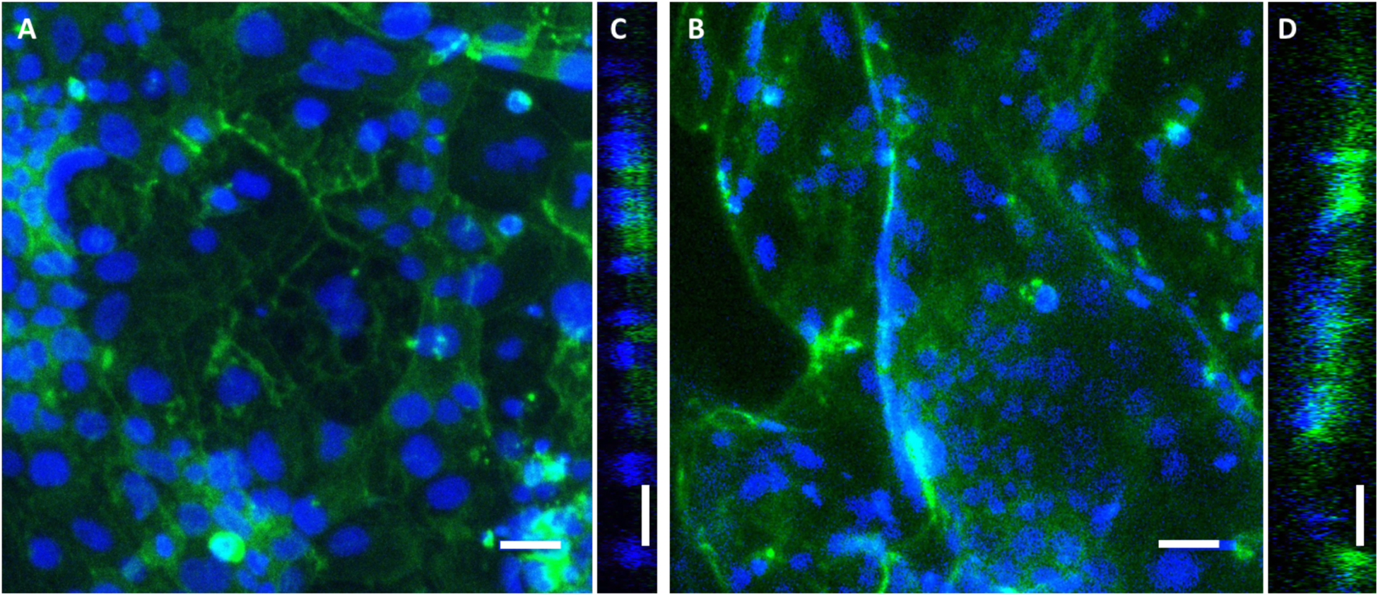
Figure 6.

Representative fluorescent images of Caco-2 monolayers cultured for 10 days on flat PDMS substrates (a) and biomimetic PDMS replicas (b). Optical cross sections of growth substrates are shown as Y-orthogonal views of the flat (c) and replica (d) substrate surfaces. Blue = Hoechst nuclei, green = filamentous actin cytoskeleton. Bars = 50 um. Confocal maximum intensity projections shown.
