PLOS ONE logoLink to PLOS ONE
. 2021 Apr 22;16(4):e0249936. doi: 10.1371/journal.pone.0249936

Suitability of IgG responses to multiple Plasmodium falciparum antigens as markers of transmission intensity and pattern

Eric Kyei-Baafour 1,2, Mavis Oppong 3, Kwadwo Asamoah Kusi 1, Abena Fremaah Frempong 1, Belinda Aculley 1, Fareed K N Arthur 2, Regis Wendpayangde Tiendrebeogo 4, Susheel K Singh 4,5, Michael Theisen 4,5, Margaret Kweku 3, Bright Adu 1, Lars Hviid 4, Michael Fokuo Ofori 1,*
Editor: Takafumi Tsuboi6
PMCID: PMC8062017  PMID: 33886601

Abstract

Detection of antibody reactivity to appropriate, specific parasite antigens may constitute a sensitive and cost-effective alternative to current tools to monitor malaria transmission across different endemicity settings. This study aimed to determine the suitability of IgG responses to a number of P. falciparum antigens as markers of transmission intensity and pattern. Antibody responses to multiple malaria antigens were determined in 905 participants aged 1–12 years from three districts with low (Keta), medium (Hohoe) and high (Krachi) transmission intensity in the Volta region of Ghana. Blood film microscopy slides and dry blood spots (DBS) were obtained for parasitaemia detection and antibody measurement, respectively. Sera were eluted from DBS and levels of IgG specific for 10 malaria antigens determined by a multiplex assay. Results were compared within and among the districts. Total IgG responses to MSPDBL1, MSPDBLLeucine, MSP2-FC27, RAMA, and PfRh2a and PfRh2b were higher in Krachi than in Hohoe and Keta. Seroprevalence of IgG specific for MSPDBLLeucine, RON4, and PfRh2b were also highest in Krachi. Responses to RALP-1, PfRh2a and PfRh2b were associated with patent but asymptomatic parasitaemia in Keta, while responses to MSPDBL1, MSPDBLLeucine, MSP2-FC27, RAMA, Rh2-2030, and PfRh2b were associated with parasite carriage in Hohoe, but not in Krachi. Using ROC analysis, only PfRh2b was found to predict patent, but asymptomatic, parasitaemia in Keta and Hohoe. Antibody breadth correlated positively with age (r = 0.29, p<0.0001) and parasitaemia (β = 3.91; CI = 1.53 to 6.29), and medium to high transmission (p<0.0001). Our findings suggest differences in malaria-specific antibody responses across the three transmission zones and that PfRh2b has potential as a marker of malaria transmission intensity and pattern. This could have implications for malaria control programs and vaccine trials.

Introduction

Malaria continues as a serious challenge to health systems in sub-Saharan Africa, despite increased efforts to control the disease. In Africa, 96% of malaria cases were due to Plasmodium falciparum in 2019. Out of the over 409,000 global deaths from malaria in 2019, 94% occurred in Africa [1].

In Ghana, malaria is endemic with the entire population at risk, and the disease accounts for about 30% of all out-patients [2]. Malaria transmission in Ghana differs among its three major ecological zones. It is lowest in the coastal shrub zone of southern Ghana, intermediate in the middle belt dominated by semi-deciduous and transitional forest, and highest in the northern part of the country, characterized by guinea savannah [3].

Malaria transmission intensity is measured using Plasmodium parasite prevalence i.e., the proportion of the population infected with the parasites. However, parasite prevalence is highly dependent on the method used to detect parasites in the blood of infected individuals [4]. Entomological inoculation rate (EIR) is another malaria transmission intensity indicator that shows the rate at which individuals are bitten by infective mosquitoes [5]. The estimation of transmission by EIR suffers from low precision as a result of temporal distributions of vectors [5,6] and from being labour-intensive [7]. Antibody responses to malaria-specific antigens have been suggested as alternative markers of malaria transmission intensity [8] and differences in transmission patterns [9,10]. Spatial heterogeneity in malaria transmission has therefore been estimated using serological tools [1113].

Malaria-specific antibodies elicited by natural infection are generally considered markers of parasite exposure and good for sero-surveillance. These antibodies can also be used to predict parasite exposure over time [4]. However, some may not be able to define properly heterogeneity in malaria transmission, because of their persistence in circulation. In addition, estimating malaria transmission reliably with methods such as EIR and microscopy are becoming increasingly difficult as the prevalence of clinical cases declines. Changes in the burden of malaria in low-transmission settings may thus not be detected [14]. There is therefore a need to characterize parasite-specific immune responses in different transmission settings to select good markers for transmission monitoring. Furthermore, the characterization of antibody responses will enhance efforts to develop more accurate tools to monitor transmission [4,9,15].

Most serology studies of malaria transmission patterns have focused on few antigens such as circumsporozoite protein (CSP), cell-traversal protein for ookinetes and sporozoites (CelTOS), apical membrane antigen 1 (AMA1), and merozoite surface protein 1 (MSP1) [8,9,16,17]. However, many other antigens need evaluation to expand the repertoire used to determine heterogeneities in malaria transmission. Ten antigens were selected for this study: merozoite surface protein Duffy binding ligand 1 (MSPDBL1, MSPDBL-Leucine), erythrocyte-binding antigen (EBA140RIII-V), merozoite surface protein 2 (MSP2FC27), rhoptry-associated like protein (RALP-1), rhoptry-associated membrane antigen RAMA, Plasmodium falciparum reticulocyte homologue (PfRh2-2030, PfRh2A, PfRh2B), and rhoptry-associated neck protein (RON4). All are bloodstage antigens, and some have been found to be associated with protection from malaria and to be good markers of exposure [18,19].

Malaria transmission studies in the eastern part of Ghana are scanty and no study has compared anti-malarial antibody responses across the three ecological zones spanned by the Volta region in Ghana. Entomological inoculation rates increase from the southern coastal belt (62.1 infective bites/person/year) [20], through the middle forest transition zone (269 infective bites/person/year) [21], to the guinea savannah zone in the north (643 infective bites/person/year) [22]. In this study, we compared antibody levels to multiple malaria antigens in three districts, each represents one of the three ecological zones in the Volta region of Ghana, to assess their suitability to determine their suitability as markers of transmission intensity and pattern.

Material and methods

Study site and population

This cross-sectional community-based survey was carried out in December 2018 in three randomly selected districts in the Volta region, each with a different ecological setting. The study involved children aged 1–12 years living in communities within the selected districts. The study site and population have been described elsewhere [23]. Briefly, the study employed a multi-stage sampling method in which the three [3] ecological zones of the Volta Region, southern, middle and northern zones reflecting the three malaria transmission zones, were used. This approach was used to ensure that the results obtained could reflect the entire population of Volta Region. One district each from the three zones was randomly picked. The communities in the selected districts were also listed and picked randomly. Children were only included in the study after informed consent has been granted by parents/guardians, and were permanently resident in the selected districts. Febrile children and those who were reported ill two weeks before sampling by parents/guardians were excluded from the study. Children with congenital defects were also not included in the study.

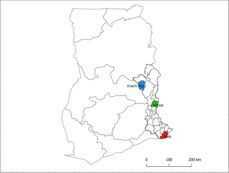
The three selected districts were Krachi (guinea savannah zone) in the north, Hohoe (forest transition zone) in the middle, and Keta (coastal shrub and grassland zone) along the coast in the south. The Volta region, like other parts of Ghana, has two major seasons: a dry season from November to April, and a rainy season, which spans the months from May to October. However, rainfall in the areas around Hohoe and Keta is bimodal with a major peak in June and a minor peak in October [24]. Krachi and the adjoining districts experience one rainy season peaking in August [25]. Malaria prevalence in the Volta region by RDT and microscopy in 2014 were 36.8% and 25.2% respectively [26]. The locations of the three study sites are shown (Fig 1).

Fig 1. Map of Ghana located in West Africa showing the region and districts where sampling was done.

Fig 1

This was the map of Ghana with the 10 regions as they existed before December 2019 when the study was performed. The red shaded area is Keta, the blue is Krachi and the green area is Hohoe. (Map was created using QGIS version 3.4.7 by Miss Jessica Asante, Department of Epidemiology, School of Public Health, University of Ghana).

Sampling

Ethical Approval for the study was obtained from the Research Ethics Committee of the University of Health and Allied Sciences (UHAS-REC A.1 (5) 18–19). Written informed consent was obtained from parents/guardians of children aged 1–12 years, and finger-prick blood spots (DBS) were collected using Whatman No. 5 filter paper (GE Healthcare, England). The spots were air-dried and stored desiccated at 4°C until ready for use. Thick and thin slides were also prepared for malaria microscopy.

Examination of blood smears

Giemsa-stained thick and thin blood smears were examined using a light microscope. A slide was considered negative if no parasites were observed after examining 200 microscopic fields at 100× magnification of the thick smear. Parasite density was estimated against 500 leucocytes, using an assumed leucocyte count of 8,000/μL of blood and expressed as parasites/μL. Polymerase Chain Reaction (PCR) data to measure sub-microscopic parasitaemia from the same cohort has already been published elsewhere [23].

Elution of serum from filter paper. Elution of serum from the filter paper was done using a protocol described by Corran et al. [27] with slight modifications. Briefly, zip-lock bags containing individually wrapped filter papers were removed from storage and allowed to warm to room temperature for 30 minutes. Dried blood spots of about 2.5 mm diameter were cut using a leather punch into 96-well microtiter plates. To elute serum from spots, 150 μL PBS with 0.05% Tween 20, and 0.05% sodium azide was added to each well containing punch-outs, after which each plate was placed on a shaker overnight at 150 rpm. Finally, eluted samples were aliquoted into separate 96-well microtiter plate and kept at -20°C until use.

Coupling of antigens to microsphere beads. The antigens used here (MSPDBL1, EBA140RIII-V, MSPDBLLeucine, MSP2FC27, RALP-1, RAMA, PfRh2-2030, PfRh2A, PfRh2B, and RON4), were expressed in Lactococcus lactis expression system using P. falciparum 3D7 variants with only MSP2FC27 cloned from P.falciparum FC27 strain, as described previously [28]. These antigens were selected based on their different localization in/on merozoites [29], to determine their usefulness as transmission monitoring markers Antigen coupling was also done as previously described [30,31], with slight modifications. Briefly, each antigen was covalently coupled to microsphere beads with each bead region noted according to the manufacturer’s protocol (Luminex). The regions used were MSPDBL1-{66}, EBA140RIII-V-{32}, MSPDBLLeucine-{52}, MSP2FC27-{35}, RALP-1-{45}, RAMA-{77}, PfRh2-2030-{33}, PfRh2a-{47}, PfRh2b-{80}, and RON4-{56}. BSA coupled to the bead region {89} was used as a control. Coupled beads were stored at 4°C until use.

Measurement of antigen-specific antibody levels by multiplex assay. Antibodies with specificity for the panel of 10 P. falciparum recombinant antigens in eluted serum were measured on the Luminex 200 x-MAP platform (Luminex Inc., Austin, TX USA) as described previously [30], with slight modifications. Nine hundred and five (905) eluted samples were analysed. Briefly, a multiscreen filter base plate (Millipore, Billerica, MA) was pre-wetted with 100 μL/well freshly prepared assay buffer (PBS, 0.05% Tween 20, 1% BSA, 0.05% sodium azide, pH 7.4) for 30 minutes. Eluted sera and negative control sera from malaria naïve individuals (individuals with no travel history to any malaria-endemic country and deemed not to have encountered any malaria antigen) were diluted 1:5 giving a final dilution of 1:500. Adult plasma samples found to have higher responses to all the antigens included in the study were pooled and used as a positive control. Approximately 1,250 coupled beads from each region were mixed in equal volumes and added to each well at 50 μL/well of the pre-wetted plates. The plates were washed three times with assay buffer and test samples and controls added at 100 μL/well. Plates were incubated in the dark for 2 hours on a shaker at a speed of 300 rpm. After three washes, plates were incubated with biotin-conjugated goat-anti-human IgG (KPL, Gaithersburg, MD, USA) (diluted at 1:500, 100 μL/well) for 1 h. This was followed by incubation with 100 μL/well streptavidin-conjugated phycoerythrin (Thermo Fisher) diluted at 1:200 dilution for 30 minutes in the dark. After three washes, 100 μL assay buffer was added and plates subsequently read with the Luminex 200 system (Luminex Corporation). Results were expressed as mean fluorescence intensity (MFI).

Data analysis. Data was normalized for inter-plate and day-to-day variation by dividing the test sample on each plate by the positive control of the assay plate. This was then multiplied by the total mean positive control for all the plates to obtain the normalized MFI values using the formula:

(Sample/Plate Positive control) x Mean positive control for all plates)

Mean fluorescence intensity of samples obtained from malaria-naïve volunteers were used to define a cut-off for seropositivity of antibodies, calculated as the mean naïve MFI plus 2 standard deviations of the mean. Seroprevalence was antigen-specific. Differences in median antibody levels across the three districts were also determined using the Kruskal-Wallis test. Antibody levels were then log10-transformed for further analysis. Age was stratified into two groups, those below 5 years and those above 5 years, and student’s t-test used to determine differences in the log10-transformed MFI in the two age groups for each district. Participants were divided into two subsets based on malaria positivity, and multiple logistic tests used to determine antibody responses associated with parasite carriage. Receiver operating characteristics (ROC) curves were fitted to predict parasite carriage using antibody levels as predictors. Antibody levels were categorized based on quartiles and assigned 0 for lowest, 1 for the second, 2 for third, and 3 for the highest quartile. The scores were summed up for the 10 antigens for each individual to generate breadth scores. The relationship between malaria transmission (defined using infective bites per person per year [2022]) and breadth scores was determined using linear regression adjusting for age and bed net usage. Analysis and graphs were done using R statistical software version 4.0.0 and GraphPad Prism 8.0.2. Statistical significance was set as p < 0.05.

Results

Description of study participants

The study recruited 938 children with a mean age (± standard deviation) of 6.4 (± 3.4) years. However, only 905 filter blots were found to be of good quality and thus used for this analysis. The three study sites contributed approximately the same number of participants, and the proportions of males and females were comparable. Children from Keta district had the lowest haemoglobin levels, compared to the other two districts. There were significant proportional differences among the districts in bed net usage. The prevalence of P. falciparum infection did not differ among the three sites, but the median parasite density was significantly higher at Krachi than the two other study sites. Parasite prevalence by PCR (already described in [23]) was higher than prevalence by microscopy. It did not however differ between the three sites (Table 1). None of the children was febrile, and all P. falciparum infections identified were asymptomatic.

Table 1. Characteristics of study participants.

Variable Keta (n = 272) Hohoe (n = 327) Krachi (n = 306) Total (n = 905) p-value
Gender
Male 134 (49.3) 146 (44.6) 147 (48.0) 427 (47.2)
Female 138 (50.7) 181 (55.4) 159 (52.0) 478 (52.8) 0.50*
Age Group (yrs)
Below 5 90 (33.1) 165 (50.5) 113 (36.9) 368 (40.7)
Above 5 182 (66.9) 162 (49.5) 193 (63.1) 537 (59.3) < 0.001*
Ethnicity
Others 1 (0.4) 159 (48.6) 127 (41.5) 287 (31.7)
Ewe 264 (97.1) 128 (39.1) 25 (8.2) 417 (46.1)
Akan 7 (2.6) 7 (2.1) 9 (2.9) 23 (2.5)
Guan 0 (0.0) 33 (10.1) 145 (47.4) 178 (19.7) < 0.001*
Bed net Usage
Yes 258(94.9) 282(86.2) 281(91.8) 821(90.7)
No 14(5.1) 45(13.8) 25(8.2) 84(9.3) 0.001*
Hbg/dL(sd) 10.5 (1.3) 11.2 (1.7) 11.6 (1.6) 11.2 (1.6) < 0.001#
Microscopy Parasite prevalence
Positive 11(4.0) 24(7.3) 14(4.6) 49(5.4)
Negative 261(96.0) 303(92.7) 292(95.4) 856(94.6) 0.15*
PCR Parasite prevalence
Positive 31(11.4) 44(13.5) 52(17.0) 127(14.0)
Negative 241(88.6) 283(86.5) 254(83.0) 778(86.0) 0.14
Median parasite density/μl (iqr) 520 (220–7600) 780 (350–3190) 4660 (1360–45525) 1280 (460–7680) 0.020$

*Proportional differences between the three districts for gender, age groups, ethnicity, bed net usage, and parasite prevalence were determined using Chi-square test.

# the difference in mean haemoglobin (Hb) between districts was determined using ANOVA, and the Kruskal-Wallis test was used to calculate the differences in median parasite density

$. (%) Numbers in brackets are percentages. Iqr in the interquartile ranges. The “other” under the ethnicity is made up of minority groups who are not the predominant ethnic grouping in the areas studied. These include Hausa, Kotokoli, Zamrama, Mossi, Chamba, Yoruba, Fulani, Ga, Waala, Nawure, Kabre, Kokomba, and Tokosi

Seroprevalence and variation in IgG titres to malaria antigens

Generally, all children had detectable levels of IgG to the antigens tested. Antibody levels against all antigens tested increased with age in all the three districts. (Fig 2).

Fig 2. Distribution of IgG levels (log-transformed MFI) as a function of age.
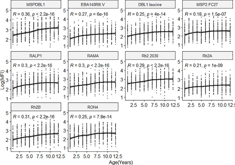
Fig 2

Overall differences of antibody levels to the ten antigens tested with age in the study population. The regression line shows the LOESS smoothed estimate of the log transformed MFI.

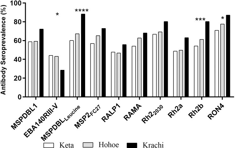
Seroprevalence was higher in Krachi for all the antigens tested, except for EBA140RIII-V, which had lower seroprevalence. The seroprevalence of MSPDBLLeucine-, PfRh2b-, and anti-RON4-specific IgG were significantly higher in Krachi compared to Hohoe and Keta (Fig 3).

Fig 3. Seroprevalence of antibodies found in the three districts.

Fig 3

Stars represent antigens with significantly higher proportions between the three districts using chi-square test. *p = <0.05, **p<0.01, ***p<0.001, ****p<0.0001. X-axis represent the antigens tested. Patterns represent study site. Seropositivity was defined as individuals whose mean total IgG levels to the antigens tested were higher than 2 standard deviations above the mean of malaria naïve individuals.

The highest antibody seroprevalence of 88.3% to MSPDBLLeucine and the lowest of 28.6% to EBA140RIII-V were observed in Krachi.

A comparison of antigen-specific antibody levels among the three districts showed that antibodies to MSPDBL1, MSPDDBL1Leucine, MSP2FC27, RAMA, PfRh2a and PfRh2b were all significantly higher in Krachi than in the other two districts. Keta had the lowest antibody levels except for PfRh2-2030 (Fig 4).

Fig 4. Total IgG levels to multiple malaria antigens in the three districts.
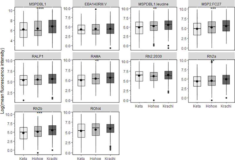
Fig 4

Box and whisker plots with a round dot in the middle, the median IgG level of the group. Difference in antibody levels between the districts are shown for each antigen tested. The x-axis represents the districts (** p<0.01, ***p<0.001, ****p<0.0001).

Antibody levels are associated with P. falciparum parasitaemia

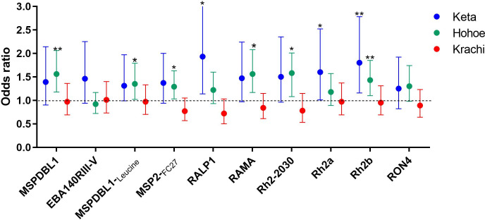
To determine whether higher levels of antibodies were associated with parasite carriage, parasitaemia was determined by microscopy. Participants were then categorized, based on the presence or absence of parasites as a binary outcome variable, and antibody levels used as the predictor variable in a multivariable logistic regression analysis adjusting for district, age, ethnicity, and bed net usage. Overall, higher antibody levels to MSPDBL1 (odds ratio (OR) = 1.41, 95% confidence interval (CI) = 1.15 to 1.73), RAMA (OR = 1.32; CI = 1.06 to 1.63), PfRh2-2030, (OR = 1.27; CI = 1.02 to 1.59), and PfRh2b (OR = 1.29; CI = 1.06 to 1.57) were associated with increased odds of P. falciparum parasite carriage. Also, a trend from low antibody levels in non-parasitaemic individuals to high antibody levels in individuals with sub-microscopic parasitaemia was observed in individuals in Keta and Hohoe while no differences were observed in Krachi except antibodies to MSPDBL1 and PfRh2-2030 (S1 Fig). Individuals with sub-microscopic parasitaemia are those with microscopy negative but are PCR positive. When antibody levels were compared between only microscopic and sub-microscopic groups, a trend of higher responses in the sub-microscopic group compared to the microscopic group was observed across the three sites. The responses were, however, not significant except MSP2-FC27 and PfRh2-2030 which were significantly high in the sub-microscopic group in Krachi.

In a site-specific analysis, higher antibody responses to RALP-1 (OR = 1.93; CI = 1.14 to 3.26), PfRh2a (OR = 1.60; CI = 1.01 to 2.52), and PfRh2b (OR = 1.80; CI = 1.16 to 2.78), were associated with increased odds of carrying microscopic parasites in Keta. In Hohoe, the responses associated with parasite carriage were MSPDBL1 (OR = 1.56; CI = 1.18 to 2.06), MSPDBLLeucine (OR = 1.35; CI = 1.02 to 1.97), MSP2FC27 (OR = 1.29; CI = 1.03 to 1.63), RAMA (OR = 1.56; CI = 1.17 to 2.08), PfRh2-2030 (OR = 1.48; CI = 1.08 to 2.01), and PfRh2b (OR = 1.43; CI = 1.10 to 1.85). However, there was no association between parasite carriage and antibody levels in Krachi (p>0.05 for all antigens tested) (Fig 5).

Fig 5. Total IgG responses are associated with parasite carriage.

Fig 5

Multivariate logistic regression was used to predict parasite presence. Models were adjusted for age, ethnicity, bed net usage and district. Circles represent odds ratios and error bars are 95% confidence intervals. The black dotted line represents an OR of 1 which indicated no association with parasitaemia (*p = <0.05, **p<0.01, ***p<0.001, ****p<0.0001).

In a linear model adjusting for age, ethnicity, bed net usage and haemoglobin levels to determine the relationship between district and parasitaemia, children in Krachi were found to have higher parasitaemia (β = 2.29; 95% CI = 0.56 to 4.02) than those in Hohoe. Also, sub-microscopic parasitaemia was associated with the high transmission district of Krachi (OR = 3.26; 95% CI = 1.39 to 7.64).

ROC curves were used to define a threshold MFI for predicting parasitaemia. The overall area under the curve (AUC) was lower than 66% for all antigens tested, however in a site-specific analysis, AUC for all the antigens in Keta was above 70% except RON4 67.6% (95% CI = 49.0 to 86.2) and MSPDBL1Leucine 68.2% (95% CI = 53.4 to 83.0), with PfRh2b having an AUC of 84.3 (95% CI = 76.3to 92.3). The AUC for all the antigens in the other districts were below 70% (Table 2). A threshold of PfRh2b levels at 639.5 MFI, was associated with a sensitivity of 88.9% (95% CI = 66.7–100) and a specificity of 79.4% (95% CI = 73.5–84.8) in predicting P. falciparum infection by microscopy with an AUC of 84.3%. The 95% CI were computed with 2,000 stratified bootstrap replicates.

Table 2. Area under the curve (AUC) following receiver operating characteristics (ROC) analysis using antigens a predictor of parasitaemia.

Antigens Keta AUC (95% CI) Hohoe AUC (95% CI) Krachi AUC (95% CI)
MSPDBL1 72.0 (56.8–87.1) 69.7 (59.3–80.1) 51.9 (41.6–62.2)
EBA140RIII-V 71.8 (50.8–92.8) 53.4 (42.5–64.2) 52.5 (37.7–67.3)
MSPDBLLeucine 68.2 (53.4–83.0) 65.3 (54.1–76.5) 52.4 (40.8–63.9)
MSP2FC27 72.0 (62.6–81.3) 64.9 (54.0–75.8) 63.9 (51.9–75.9)
RALP1 76.7 (66.3–87.1) 59.7 (47.7–71.6) 60.8 (48.4–73.2)
RAMA 76.7 (59.1–94.3) 69.0 (56.7–79.3) 57.0 (45.0–69.0)
PfRh22030 74.3 (58.0–90.0) 66.3 (55.7–76.9) 56.4 (44.7–68.7)
PfRh2a 75.5 (62.2–89.3) 60.4 (46.4–74.4) 52.9 (40.1–65.7)
PfRh2b 84.3 (76.3–92.3) 67.6 (56.9–78.4) 51.6 (40.4–62.8)
RON4 67.6 (49.0–86.2) 62.0 (50.5–73.6) 58.4 (46.0–70.7)

The 95% CI for the area under the curve was calculated with 2000 stratified bootstrap replicates.

Association between breadth of antibody response and malaria transmission

Antibody responses to all the 10 antigens were scored depending on the quartiles for each individual and summed up to give the breadth score (number of antigens recognized). The breadth of antibody response in this study was between zero (0) and 30 with a breadth of 30 being the highest. Age, ethnicity and parasitaemia (by microscopy and PCR) factors were used in a linear model to study their relationship with breadth score. Parasitaemia was positively associated with high breadth score, while children above 5years of age had higher breadth score. Ethnicity (Ewe) was also negatively correlated with breadth score (Table 3).

Table 3. Factors associated with antibody breadth score.

Variable β Coefficient CI p-value
Age
Below 5 yrs Ref
Above 5 yrs 4.67 3.58 to 5.77 < 0.0001
Ethnicity
Akan Ref
Ewe -4.27 -7.73 to -0.81 0.0158
Guan -3.10 -6.69 to 0.48 0.0899
others -3.37 -6.87 to 0.14 0.0599
Parasitaemia
Negative Ref
Positive 3.91 1.53 to 6.29 0.0013
PCR
Negative Ref
Positive 4.46 2.55 to 6.37 < 0.0001

Multiple linear regression was used to generate coefficient and CI.

To determine if transmission intensity was associated with antibody breadth score, the relationship between breadth score and malaria transmission (districts) was assessed in a linear model adjusting for age and ethnicity. Breadth score was positively associated with areas of higher malaria transmission (Krachi and Hohoe) compared to the low transmission area (Keta) (Table 4).

Table 4. Association between malaria transmission and antibody breadth score.

District β Coefficient CI p-value
Keta Ref
Hohoe 4.18 2.56–5.79 < 0.0001
Krachi 4.53 2.56–6.50 < 0.0001

Multiple linear regression adjusting for age and ethnicity was used to generate coefficient and CI.

Discussion

P. falciparum antigen-specific serology has been proposed as a tool to reduce the challenges of malaria transmission monitoring [4,8,32]. A key advantage of serology in the estimation of malaria transmission intensity is the ability to test large populations using dried spot samples with responses to multiple antigens using a multiplex assay approach [27,33]. To examine their suitability for this purpose, we determined antibody responses to multiple malaria antigens in three ecologically distinct districts of eastern Ghana with varying malaria transmission intensities.

The overall parasite prevalence (5.4%) was not significantly different among the three districts. The observed prevalence in Hohoe (7.3%) was lower than reported recently (16%) [34], which may be a result of ongoing malaria interventions in the area [35]. The observed prevalence (4.0%) in Keta was similar to that reported previously (3.7%) from a nearby site in southern Ghana with similar transmission pattern [36]. The similar prevalence in all the three study sites could be the due to the various malaria prevention interventions, which may have erased previous differences. The higher parasite densities of asymptomatic infections in Krachi than at the other sites may reflect better immunological control of disease-causing mechanisms (anti-disease immunity) in the high-endemicity setting [37].

Generally, participants in the study responded to all ten antigens tested and the antigens positively correlated with each other (r2>0.4, p<0.001 for all the antigens tested). Six (MSPDBL1, MSPDBLLeucine, MSP2FC27, RAMA, PfRh2a, and PfRh2b), (Table 2) elicited responses that were higher in Krachi compared to the other two districts. Variation in malaria transmission in the different ecological zones in Ghana has been reported [3]. The observed differences in the antibody levels to some of the antigens measured within the three districts reflected their pattern of malaria transmission, supporting the proposed use of serology to monitor transmission [4]. Antibody levels increased with age, reflecting the accumulation of IgG to an increasing number of different parasite antigens with time, thereby increasing antibody breadth [38,39]. However, levels of IgG specific for EBA140RIII-V, PfRh2-2030, RALP-1, and RON4, did not differ among the three districts, indicating that they may be less suitable for monitoring transmission intensity. Since sampling was done in the dry season, the antibodies to these antigens may have waned since responses to some antigens have shorter half-life [40,41]. Also, these IgG responses much promptly react to the transmission change, and predict more recent transmission status. Seroprevalence of IgG to EBA140RIII-V was significantly lower in the high transmission area of Krachi, which is in contrast to earlier reports from Ghana [42]. However, it must be noted that differences in IgG quantification methods (bead-based array versus ELISA) may have accounted for the observed differences. Also, polymorphisms in EBA140III-V in parasite strains in the district may alter host receptor binding [43] and thus reduce their recognition in the district.

Infection with P. falciparum is generally thought to boost parasite-specific antibody responses by about 20% [44]. In the current study, parasitaemic (microscopic and sub-microscopic individuals) in both Hohoe and Keta districts had higher antibody levels compared to the non-parasitaemic individuals (S1 Fig) indicating that persisting parasitaemia does trigger continuous antibody production [45,46]. Also, sub-microscopic individuals with higher antibody levels may have a better control of parasitaemia reflecting in the low parasite levels. The lower antibody levels in the microscopic individuals compared to the sub-microscopic may indicate a low threshold not enough to control parasitaemia hence the high parasite levels diagnosed with microscopy. Since sub-microscopic parasitaemia is responsible for about 20% to 50% of human to mosquito infections [47], our finding that sub-microscopic parasitaemia is associated with Krachi which is a high transmission district corroborate earlier reports that sub-microscopic parasitaemia in asymptomatics could be a major factor in malaria transmission. It has also been suggested that polyclonal infections increase the breadth of antibody responses reflecting exposure and thus reduced risk of disease [48,49]. Thus, the high antibody responses in individuals from Krachi may be as a result of immune tolerance. These may indicate that a threshold of antibody level is needed to control parasitaemia and thus protection [50]. In a study conducted in Uganda, higher antibody levels against blood-stage antigens were found to be protective against malaria symptoms once the subjects were parasitaemic [51]. Although the current study was a cross-sectional study, with the limitation that we could not follow the participants to know the exact point at which infection occurred, we believe higher antibody levels in the parasitaemic children could reflect higher transmission. The similar antibody levels between parasitaemic and non-parasitaemic children in Krachi may be as a result of the high transmission reflecting in high parasite density which is associated with high sub-microscopic parasitaemia and polyclonality often expressed as multiplicity of infection (MOI) [52,53]. Antibody levels expressed by both parasitaemic and non-parasitaemic but high MOI individuals may be almost the same since high sub-microscopic parasitaemia may also induce higher responses. Sub-microscopic parasitaemia is about 50% higher than microscopy [54]. It could also be as a result of maintenance of antibodies from the rainy season. We could not collect data for entomological inoculation rate (EIR) during sampling, which is another limitation but the low to high antibody levels from Keta to Krachi, respectively, may indicate transmission differences which reflects antibody levels in the three districts.

This study also found site-specific antibody responses to associate with P. falciparum carriage (Fig 5). Different responses were associated with P. falciparum carriage in both Keta and Hohoe, although levels of PfRh2b-specific IgG were associated with parasite carriage in both districts. Keta and Hohoe districts both have lower malaria transmission than Krachi, and the association of PfRh2b with parasitaemia indicates a possible use of that antigen to monitor malaria transmission and pattern in low transmission setting. The different antigens predicting parasite carriage in the two districts with different transmission intensities reflect possible differences in responses of individuals within each transmission zone. We used ROC analysis to confirm if indeed PfRh2b and any other antigen could be used to monitor transmission and pattern in a low transmission setting. We found that IgG responses to PfRh2b at a level of 639.5 MFI in low transmission settings are likely to predict P. falciparum carriage by microscopy further confirming the possible use of PfRh2b to monitor transmission and pattern.

Several reports have linked breadth of antibody responses to malaria immunity and that individuals with higher antibody breadth have better protection from malaria [18,55]. A study in three highly endemic districts in Ghana identified age and endemicity as predictors of antibody breadth [38]. Thus, the relationship between age, ethnicity and parasitaemia was explored. Our observation of a low to high trend of antibody breadth from keta to Krachi suggests that breadth may be linked to transmission intensity and pattern. The low antibody breadth score in children living in Keta could be linked to the low transmission intensity resulting in low antibody responses in the district.

Though antibody levels may be higher in the rainy season, the data presented here demonstrate variations in antibody response to multiple malarial antigens in children across different ecological regions with varying transmission intensities, confirming previous reports of serology as an alternative malaria transmission monitoring tool [4,8,9].

In conclusion, the study highlights the potential of antibodies against PfRh2b as a useful marker for predicting malaria transmission intensity and pattern in low malaria transmission setting. Antibody levels and seroprevalence reflects changes in transmission between the three ecological zones. Low breadth score was found to be associated with low malaria transmission. The study findings have implications for malaria control interventions. Also, the design and testing of vaccines must take into consideration the heterogeneity of immune responses in the different ecological zones.

Supporting information

S1 Fig. Total IgG responses between parasitaemic and non-parasitaemic individuals.

Box and whisker plots with a round dot in the middle, the median IgG level of the group. Differences in antibody levels between non-parasitaemic, microscopic, and sub-microscopic individuals for each antigen are shown for each district. The x-axis represents the districts, and the y-axis represent the log-transformed antibody levels (** p<0.01, ***p<0.001, ****p<0.0001).

(TIF)

Acknowledgments

The Authors are grateful to the parents and guardians of the children involved in the study. We also thank the District and Municipal Directors of Health in Keta, Hohoe, and Krachi West Districts for supporting the team during sampling. We are also thankful to Mr Jones Amo Amponsah and Alex Danso-Coffie of the Immunology Department, NMIMR for his technical assistance.

Data Availability

Data are available from figshare (DOI: 10.6084/m9.figshare.14046911.v1).

Funding Statement

This study was supported by the Danish Research Council for Development Research (Grant No. 17-02-KU) awarded to LH and MFO. BAdu and MT are supported by Ministry of Foreign Affairs of Denmark (DFC file no.14-P01-GHA) and administered by DANIDA Fellowship Centre. The funders had no role in study design, data collection and interpretation, or the decision to submit the work for publication.

References

  • 1.WHO. World malaria report 2020: 20 years of global progress and challenges. WHO, Geneva.2020.
  • 2.NMCP. 2015 ANNUAL REPORT Ghana Health Service, Accra. 2016.
  • 3.Afari EA, Akanmori BD, Nakano T, Ofori-Adjei D. Plasmodium falciparum: sensitivity to chloroquine in vivo in three ecological zones in Ghana. Trans R Soc Trop Med Hyg. 1992; 86:231–232. 10.1016/0035-9203(92)90285-k [DOI] [PubMed] [Google Scholar]
  • 4.Corran P, Coleman P, Riley E, Drakeley C. Serology: a robust indicator of malaria transmission intensity? Trends Parasitol 2007; 23 (12):575–582. 10.1016/j.pt.2007.08.023 [DOI] [PubMed] [Google Scholar]
  • 5.Hay SI, Rogers DJ, Toomer JF, Snow RW. Annual Plasmodium falciparum entomological inoculation rates (EIR) across Africa: literature survey, Internet access and review. Trans R Soc Trop Med Hyg. 2000; 94:113–127. 10.1016/s0035-9203(00)90246-3 [DOI] [PMC free article] [PubMed] [Google Scholar]
  • 6.Magbity E, Lines J. Spatial and temporal distribution of Anopheles gambiae sl (Diptera: Culicidae) in two Tanzanian villages: implication for designing mosquito sampling routines. Bull. Entomol. Res. 2002;92:483–488. 10.1079/ber2002200 [DOI] [PubMed] [Google Scholar]
  • 7.Dye C. The analysis of parasite transmission by bloodsucking insects. Annu Rev Entomol. 1992;37:1–19. 10.1146/annurev.en.37.010192.000245 [DOI] [PubMed] [Google Scholar]
  • 8.Kusi KA, Bosomprah S, Dodoo D, Kyei-Baafour E, Dickson EK, Mensah D, et al. Anti-sporozoite antibodies as alternative markers for malaria transmission intensity estimation. Malar J 2014;13:103. 10.1186/1475-2875-13-103 [DOI] [PMC free article] [PubMed] [Google Scholar]
  • 9.Kusi KA, Bosomprah S, Kyei-Baafour E, Dickson EK, Tornyigah B, Angov E, et al. Seroprevalence of Antibodies against Plasmodium falciparum Sporozoite Antigens as Predictive Disease Transmission Markers in an Area of Ghana with Seasonal Malaria Transmission. PLoS ONE 2016; 11:e0167175. 10.1371/journal.pone.0167175 [DOI] [PMC free article] [PubMed] [Google Scholar]
  • 10.Supargiyono S, Bretscher MT, Wijayanti MA, Sutanto I, Nugraheni D, Rozqie R, et al. Seasonal changes in the antibody responses against Plasmodium falciparum merozoite surface antigens in areas of differing malaria endemicity in Indonesia. Malar J. 2013; 12:444. 10.1186/1475-2875-12-444 [DOI] [PMC free article] [PubMed] [Google Scholar]
  • 11.Biggs J, Raman J, Cook J, Hlongwana K, Drakeley C, Morris N, et al. Serology reveals heterogeneity of Plasmodium falciparum transmission in northeastern South Africa: implications for malaria elimination. Malar J. 2017;16:48. 10.1186/s12936-017-1701-7 [DOI] [PMC free article] [PubMed] [Google Scholar]
  • 12.Baidjoe AY, Stevenson J, Knight P, Stone W, Stresman G, Osoti V, et al. Factors associated with high heterogeneity of malaria at fine spatial scale in the Western Kenyan highlands. Malar J. 2016.15:307. 10.1186/s12936-016-1362-y [DOI] [PMC free article] [PubMed] [Google Scholar]
  • 13.Arnold BF, van der Laan MJ, Hubbard AE, Steel C, Kubofcik J, Hamlin KL, et al. Measuring changes in transmission of neglected tropical diseases, malaria, and enteric pathogens from quantitative antibody levels. PLOS Negl. Trop. Dis. 2017. 11:e0005616. 10.1371/journal.pntd.0005616 [DOI] [PMC free article] [PubMed] [Google Scholar]
  • 14.Ochola L, Vounatsou P, Smith T, Mabaso M, Newton C. The reliability of diagnostic techniques in the diagnosis and management of malaria in the absence of a gold standard. Lancet Infect Dis. 2006;6(9):582–588. 10.1016/S1473-3099(06)70579-5 [DOI] [PubMed] [Google Scholar]
  • 15.Cook J, Reid H, Iavro J, Kuwahata M, Taleo G, Clements A, et al. Using serological measures to monitor changes in malaria transmission in Vanuatu. Malar J. 2010; 9: 169. 10.1186/1475-2875-9-169 [DOI] [PMC free article] [PubMed] [Google Scholar]
  • 16.Kwenti TE, Moye AL, Wiylanyuy AB, Njunda LA, Nkuo-Akenji T. Variation in the immune responses against Plasmodium falciparum merozoite surface protein-1 and apical membrane antigen-1 in children residing in the different epidemiological strata of malaria in Cameroon. Malar J. 2017. 16:453. 10.1186/s12936-017-2105-4 [DOI] [PMC free article] [PubMed] [Google Scholar]
  • 17.Idris ZM, Chan CW, Kongere J, Hall T, Logedi J, Gitaka J, et al. Naturally acquired antibody response to Plasmodium falciparum describes heterogeneity in transmission on islands in Lake Victoria. Sci. Rep. 2017;7:9123. 10.1038/s41598-017-09585-4 [DOI] [PMC free article] [PubMed] [Google Scholar]
  • 18.Kana IH, Garcia-Senosiain A, Singh SK, Tiendrebeogo RgW, Chourasia BK, Malhotra P, et al. Cytophilic Antibodies Against Key Plasmodium falciparum Blood Stage Antigens Contribute to Protection Against Clinical Malaria in a High Transmission Region of Eastern India. J.infect Dis. 2018; 218:956–965. 10.1093/infdis/jiy258 [DOI] [PubMed] [Google Scholar]
  • 19.Garcia-Senosiain A, Kana IH, Singh SK, Chourasia BK, Das MK, Dodoo D, et al. Peripheral Merozoite Surface Proteins Are Targets of Naturally Acquired Immunity against Malaria in both India and Ghana. Infection and Immunity 2020.88 (4):e00778–19. 10.1128/IAI.00778-19 [DOI] [PMC free article] [PubMed] [Google Scholar]
  • 20.Tchouassi DP, Quakyi IA, Addison EA, Bosompem KM, Wilson MD, Appawu MA, et al. Characterization of malaria transmission by vector populations for improved interventions during the dry season in the Kpone-on-Sea area of coastal Ghana. Parasit vectors. 2012; 5:212. 10.1186/1756-3305-5-212 [DOI] [PMC free article] [PubMed] [Google Scholar]
  • 21.Dery DB, Brown C, Asante KP, Adams M, Dosoo D, Amenga-Etego S, et al. Patterns and seasonality of malaria transmission in the forest-savannah transitional zones of Ghana. Malar J. 2010; 9:314. 10.1186/1475-2875-9-314 [DOI] [PMC free article] [PubMed] [Google Scholar]
  • 22.Appawu M, Owusu-Agyei S, Dadzie S, Asoala V, Anto F, Koram K, et al. Malaria transmission dynamics at a site in northern Ghana proposed for testing malaria vaccines. Trop Med Int Health 2004; 9:164–170. 10.1046/j.1365-3156.2003.01162.x [DOI] [PubMed] [Google Scholar]
  • 23.Oppong M, Lamptey H, Kyei-Baafour E, Aculley B, Ofori EA, Tornyigah B, et al. Prevalence of sickle cell disorders and malaria infection in children aged 1–12 years in the Volta Region, Ghana: a community-based study. Malar J. 2020.19:426. 10.1186/s12936-020-03500-5 [DOI] [PMC free article] [PubMed] [Google Scholar]
  • 24.Ofori MF, Lamptey H, Dickson EK, Kyei-Baafour E, Hviid L. Etiology of Placental Plasmodium falciparum Malaria in African Women. J Infect Dis. 2018;218:277–281. 10.1093/infdis/jiy168 [DOI] [PMC free article] [PubMed] [Google Scholar]
  • 25.Ferrer BE, Webster J, Bruce J, Narh- Bana SA, Narh CT, Allotey N-K, et al. Integrated community case management and community-based health planning and services: a cross sectional study onthe effectiveness of the national implementation for the treatment of malaria, diarrhoea and pneumonia. Malar J. 2016; 15:340. 10.1186/s12936-016-1380-9 [DOI] [PMC free article] [PubMed] [Google Scholar]
  • 26.Ghana Statistical Service GHS, ICF International. 2014 Ghana Demographic and Health Survey. 2015.
  • 27.Corran PH, Cook J, Lynch C, Leendertse H, Manjurano A, Griffin J, et al. Dried blood spots as a source of anti-malarial antibodies for epidemiological studies. Malar J. 2008: 7:195. 10.1186/1475-2875-7-195 [DOI] [PMC free article] [PubMed] [Google Scholar]
  • 28.Singh SK, Tiendrebeogo RW, Chourasia BK, Kana IH, Singh S, Theisen M. Lactococcus lactis provides an efficient platform for production of disulfide-rich recombinant proteins from Plasmodium falciparum. Microb Cell Fact. 2018; 17:55. 10.1186/s12934-018-0902-2 [DOI] [PMC free article] [PubMed] [Google Scholar]
  • 29.Beeson JG, Drew DR, Boyle MJ, Feng G, Fowkes FJ, Richards JS. Merozoite surface proteins in red blood cell invasion, immunity and vaccines against malaria. FEMS Microbiol Revi. 2016; 40:343–372. [DOI] [PMC free article] [PubMed] [Google Scholar]
  • 30.Kusi KA, Manu EA, Gwira TM, Kyei-Baafour E, Dickson EK, Amponsah JA, et al. Variations in the quality of malaria-specific antibodies with transmission intensity in a seasonal malaria transmission area of Northern Ghana. PLoS One. 2017; 12(9):e0185303. 10.1371/journal.pone.0185303 [DOI] [PMC free article] [PubMed] [Google Scholar]
  • 31.Jepsen MPG, Röser D, Christiansen M, Larsen SO, Cavanagh DR, Dhanasarnsombut K, et al. Development and evaluation of a multiplex screening assay for Plasmodium falciparum exposure. J. Immunol Methods. 2012; 384:62–70. 10.1016/j.jim.2012.07.009 [DOI] [PubMed] [Google Scholar]
  • 32.Drakeley CJ, Corran PH, Coleman PG, Tongren JE, McDonald SL, Carneiro I, et al. Estimating medium- and long-term trends in malaria transmission by using serological markers of malaria exposure. Proc Natl Acad Sci U S A. 2005; 102.(14):5108–5113. 10.1073/pnas.0408725102 [DOI] [PMC free article] [PubMed] [Google Scholar]
  • 33.Assefa A, Ali Ahmed A, Deressa W, Sime H, Mohammed H, Kebede A, et al. Multiplex serology demonstrate cumulative prevalence and spatial distribution of malaria in Ethiopia. Malar J. 2019; 18:246. 10.1186/s12936-019-2874-z [DOI] [PMC free article] [PubMed] [Google Scholar]
  • 34.Takramah W, Kweku M, Axame W, Owusu R, Takase M, Tarkang E, et al. Prevalence and factors associated with malaria infection among children under five in urban and rural communities in the Hohoe Municipality of Ghana. 2017; IJCC 1: 78–82. [Google Scholar]
  • 35.Kweku M, Appiah EK, Takramah W, Enuameh Y, Norman I, Binka F. Effect of a malaria control program on the prevalence of malaria, fever and Anaemia in children under five years in the Hohoe municipality of Ghana: a comparative analysis of cross-sectional surveys. Adv Infect Dis. 2015; 5: 180. [Google Scholar]
  • 36.Abukari Z, Okonu R, Nyarko SB, Lo AC, Dieng CC, Salifu SP, et al. The diversity, multiplicity of infection and population structure of P. falciparum parasites circulating in asymptomatic carriers living in high and low malaria transmission settings of Ghana. Genes 2019; 10:434. 10.3390/genes10060434 [DOI] [PMC free article] [PubMed] [Google Scholar]
  • 37.Snow RW, Omumbo JA, Lowe B, Molyneux CS, Obiero JO, Palmer A, et al. Relation between severe malaria morbidity in children and level of Plasmodium falciparum transmission in Africa. Lancet. 1997; 349:1650–1654. 10.1016/S0140-6736(97)02038-2 [DOI] [PubMed] [Google Scholar]
  • 38.Stanisic DI, Fowkes FJ, Koinari M, Javati S, Lin E, Kiniboro B, Ret al. Acquisition of antibodies against Plasmodium falciparum merozoites and malaria immunity in young children and the influence of age, force of infection, and magnitude of response. Infect Immun. 2015; 83: 646–660. 10.1128/IAI.02398-14 [DOI] [PMC free article] [PubMed] [Google Scholar]
  • 39.Nebie I, Diarra A, Ouedraogo A, Soulama I, Bougouma EC, Tiono AB, et al. Humoral responses to Plasmodium falciparum blood-stage antigens and association with incidence of clinical malaria in children living in an area of seasonal malaria transmission in Burkina Faso, West Africa. Infect Immun. 2008; 76: 759–766. 10.1128/IAI.01147-07 [DOI] [PMC free article] [PubMed] [Google Scholar]
  • 40.Kinyanjui SM, Conway DJ, Lanar DE, Marsh K. IgG antibody responses to Plasmodium falciparum merozoite antigens in Kenyan children have a short half-life. Malar J. 2007; 6:82–82. 10.1186/1475-2875-6-82 [DOI] [PMC free article] [PubMed] [Google Scholar]
  • 41.White MT, Griffin JT, Akpogheneta O, Conway DJ, Koram KA, Riley EM, et al. Dynamics of the antibody response to Plasmodium falciparum infection in African children. J Infect Dis. 2014; 210:1115–22. 10.1093/infdis/jiu219 [DOI] [PubMed] [Google Scholar]
  • 42.Mensah-Brown HE, Aspeling-Jones H, Delimini RK, Asante KP, Amlabu E, Bah SY, et al. Antibody Reactivity to Merozoite Antigens in Ghanaian Adults Correlates With Growth Inhibitory Activity Against Plasmodium falciparum in Culture. Open Forum Infect Dis. 2019; 28;6(7): ofz254. 10.1093/ofid/ofz254 [DOI] [PMC free article] [PubMed] [Google Scholar]
  • 43.Mayer DG, Mu J-B, Feng X, Su X-z, Miller LH. Polymorphism in a Plasmodium falciparum erythrocyte-binding ligand changes its receptor specificity. J Ext Med. 2002; 196:1523–1528. [DOI] [PMC free article] [PubMed] [Google Scholar]
  • 44.Kaddumukasa M, Lwanira C, Lugaajju A, Katabira E, Persson KEM, Wahlgren M, et al. Parasite Specific Antibody Increase Induced by an Episode of Acute P. falciparum Uncomplicated Malaria. PLOS ONE. 2015; 10: e0124297. 10.1371/journal.pone.0124297 [DOI] [PMC free article] [PubMed] [Google Scholar]
  • 45.Smith T, Felger I, Tanner M, Beck HP. Premunition in Plasmodium falciparum infection: insights from the epidemiology of multiple infections. Trans R Soc Trop Med Hyg. 1999; 93 Suppl 1:59–64. 10.1016/s0035-9203(99)90329-2 [DOI] [PubMed] [Google Scholar]
  • 46.Baird JK. Age-dependent characteristics of protection v. susceptibility to Plasmodium falciparum. Ann Trop Med Parasitol. 1998; 92:367–390. 10.1080/00034989859366 [DOI] [PubMed] [Google Scholar]
  • 47.Okell LC, Bousema T, Griffin JT, Ouedraogo AL, Ghani AC, Drakeley CJ. Factors determining the occurrence of submicroscopic malaria infections and their relevance for control. Nat Commun 2012; 3:1237. 10.1038/ncomms2241 [DOI] [PMC free article] [PubMed] [Google Scholar]
  • 48.Färnert A, Williams TN, Mwangi TW, Ehlin A, Fegan G, Macharia A, et al. Transmission-dependent tolerance to multiclonal Plasmodium falciparum infection. J Infect Dis. 2009; 200: 1166–1175. 10.1086/605652 [DOI] [PMC free article] [PubMed] [Google Scholar]
  • 49.Bereczky S, Liljander A, Rooth I, Faraja L, Granath F, Montgomery SM, et al. Multiclonal asymptomatic Plasmodium falciparum infections predict a reduced risk of malaria disease in a Tanzanian population. Microbes and Infect. 2007; 9:103–110. [DOI] [PubMed] [Google Scholar]
  • 50.Murungi LM, Kamuyu G, Lowe B, Bejon P, Theisen M, Kinyanjui SM, et al. A threshold concentration of anti-merozoite antibodies is required for protection from clinical episodes of malaria. Vaccine. 2013; 31: 3936–3942. 10.1016/j.vaccine.2013.06.042 [DOI] [PMC free article] [PubMed] [Google Scholar]
  • 51.Greenhouse B, Ho B, Hubbard A, Njama-Meya D, Narum DL, Lanar DE, Det al. Antibodies to Plasmodium falciparum antigens predict a higher risk of malaria but protection from symptoms once parasitemic. J Infect Dis. 2011; 204:19–26. 10.1093/infdis/jir223 [DOI] [PMC free article] [PubMed] [Google Scholar]
  • 52.Sondo P, Derra K, Rouamba T, Nakanabo Diallo S, Taconet P, Kazienga A, et al. Determinants of Plasmodium falciparum multiplicity of infection and genetic diversity in Burkina Faso. Parasit Vectors. 2020; 13:427. 10.1186/s13071-020-04302-z [DOI] [PMC free article] [PubMed] [Google Scholar]
  • 53.Diouf B, Diop F, Dieye Y, Loucoubar C, Dia I, Faye J, et al. Association of high Plasmodium falciparum parasite densities with polyclonal microscopic infections in asymptomatic children from Toubacouta, Senegal. Malar J. 2019; 18:48. 10.1186/s12936-019-2684-3 [DOI] [PMC free article] [PubMed] [Google Scholar]
  • 54.Okell LC, Ghani AC, Lyons E, Drakeley CJ. Submicroscopic infection in Plasmodium falciparum-endemic populations: a systematic review and meta-analysis. J Infect Dis. 2009; 200: 1509–1517. 10.1086/644781 [DOI] [PubMed] [Google Scholar]
  • 55.Osier FH, Fegan G, Polley SD, Murungi L, Verra F, Tetteh KK, et al. Breadth and magnitude of antibody responses to multiple Plasmodium falciparum merozoite antigens are associated with protection from clinical malaria. Infect Immun. 2008; 76: 2240–2248. 10.1128/IAI.01585-07 [DOI] [PMC free article] [PubMed] [Google Scholar]

Decision Letter 0

Takafumi Tsuboi

8 Jan 2021

PONE-D-20-38844

Suitability of IgG responses to multiple Plasmodium falciparum antigens as markers of transmission intensity and pattern

PLOS ONE

Dear Dr. Ofori,

Thank you for submitting your manuscript to PLOS ONE. After careful consideration, we feel that it has merit but does not fully meet PLOS ONE’s publication criteria as it currently stands. Therefore, we invite you to submit a revised version of the manuscript that addresses the points raised during the review process.

Please respond to all of the comments from the Reviewers and revise the manuscript.

Please submit your revised manuscript by Feb 13 2021 11:59PM. If you will need more time than this to complete your revisions, please reply to this message or contact the journal office at plosone@plos.org. When you're ready to submit your revision, log on to https://www.editorialmanager.com/pone/ and select the 'Submissions Needing Revision' folder to locate your manuscript file.

Please include the following items when submitting your revised manuscript:

  • A rebuttal letter that responds to each point raised by the academic editor and reviewer(s). You should upload this letter as a separate file labeled 'Response to Reviewers'.

  • A marked-up copy of your manuscript that highlights changes made to the original version. You should upload this as a separate file labeled 'Revised Manuscript with Track Changes'.

  • An unmarked version of your revised paper without tracked changes. You should upload this as a separate file labeled 'Manuscript'.

If you would like to make changes to your financial disclosure, please include your updated statement in your cover letter. Guidelines for resubmitting your figure files are available below the reviewer comments at the end of this letter.

If applicable, we recommend that you deposit your laboratory protocols in protocols.io to enhance the reproducibility of your results. Protocols.io assigns your protocol its own identifier (DOI) so that it can be cited independently in the future. For instructions see: http://journals.plos.org/plosone/s/submission-guidelines#loc-laboratory-protocols

We look forward to receiving your revised manuscript.

Kind regards,

Takafumi Tsuboi

Academic Editor

PLOS ONE

Journal Requirements:

When submitting your revision, we need you to address these additional requirements.

1. Please ensure that your manuscript meets PLOS ONE's style requirements, including those for file naming. The PLOS ONE style templates can be found at

https://journals.plos.org/plosone/s/file?id=wjVg/PLOSOne_formatting_sample_main_body.pdf and

https://journals.plos.org/plosone/s/file?id=ba62/PLOSOne_formatting_sample_title_authors_affiliations.pdf

2. In your Methods section, please provide additional information about the participant recruitment method and the demographic details of your participants. Please ensure you have provided sufficient details to replicate the analyses such as:

a) a description of any inclusion/exclusion criteria that were applied to participant recruitment,

b) a statement as to whether your sample can be considered representative of a larger population, and

c) a description of how participants were recruited.

3.  We note that Figure 1 in your submission contain map images which may be copyrighted. All PLOS content is published under the Creative Commons Attribution License (CC BY 4.0), which means that the manuscript, images, and Supporting Information files will be freely available online, and any third party is permitted to access, download, copy, distribute, and use these materials in any way, even commercially, with proper attribution. For these reasons, we cannot publish previously copyrighted maps or satellite images created using proprietary data, such as Google software (Google Maps, Street View, and Earth). For more information, see our copyright guidelines: http://journals.plos.org/plosone/s/licenses-and-copyright.

We require you to either (1) present written permission from the copyright holder to publish these figures specifically under the CC BY 4.0 license, or (2) remove the figures from your submission:

3.1.    You may seek permission from the original copyright holder of Figure 1 to publish the content specifically under the CC BY 4.0 license. 

We recommend that you contact the original copyright holder with the Content Permission Form (http://journals.plos.org/plosone/s/file?id=7c09/content-permission-form.pdf) and the following text:

“I request permission for the open-access journal PLOS ONE to publish XXX under the Creative Commons Attribution License (CCAL) CC BY 4.0 (http://creativecommons.org/licenses/by/4.0/). Please be aware that this license allows unrestricted use and distribution, even commercially, by third parties. Please reply and provide explicit written permission to publish XXX under a CC BY license and complete the attached form.”

Please upload the completed Content Permission Form or other proof of granted permissions as an "Other" file with your submission.

In the figure caption of the copyrighted figure, please include the following text: “Reprinted from [ref] under a CC BY license, with permission from [name of publisher], original copyright [original copyright year].”

3.2.    If you are unable to obtain permission from the original copyright holder to publish these figures under the CC BY 4.0 license or if the copyright holder’s requirements are incompatible with the CC BY 4.0 license, please either i) remove the figure or ii) supply a replacement figure that complies with the CC BY 4.0 license. Please check copyright information on all replacement figures and update the figure caption with source information. If applicable, please specify in the figure caption text when a figure is similar but not identical to the original image and is therefore for illustrative purposes only.

The following resources for replacing copyrighted map figures may be helpful:

USGS National Map Viewer (public domain): http://viewer.nationalmap.gov/viewer/

The Gateway to Astronaut Photography of Earth (public domain): http://eol.jsc.nasa.gov/sseop/clickmap/

Maps at the CIA (public domain): https://www.cia.gov/library/publications/the-world-factbook/index.html and https://www.cia.gov/library/publications/cia-maps-publications/index.html

NASA Earth Observatory (public domain): http://earthobservatory.nasa.gov/

Landsat: http://landsat.visibleearth.nasa.gov/

USGS EROS (Earth Resources Observatory and Science (EROS) Center) (public domain): http://eros.usgs.gov/#

Natural Earth (public domain): http://www.naturalearthdata.com/

[Note: HTML markup is below. Please do not edit.]

Reviewers' comments:

Reviewer's Responses to Questions

Comments to the Author

1. Is the manuscript technically sound, and do the data support the conclusions?

The manuscript must describe a technically sound piece of scientific research with data that supports the conclusions. Experiments must have been conducted rigorously, with appropriate controls, replication, and sample sizes. The conclusions must be drawn appropriately based on the data presented.

Reviewer #1: Yes

Reviewer #2: Partly

**********

2. Has the statistical analysis been performed appropriately and rigorously?

Reviewer #1: Yes

Reviewer #2: Yes

**********

3. Have the authors made all data underlying the findings in their manuscript fully available?

The PLOS Data policy requires authors to make all data underlying the findings described in their manuscript fully available without restriction, with rare exception (please refer to the Data Availability Statement in the manuscript PDF file). The data should be provided as part of the manuscript or its supporting information, or deposited to a public repository. For example, in addition to summary statistics, the data points behind means, medians and variance measures should be available. If there are restrictions on publicly sharing data—e.g. participant privacy or use of data from a third party—those must be specified.

Reviewer #1: Yes

Reviewer #2: Yes

**********

4. Is the manuscript presented in an intelligible fashion and written in standard English?

PLOS ONE does not copyedit accepted manuscripts, so the language in submitted articles must be clear, correct, and unambiguous. Any typographical or grammatical errors should be corrected at revision, so please note any specific errors here.

Reviewer #1: Yes

Reviewer #2: Yes

**********

5. Review Comments to the Author

Please use the space provided to explain your answers to the questions above. You may also include additional comments for the author, including concerns about dual publication, research ethics, or publication ethics. (Please upload your review as an attachment if it exceeds 20,000 characters)

Reviewer #1: This study “Suitability of IgG responses to multiple Plasmodium falciparum antigens as markers of transmission intensity and pattern” by Kyei-Baafour et al sought to assess the suitability of P. falciparum parasite specific antibodies as markers of transmission intensity and pattern. They used a multiplex assay to evaluate Antibodies (Abs) responses to 10 antigens and examined their relationships with age, parasitemia, and the study sites. They concluded that PfRh2b has potential as a marker of malaria transmission intensity and pattern.

The manuscript is well written with few errors. Although the conclusions are largely valid they are not novel. Nevertheless, there are several issues that authors need to address in order to improve the findings.

Introduction

1. The authors should consider making line 77-87 as a stand-alone paragraph to explain the what constitute a good Abs marker of transmission intensity and pattern.

2. Lines 57-60 would best be moved and combined with line 88 to clearly show that existing gap.

3. Line 48 needs to be corrected. ..“In Africa, 96% of malaria cases are due to Plasmodium falciparum of the cases in 2019”.

Methods

1. In general, the authors should make it clear – at least briefly - to the readers what was done without having to do a lot of cross-referencing with previous study.

2. Where were the DBS stored? A-20°C or at 4oC as previously reported?

3. This study is well meaning, however, between 6 and 14 years had passed since the transmission intensities (as referenced 5, 6 &7) were documented and the time of samples collection. In these years, a lots of malaria control interventions were conducted in the same regions mainly driven by the EIR data. This would translate that the EIR were very different or even opposite. The conclusions would be more informative if accompanied by the corresponding EIR taken during sample collection.

4. The study lacks a clear rationale of why the 10 proteins were selected over other parasites >5000 proteins and why other more common protein families such as VSAs were not considered.

5. And, how were the 10 proteins were produced, and which parasite variant(s) were they based on? This is important for discussion study.

6. Which QC measures were put in place to make sure same amount of serum was collected and eluted from each DBS between individuals and across the three different site?

7. How did the authors adjust or normalize the amount of hemoglobin co-eluted with serum since high levels of hemoglobin, as expected in some samples, may disrupt antibody binding or clog the probe during Luminex resulting in lower signals in those samples? Lack of this normalization introduces uncertainty into the data.

8. What was the source of the malaria-naïve control samples ? How was the seropositivity cut-off determined? Was it antigen- specific or study specific? This need to be clarified.

Results

1. Table 1. the presentation of P values needs to be standardized i.e. < 0.0001 or < 1e-04.

2. The tense in line 187 need to be in line with other results.

3. Fig 2. Standardize the y-axis i.e. 10 or 10.0.

4. Fig 2. I think it would be more informative to show a matrix of correlation scatter plots each with a regression line between age and MFI since the two are continues variables. Ages 1-5 and 5-15 are quite diverse and a lot may be hidden by the current presentation.

190. In this population, we would expect that a great percentage of the older children would have submicroscopic infection and all P. falciparum infections identified were asymptomatic. The authors seems not to have considered the effect of this submicroscopic infections on antibody responses. In addition, they should further evaluate the effect of Abs on increasing parasite density to access the ability of individuals to control the parasitemia.

Discussion

1. Since the main objective of this study was to identify potential markets of malaria transmission, submicroscopic infections should be considered and discussed throughout the study. This is expected to exist in a region of high malaria transmission, and is considered to be a major contributor of gametocytes necessary for transmission. Several researchers have already reported this.

https://malariajournal.biomedcentral.com/articles/10.1186/s12936-016-1482-4

https://malariajournal.biomedcentral.com/articles/10.1186/s12936-018-2479-y

https://www.nature.com/articles/s41598-019-53386-w

2. Line: 359 – 366: Again, the authors would like to associate the Abs data with EIR collected many years earlier. This may be very misleading and needs to be further explained or removed.

Reviewer #2: [Remarks to the Author]

This manuscript described on the results of seroepidemiological survey targeting for multiple malaria antigens in the 1-12 years old participants in the Volta region of Ghana. By comparing the antibody level, seroprevalence, and the breadth of antibody response to 10 malaria blood stage antigens with age, endemicity, parasitic status, the authors characterized differences in malaria-specific antibody responses. Furthermore receiver operating characteristics analysis was performed using antigens a predictor of parasitemia and proposed that PfRh2b has potential as a malaria of malaria transmission intensity. The serological analysis has been performed in an appropriate manner, however I think the authors can revisit the analysis purpose and discussion once again as indicating in the following comments.

[Major comments]

1. One of the aim of this study is to test whether the reaction to these antibody can guide us to predict the difference of malaria transmission, and the authors tried to investigate it by focusing on three different areas in the Volta region of Ghana. The authors claimed that these three categorized in three different ecological zones having different malaria transmission intensity. However the provided results of malaria prevalence showed pretty much similar level of it in the all areas. Although the authors discussed that this might be due to the various malaria prevention interventions and that the difference in the parasite densities might reflect the difference of the previous transmission, it still supports the transmission intensity has changed recently. If we want to discuss the usefulness of serology as a prediction of the transmission, I think we cannot ignore this fact. In other way around, some antibodies which the authors did not find the difference between the area may much sensitively reflect the change of the transmission. Furthermore the antibodies have a variation in the period of remaining in the blood stream, thus (a) the clarification of transmission dynamics in a time course, (b) the clarification of focus (which time point the authors focus on and what kind of prediction model the authors have in mind) and (c) discussion taking into account for the antibody lasting period with transmission course may need to make the discussion sense.

2. The authors concluded PfRh2b as a potential marker of malaria transmission intensity and pattern based on the association of it with parasite carriage. However I think this is a discussion confusing the prediction of individuals and population. The analysis performed here is to see that the antibody reaction can distinguish malaria parasite carrier from no-carrier, and even though the high transmission intensity is correlated to the accumulation of parasite carriers, these are different parameter, thus we cannot conclude in that way. Furthermore the parasitic status is a single point evaluation, thus linking the serological value with this is logically unexplainable even though there are correlation.

[Minor comments]

l.56 Bringing parasite prevalence prior to EIR may be more understandable as a general discussion. Or basically, to predict the prevalence, we try to use EIR or serology.

l.81 The reason why these antigens were selected are not clear, especially in the light of research objectives. This helps us to understand the objectives of study.

l.96 How were “random” selection performed?

Fig.1 The legend should also mention sub-region.

l.123 I think it is “1000x magnification”. Did you exam with thick smear or thin smear?

l.151 What is the definition of the malaria naïve individuals?

l.165 How many naïve samples were included to get the mean?

l.177 Typo of the end of the parenthesis

Table.1 What is the number in the bracket of Hb?

Figure.3 MSP1DBL-Leucine> MSPDBL-Leucine

l.264 What is the purpose of performing this linear model analysis?

l.317 I believe the advantage of serology is that it can predict and study the trend and/or history of the infection in the target population from a single point survey. Estimating the prevalence of that timing by testing large population with DBS can be done with normal prevalence survey.

l.339 As mentioned major comment #1, probably we could rather say that these IgG much promptly react to the transmission change, and predict more recent transmission status.

l.339 duplicated “however”

l.355 Why this hypothesis only applied for Krachi? These phenomenon may occur in other area as well, thus this explanation may not be appropriate for.

l.371 The key message from this and the following paragraph was not clear to me.

**********

6. PLOS authors have the option to publish the peer review history of their article (what does this mean?). If published, this will include your full peer review and any attached files.

If you choose “no”, your identity will remain anonymous but your review may still be made public.

Do you want your identity to be public for this peer review? For information about this choice, including consent withdrawal, please see our Privacy Policy.

Reviewer #1: No

Reviewer #2: Yes: Wataru Kagaya

[NOTE: If reviewer comments were submitted as an attachment file, they will be attached to this email and accessible via the submission site. Please log into your account, locate the manuscript record, and check for the action link "View Attachments". If this link does not appear, there are no attachment files.]

While revising your submission, please upload your figure files to the Preflight Analysis and Conversion Engine (PACE) digital diagnostic tool, https://pacev2.apexcovantage.com/. PACE helps ensure that figures meet PLOS requirements. To use PACE, you must first register as a user. Registration is free. Then, login and navigate to the UPLOAD tab, where you will find detailed instructions on how to use the tool. If you encounter any issues or have any questions when using PACE, please email PLOS at figures@plos.org. Please note that Supporting Information files do not need this step.

PLoS One. 2021 Apr 22;16(4):e0249936. doi: 10.1371/journal.pone.0249936.r002

Author response to Decision Letter 0


18 Feb 2021

Title of paper:

Suitability of IgG responses to multiple Plasmodium falciparum antigens as markers of transmission intensity and pattern

Kyei-Baafour et al.

Dear Editor,

Thank you for your email on 8th January 2021, in which you informed us that our manuscript has been peer reviewed and that it may be considered for publication after we have revised the manuscript as suggested by the reviewers (Manuscript ID 295277), title above. We thank the Editor and reviewers for their critical review and additional comments which we know when addressed will improve the manuscript.

We hereby submit for your consideration a revised version, in which we have carefully considered all the comments and suggestions made by the reviewers.

Please find below our point-by-point response to editorial corrections and reviewers’ comments, and a tracked changes version of the revised manuscript.

Thank you.

Yours Sincerely,

Michael F. Ofori

1. Please ensure that your manuscript meets PLOS ONE’s style requirements, including those for file naming.

Response: The manuscript has been reformatted to meet the PLOS ONE’s style including the

naming of files

2. In your Methods section, please provide additional information about the participant recruitment method and the demographic details of your participants. Please ensure you have provided sufficient details to replicate the analyses such as:

a) a description of any inclusion/exclusion criteria that were applied to participant recruitment,

b) a statement as to whether your sample can be considered representative of a larger population, and

c) a description of how participants were recruited.

Response: Statements have been added from Line 100-108 describing how the three districts were selected to reflect the volta region, and how the participants were recruited with the inclusion and exclusion criteria.

3. We note that Figure 1 in your submission contain map images which may be copyrighted. All PLOS content is published under the Creative Commons Attribution License (CC BY 4.0), which means that the manuscript, images, and Supporting Information files will be freely available online, and any third party is permitted to access, download, copy, distribute, and use these materials in any way, even commercially, with proper attribution. For these reasons, we cannot publish previously copyrighted maps or satellite images created using proprietary data, such as Google software (Google Maps, Street View, and Earth). For more information, see our copyright guidelines: http://journals.plos.org/plosone/s/licenses-and-copyright.

We require you to either (1) present written permission from the copyright holder to publish these figures specifically under the CC BY 4.0 license, or (2) remove the figures from your submission:

3.1. You may seek permission from the original copyright holder of Figure 1 to publish the content specifically under the CC BY 4.0 license.

We recommend that you contact the original copyright holder with the Content Permission Form (http://journals.plos.org/plosone/s/file?id=7c09/content-permission-form.pdf) and the following text:

“I request permission for the open-access journal PLOS ONE to publish XXX under the Creative Commons Attribution License (CCAL) CC BY 4.0 (http://creativecommons.org/licenses/by/4.0/). Please be aware that this license allows unrestricted use and distribution, even commercially, by third parties. Please reply and provide explicit written permission to publish XXX under a CC BY license and complete the attached form.”

Please upload the completed Content Permission Form or other proof of granted permissions as an "Other" file with your submission.

In the figure caption of the copyrighted figure, please include the following text: “Reprinted from [ref] under a CC BY license, with permission from [name of publisher], original copyright [original copyright year].”

3.2. If you are unable to obtain permission from the original copyright holder to publish these figures under the CC BY 4.0 license or if the copyright holder’s requirements are incompatible with the CC BY 4.0 license, please either i) remove the figure or ii) supply a replacement figure that complies with the CC BY 4.0 license. Please check copyright information on all replacement figures and update the figure caption with source information. If applicable, please specify in the figure caption text when a figure is similar but not identical to the original image and is therefore for illustrative purposes only.

Response: The map image was design in-house by Jessica Asante using QGIS version 3.4.7 and a line has been inserted in Line 120-121 crediting her.

Reviewer #1: This study “Suitability of IgG responses to multiple Plasmodium falciparum antigens as markers of transmission intensity and pattern” by Kyei-Baafour et al sought to assess the suitability of P. falciparum parasite specific antibodies as markers of transmission intensity and pattern. They used a multiplex assay to evaluate Antibodies (Abs) responses to 10 antigens and examined their relationships with age, parasitemia, and the study sites. They concluded that PfRh2b has potential as a marker of malaria transmission intensity and pattern.

The manuscript is well written with few errors. Although the conclusions are largely valid they are not novel. Nevertheless, there are several issues that authors need to address in order to improve the findings.

Introduction

1. The authors should consider making line 77-87 as a stand-alone paragraph to explain the what constitute a good Abs marker of transmission intensity and pattern.

Response: A paragraph has been created as suggested by reviewer on Line 75-85

2. Lines 57-60 would best be moved and combined with line 88 to clearly show that existing gap.

Response: The paragraph (Lines 57-60) has been moved and has now been combined with the last paragraph (Lines 86 -93)

3. Line 48 needs to be corrected. ..“In Africa, 96% of malaria cases are due to Plasmodium falciparum of the cases in 2019”.

Response: Line 49 has been corrected to read “In Africa, 96% of malaria cases were due to Plasmodium falciparum in 2019”

Methods

1. In general, the authors should make it clear – at least briefly - to the readers what was done without having to do a lot of cross-referencing with previous study.

Response: the sampling method employed and inclusion/exclusion criteria have been inserted on lines 100-111

2. Where were the DBS stored? A-20°C or at 4oC as previously reported?

Response: Where the DBS were stored has been indicated (line 129)

3. This study is well meaning, however, between 6 and 14 years had passed since the transmission intensities (as referenced 5, 6 &7) were documented and the time of samples collection. In these years, a lots of malaria control interventions were conducted in the same regions mainly driven by the EIR data. This would translate that the EIR were very different or even opposite. The conclusions would be more informative if accompanied by the corresponding EIR taken during sample collection.

Response: A limitation was the lack of EIR data in the current study. However, the antibody levels to almost all the antigens tested clearly shows a pattern of low responses from Keta to high responses in Krachi which may reflect differences in transmission intensity and a statement has been inserted in Line 378-381 to reflect this limitation.

4. The study lacks a clear rationale of why the 10 proteins were selected over other parasites >5000 proteins and why other more common protein families such as VSAs were not considered.

Response: They were selected to test our hypothesis about whether the position of a protein on the merozoite could help in determining its usefulness as a transmission monitoring marker. Some of these antigens have also been associated with protection by Ghanaian and Indian cohorts. A statement has been inserted that describes the rationale for selecting these proteins (Line 152 – 153).

5. And, how were the 10 proteins were produced, and which parasite variant(s) were they based on? This is important for discussion study.

Response: A statement has been inserted explaining how all the proteins were produced and the variants from which they were based. Line 150-152.

6. Which QC measures were put in place to make sure same amount of serum was collected and eluted from each DBS between individuals and across the three different site?

Response: For each DBS, the same 2.5 mm diameter of cuts were made (on line 143) and the same volume of elution buffer (150µl) was added to the cut DBS (on line 144). This gave a dilution of about 1:100 and this dilution was taken into consideration in determining the dilution for the final Luminex assay. A line has been inserted (Line 168) to reflect the final dilution in the Luminex assay.

7. How did the authors adjust or normalize the amount of hemoglobin co-eluted with serum since high levels of hemoglobin, as expected in some samples, may disrupt antibody binding or clog the probe during Luminex resulting in lower signals in those samples? Lack of this normalization introduces uncertainty into the data.

Response: data was normalized for inter-plate and day-to-day variation by dividing the test sample on each plate by the mean positive control of the assay plate. This was multiplied by the total mean positive control for all the plates to obtain the normalized value for the sample using the formula:

(Sample/Plate Positive control) x Mean positive control for all plates

A statement has been added that explains how the normalization was done (Line 181-184)

8. What was the source of the malaria-naïve control samples ? How was the seropositivity cut-off determined? Was it antigen- specific or study specific? This need to be clarified.

Response: The source of the malaria naïve control sera has been provided (Line 166 -167). Seropositivity was antigen-specific and was determined before all the analysis.

A statement has been inserted that explains how the seropositivity cut-off was determined and the fact that the seroprevalence was antigen-specific (Lines 185-187).

Results

1. Table 1. the presentation of P values needs to be standardized i.e. < 0.0001 or < 1e-04.

Response: P-values have been standardized in Table 1

2. The tense in line 187 need to be in line with other results.

Response: The tense has been corrected to reflect the suggestion (Line 208).

3. Fig 2. Standardize the y-axis i.e. 10 or 10.0.

Response: the y-axis of Figure 2 has been log transformed and standardized

4. Fig 2. I think it would be more informative to show a matrix of correlation scatter plots each with a regression line between age and MFI since the two are continues variables. Ages 1-5 and 5-15 are quite diverse and a lot may be hidden by the current presentation.

Response: Figure 2 has been replaced with a correlation scatter plot for each antigen tested with a regression line showing the coefficient and p-value as suggested by Reviewer

190. In this population, we would expect that a great percentage of the older children would have submicroscopic infection and all P. falciparum infections identified were asymptomatic. The authors seems not to have considered the effect of this submicroscopic infections on antibody responses. In addition, they should further evaluate the effect of Abs on increasing parasite density to access the ability of individuals to control the parasitemia.

Response: A statement has been inserted that considers the effect of submicroscopic infection on antibody responses. (Line 270-274).

Discussion

1. Since the main objective of this study was to identify potential markets of malaria transmission, submicroscopic infections should be considered and discussed throughout the study. This is expected to exist in a region of high malaria transmission, and is considered to be a major contributor of gametocytes necessary for transmission. Several researchers have already reported this.

https://malariajournal.biomedcentral.com/articles/10.1186/s12936-016-1482-4

https://malariajournal.biomedcentral.com/articles/10.1186/s12936-018-2479-y

https://www.nature.com/articles/s41598-019-53386-w

Response: Analysis of the parasitaemic individuals taking into consideration sub-microscopic has been included in the manuscript. Tables 1 and 3 have been updated with PCR parasitaemia, supplementary figure 1 has also been updated to include effects on sub-microscopic parasitaemia on antibody levels across the three districts.

The results section has been updated (Line 270-274) likewise the discussion (lines 387 and 390 - 394) also updated.

2. Line: 359 – 366: Again, the authors would like to associate the Abs data with EIR collected many years earlier. This may be very misleading and needs to be further explained or removed.

Response: Response: A statement of limitation has been inserted that explains that data was not collected on entomological inoculation rates in this study (Lines 405-408)

Reviewer #2: [Remarks to the Author]

This manuscript described on the results of seroepidemiological survey targeting for multiple malaria antigens in the 1-12 years old participants in the Volta region of Ghana. By comparing the antibody level, seroprevalence, and the breadth of antibody response to 10 malaria blood stage antigens with age, endemicity, parasitic status, the authors characterized differences in malaria-specific antibody responses. Furthermore receiver operating characteristics analysis was performed using antigens a predictor of parasitemia and proposed that PfRh2b has potential as a malaria of malaria transmission intensity. The serological analysis has been performed in an appropriate manner, however I think the authors can revisit the analysis purpose and discussion once again as indicating in the following comments.

[Major comments]

1. One of the aim of this study is to test whether the reaction to these antibody can guide us to predict the difference of malaria transmission, and the authors tried to investigate it by focusing on three different areas in the Volta region of Ghana. The authors claimed that these three categorized in three different ecological zones having different malaria transmission intensity. However the provided results of malaria prevalence showed pretty much similar level of it in the all areas. Although the authors discussed that this might be due to the various malaria prevention interventions and that the difference in the parasite densities might reflect the difference of the previous transmission, it still supports the transmission intensity has changed recently. If we want to discuss the usefulness of serology as a prediction of the transmission, I think we cannot ignore this fact. In other way around, some antibodies which the authors did not find the difference between the area may much sensitively reflect the change of the transmission. Furthermore the antibodies have a variation in the period of remaining in the blood stream, thus (a) the clarification of transmission dynamics in a time course, (b) the clarification of focus (which time point the authors focus on and what kind of prediction model the authors have in mind) and (c) discussion taking into account for the antibody lasting period with transmission course may need to make the discussion sense.

Response A: The rainfall pattern that reflects transmission dynamics in the study areas have been described fully under the methods section. (Lines 109-116)

Response: B, the study was conducted in December and that constituted the time point for the prediction (Line 97).

Response C: A statement has been made to reflect the suggestion made by the reviewer ( 378-380)

2. The authors concluded PfRh2b as a potential marker of malaria transmission intensity and pattern based on the association of it with parasite carriage. However, I think this is a discussion confusing the prediction of individuals and population. The analysis performed here is to see that the antibody reaction can distinguish malaria parasite carrier from no-carrier, and even though the high transmission intensity is correlated to the accumulation of parasite carriers, these are different parameter, thus we cannot conclude in that way. Furthermore, the parasitic status is a single point evaluation, thus linking the serological value with this is logically unexplainable even though there are correlation.

Response: the analysis was to distinguish which antibody can distinguish or predict Plasmodium carrier from a non-carrier by microscopy. There were a number of them that came out (Fig 5), however a more robust analysis (Roc analysis) was done to confirm the findings in figure 5 and only Rh2b came out clearly to distinguish the two. This was stated clearly in the results section and discussed as well. We believe the analysis done suited our research questions.

[Minor comments]

l.56 Bringing parasite prevalence prior to EIR may be more understandable as a general discussion. Or basically, to predict the prevalence, we try to use EIR or serology.

Response: The statement has been modified as suggested by reviewer to read (Line 56-58).

l.81 The reason why these antigens were selected are not clear, especially in the light of research objectives. This helps us to understand the objectives of study.

Response: They were selected to test our hypothesis about whether the position of a protein on the merozoite could help in determining its usefulness as a transmission monitoring marker. Some of these antigens have also been associated with protection by Ghanaian and Indian cohorts. A statement has been inserted that describes the rationale for selecting these proteins (Line 152 – 153).

l.96 How were “random” selection performed?

Response: The sampling and selection method employed has been explained (Lines 103-111

Fig.1 The legend should also mention sub-region.

Response: The legend of Figure 1 has been updated to include the sub-region as suggested

(Line 117)

l.123 I think it is “1000x magnification”. Did you exam with thick smear or thin smear?

Response: It was 100X magnification (Line 134) and the statement on line 135 has also been modified that states the use of the thick smear in the estimation.

l.151 What is the definition of the malaria naïve individuals?

Response: Astaement has been inserted that describe what exactly naïve individuals mean (Line 166-168

l.165 How many naïve samples were included to get the mean?

Response: The total number of naïve samples has been specified (Line 185)

l.177 Typo of the end of the parenthesis

Response: Typo has been corrected (Line197)

Table.1 What is the number in the bracket of Hb?

Response: the number in the bracket of Hb is the standard deviation and Table 1 has been updated to reflect that.

Figure.3 MSP1DBL-Leucine> MSPDBL-Leucine

Response: Table 3 has been updated to reflect the actual name of the protein MSPDBL-Leucine

l.264 What is the purpose of performing this linear model analysis?

Response: The Linear model analysis was performed to determine the correlation between parasitaemia and the districts

l.317 I believe the advantage of serology is that it can predict and study the trend and/or history of the infection in the target population from a single point survey. Estimating the prevalence of that timing by testing large population with DBS can be done with normal prevalence survey.

Response: One advantage of serology in studying malaria transmission pattern using DBS is that some antibodies are short lived therefore any antibody signal detected as a result of recent exposure/infection and may not be affected by transmission. Normal prevalence survey using microscopy may not be sensitive in low transmission setting and that will also be labor intensive.

l.339 As mentioned major comment #1, probably we could rather say that these IgG much promptly react to the transmission change, and predict more recent transmission status.

l.339 duplicated “however”

Response: duplicated word has been removed

l.355 Why this hypothesis only applied for Krachi? These phenomenon may occur in other area as well, thus this explanation may not be appropriate for.

Response: The paragraph has been rephrased to address the concern raised by the reviewer (Line 400-408).

l.371 The key message from this and the following paragraph was not clear to me.

Response: The paragraph has been updated with a statement to make it clearer as suggested by the reviewer: (Line 424-426)

Attachment

Submitted filename: Response to Reviewers.docx

Decision Letter 1

Takafumi Tsuboi

15 Mar 2021

PONE-D-20-38844R1

Suitability of IgG responses to multiple Plasmodium falciparum antigens as markers of transmission intensity and pattern

PLOS ONE

Dear Dr. Ofori,

Thank you for submitting your manuscript to PLOS ONE. After careful consideration, we feel that it has merit but does not fully meet PLOS ONE’s publication criteria as it currently stands. Therefore, we invite you to submit a revised version of the manuscript that addresses the points raised during the review process.

Please take the minor comments from the Review 1 into consideration on the final revision.

Please submit your revised manuscript by Apr 29 2021 11:59PM. If you will need more time than this to complete your revisions, please reply to this message or contact the journal office at plosone@plos.org. When you're ready to submit your revision, log on to https://www.editorialmanager.com/pone/ and select the 'Submissions Needing Revision' folder to locate your manuscript file.

Please include the following items when submitting your revised manuscript:

  • A rebuttal letter that responds to each point raised by the academic editor and reviewer(s). You should upload this letter as a separate file labeled 'Response to Reviewers'.

  • A marked-up copy of your manuscript that highlights changes made to the original version. You should upload this as a separate file labeled 'Revised Manuscript with Track Changes'.

  • An unmarked version of your revised paper without tracked changes. You should upload this as a separate file labeled 'Manuscript'.

If you would like to make changes to your financial disclosure, please include your updated statement in your cover letter. Guidelines for resubmitting your figure files are available below the reviewer comments at the end of this letter.

If applicable, we recommend that you deposit your laboratory protocols in protocols.io to enhance the reproducibility of your results. Protocols.io assigns your protocol its own identifier (DOI) so that it can be cited independently in the future. For instructions see: http://journals.plos.org/plosone/s/submission-guidelines#loc-laboratory-protocols

We look forward to receiving your revised manuscript.

Kind regards,

Takafumi Tsuboi

Academic Editor

PLOS ONE

Journal Requirements:

Please review your reference list to ensure that it is complete and correct. If you have cited papers that have been retracted, please include the rationale for doing so in the manuscript text, or remove these references and replace them with relevant current references. Any changes to the reference list should be mentioned in the rebuttal letter that accompanies your revised manuscript. If you need to cite a retracted article, indicate the article’s retracted status in the References list and also include a citation and full reference for the retraction notice.

[Note: HTML markup is below. Please do not edit.]

Reviewers' comments:

Reviewer's Responses to Questions

Comments to the Author

1. If the authors have adequately addressed your comments raised in a previous round of review and you feel that this manuscript is now acceptable for publication, you may indicate that here to bypass the “Comments to the Author” section, enter your conflict of interest statement in the “Confidential to Editor” section, and submit your "Accept" recommendation.

Reviewer #1: (No Response)

Reviewer #2: All comments have been addressed

**********

2. Is the manuscript technically sound, and do the data support the conclusions?

The manuscript must describe a technically sound piece of scientific research with data that supports the conclusions. Experiments must have been conducted rigorously, with appropriate controls, replication, and sample sizes. The conclusions must be drawn appropriately based on the data presented.

Reviewer #1: Yes

Reviewer #2: (No Response)

**********

3. Has the statistical analysis been performed appropriately and rigorously?

Reviewer #1: Yes

Reviewer #2: (No Response)

**********

4. Have the authors made all data underlying the findings in their manuscript fully available?

The PLOS Data policy requires authors to make all data underlying the findings described in their manuscript fully available without restriction, with rare exception (please refer to the Data Availability Statement in the manuscript PDF file). The data should be provided as part of the manuscript or its supporting information, or deposited to a public repository. For example, in addition to summary statistics, the data points behind means, medians and variance measures should be available. If there are restrictions on publicly sharing data—e.g. participant privacy or use of data from a third party—those must be specified.

Reviewer #1: Yes

Reviewer #2: (No Response)

**********

5. Is the manuscript presented in an intelligible fashion and written in standard English?

PLOS ONE does not copyedit accepted manuscripts, so the language in submitted articles must be clear, correct, and unambiguous. Any typographical or grammatical errors should be corrected at revision, so please note any specific errors here.

Reviewer #1: Yes

Reviewer #2: (No Response)

**********

6. Review Comments to the Author

Please use the space provided to explain your answers to the questions above. You may also include additional comments for the author, including concerns about dual publication, research ethics, or publication ethics. (Please upload your review as an attachment if it exceeds 20,000 characters)

Reviewer #1: Review.

The authors adequately responded to all comments. Inclusion of the sub-microscopic data clearly improves the impact of this manuscript but needs to be well discussed.

Just some minor comments

Line 270: 274: Was there any antibody difference between microscopic and submicroscopic groups? Since this data is available it’s should be mentioned as well as discussed for Keta and Hohoe vis a vis Krachi. And how does this affect or influence transmission?, and relate with the authors conclusion that, “These indicate that a threshold of antibody level is needed to control parasitaemia and thus protection”.

Line 309: MSPDBL_Leucine was not consistently revised correctly. Line 302 vs line 285 reads MSPDBL1_Leucine. Generally, all antigen names should be checked again.

Reviewer #2: (No Response)

**********

7. PLOS authors have the option to publish the peer review history of their article (what does this mean?). If published, this will include your full peer review and any attached files.

If you choose “no”, your identity will remain anonymous but your review may still be made public.

Do you want your identity to be public for this peer review? For information about this choice, including consent withdrawal, please see our Privacy Policy.

Reviewer #1: Yes: Bernard N. Kanoi

Reviewer #2: No

[NOTE: If reviewer comments were submitted as an attachment file, they will be attached to this email and accessible via the submission site. Please log into your account, locate the manuscript record, and check for the action link "View Attachments". If this link does not appear, there are no attachment files.]

While revising your submission, please upload your figure files to the Preflight Analysis and Conversion Engine (PACE) digital diagnostic tool, https://pacev2.apexcovantage.com/. PACE helps ensure that figures meet PLOS requirements. To use PACE, you must first register as a user. Registration is free. Then, login and navigate to the UPLOAD tab, where you will find detailed instructions on how to use the tool. If you encounter any issues or have any questions when using PACE, please email PLOS at figures@plos.org. Please note that Supporting Information files do not need this step.

PLoS One. 2021 Apr 22;16(4):e0249936. doi: 10.1371/journal.pone.0249936.r004

Author response to Decision Letter 1


24 Mar 2021

Title of paper: PONE-D-20-38844R1

Suitability of IgG responses to multiple Plasmodium falciparum antigens as markers of transmission intensity and pattern

Kyei-Baafour et al.

Dear Editor,

Thank you for your email on 15 March 2021, in which you informed us that our manuscript after careful consideration, you feel that it has merit but does not fully meet PLOS ONE’s publication criteria as it currently stands. Therefore, you invited us to submit a revised version of the manuscript that addresses the points raised during the review process. (Manuscript ID PONE-D-20-38844R1)), title above.

We thank the Editor and reviewers for their critical review and additional comments which we know when addressed will improve the manuscript.

We hereby submit for your consideration a revised version, in which we have carefully considered all the comments and suggestions made by the reviewer and the editorial team.

Please find below our point-by-point response to editorial corrections and reviewers’ comments, and a tracked changes version of the revised manuscript.

Thank you.

Yours Sincerely,

Michael F. Ofori

EDITORIAL COMMENT:

1. Please review your reference list to ensure that it is complete and correct. If you have cited papers that have been retracted, please include the rationale for doing so in the manuscript text, or remove these references and replace them with relevant current references. Any changes to the reference list should be mentioned in the rebuttal letter that accompanies your revised manuscript. If you need to cite a retracted article, indicate the article’s retracted status in the References list and also include a citation and full reference for the retraction notice.

Response: ” Reference # 32” (Aase A, Sandlie I, Norderhaug L, Brekke OH, Michaelsen TE. The extended hinge region of IgG3 is not required for high phagocytic capacity mediated by Fc gamma receptors, but the heavy chains must be disulfide bonded. Eur J Immunol. 1993; 23:1546-51) on page 7 line 156 of the manuscript has been removed because it was a wrong citation. It was supposed to be EBA140RIII-V-{32} and not as Ref (32)

We have also added 4 new references (47-49 and 53) as a result of the recommendation by Reviewer #1 to discuss the results on sub-microscopic parasitaemia

Reviewer #1 Comments

2. The authors adequately responded to all comments. Inclusion of the sub-microscopic data clearly improves the impact of this manuscript but needs to be well discussed.

Just some minor comments

Line 270: 274: Was there any antibody difference between microscopic and submicroscopic groups? Since this data is available it’s should be mentioned as well as discussed for Keta and Hohoe vis a vis Krachi. And how does this affect or influence transmission?, and relate with the authors conclusion that, “These indicate that a threshold of antibody level is needed to control parasitaemia and thus protection”.

Response: Generally, there was a trend of higher responses in the sub-microscopic group compared to the microscopic group though not significant across the three sites except MSP2-FC27 and Rh2-2030 which were statistically high in the sub-microscopic group in Krachi, we have therefore inserted a statement describing these results under the results section (Page 14 lines 276-279) to reflect the new addition. In addition, the relationship between sub-microscopic parasitaemia and transmission has been discussed (Page 21 line 416-422). Slight modifications have been made on page 21 lines 413 -415.

3. Line 309: MSPDBL_Leucine was not consistently revised correctly. Line 302 vs line 285 reads MSPDBL1_Leucine. Generally, all antigen names should be checked again.

Response: All antigen names have been corrected throughout the text to be consistent ( Eg MSPDBL_Leucine has been changed to MSPDBLLeucine, in Table 2, RH22030 to Pf Rh22030, in Table 2 and throughout the text, etc)

Attachment

Submitted filename: Responses Reviewers 38844R1.docx

Decision Letter 2

Takafumi Tsuboi

29 Mar 2021

Suitability of IgG responses to multiple Plasmodium falciparum antigens as markers of transmission intensity and pattern

PONE-D-20-38844R2

Dear Dr. Ofori,

We’re pleased to inform you that your manuscript has been judged scientifically suitable for publication and will be formally accepted for publication once it meets all outstanding technical requirements.

Within one week, you’ll receive an e-mail detailing the required amendments. When these have been addressed, you’ll receive a formal acceptance letter and your manuscript will be scheduled for publication.

An invoice for payment will follow shortly after the formal acceptance. To ensure an efficient process, please log into Editorial Manager at http://www.editorialmanager.com/pone/, click the 'Update My Information' link at the top of the page, and double check that your user information is up-to-date. If you have any billing related questions, please contact our Author Billing department directly at authorbilling@plos.org.

If your institution or institutions have a press office, please notify them about your upcoming paper to help maximize its impact. If they’ll be preparing press materials, please inform our press team as soon as possible -- no later than 48 hours after receiving the formal acceptance. Your manuscript will remain under strict press embargo until 2 pm Eastern Time on the date of publication. For more information, please contact onepress@plos.org.

Kind regards,

Takafumi Tsuboi

Academic Editor

PLOS ONE

Additional Editor Comments (optional):

Reviewers' comments:

Reviewer's Responses to Questions

Comments to the Author

1. If the authors have adequately addressed your comments raised in a previous round of review and you feel that this manuscript is now acceptable for publication, you may indicate that here to bypass the “Comments to the Author” section, enter your conflict of interest statement in the “Confidential to Editor” section, and submit your "Accept" recommendation.

Reviewer #1: All comments have been addressed

**********

2. Is the manuscript technically sound, and do the data support the conclusions?

The manuscript must describe a technically sound piece of scientific research with data that supports the conclusions. Experiments must have been conducted rigorously, with appropriate controls, replication, and sample sizes. The conclusions must be drawn appropriately based on the data presented.

Reviewer #1: (No Response)

**********

3. Has the statistical analysis been performed appropriately and rigorously?

Reviewer #1: (No Response)

**********

4. Have the authors made all data underlying the findings in their manuscript fully available?

The PLOS Data policy requires authors to make all data underlying the findings described in their manuscript fully available without restriction, with rare exception (please refer to the Data Availability Statement in the manuscript PDF file). The data should be provided as part of the manuscript or its supporting information, or deposited to a public repository. For example, in addition to summary statistics, the data points behind means, medians and variance measures should be available. If there are restrictions on publicly sharing data—e.g. participant privacy or use of data from a third party—those must be specified.

Reviewer #1: (No Response)

**********

5. Is the manuscript presented in an intelligible fashion and written in standard English?

PLOS ONE does not copyedit accepted manuscripts, so the language in submitted articles must be clear, correct, and unambiguous. Any typographical or grammatical errors should be corrected at revision, so please note any specific errors here.

Reviewer #1: (No Response)

**********

6. Review Comments to the Author

Please use the space provided to explain your answers to the questions above. You may also include additional comments for the author, including concerns about dual publication, research ethics, or publication ethics. (Please upload your review as an attachment if it exceeds 20,000 characters)

Reviewer #1: (No Response)

**********

7. PLOS authors have the option to publish the peer review history of their article (what does this mean?). If published, this will include your full peer review and any attached files.

If you choose “no”, your identity will remain anonymous but your review may still be made public.

Do you want your identity to be public for this peer review? For information about this choice, including consent withdrawal, please see our Privacy Policy.

Reviewer #1: No

Acceptance letter

Takafumi Tsuboi

13 Apr 2021

PONE-D-20-38844R2

Suitability of IgG responses to multiple Plasmodium falciparum antigens as markers of transmission intensity and pattern

Dear Dr. Ofori:

I'm pleased to inform you that your manuscript has been deemed suitable for publication in PLOS ONE. Congratulations! Your manuscript is now with our production department.

If your institution or institutions have a press office, please let them know about your upcoming paper now to help maximize its impact. If they'll be preparing press materials, please inform our press team within the next 48 hours. Your manuscript will remain under strict press embargo until 2 pm Eastern Time on the date of publication. For more information please contact onepress@plos.org.

If we can help with anything else, please email us at plosone@plos.org.

Thank you for submitting your work to PLOS ONE and supporting open access.

Kind regards,

PLOS ONE Editorial Office Staff

on behalf of

Prof. Takafumi Tsuboi

Academic Editor

PLOS ONE

Associated Data

    This section collects any data citations, data availability statements, or supplementary materials included in this article.

    Supplementary Materials

    S1 Fig. Total IgG responses between parasitaemic and non-parasitaemic individuals.

    Box and whisker plots with a round dot in the middle, the median IgG level of the group. Differences in antibody levels between non-parasitaemic, microscopic, and sub-microscopic individuals for each antigen are shown for each district. The x-axis represents the districts, and the y-axis represent the log-transformed antibody levels (** p<0.01, ***p<0.001, ****p<0.0001).

    (TIF)

    Attachment

    Submitted filename: Response to Reviewers.docx

    Attachment

    Submitted filename: Responses Reviewers 38844R1.docx

    Data Availability Statement

    Data are available from figshare (DOI: 10.6084/m9.figshare.14046911.v1).


    Articles from PLoS ONE are provided here courtesy of PLOS

    RESOURCES