PLOS ONE logoLink to PLOS ONE
. 2021 Apr 22;16(4):e0250499. doi: 10.1371/journal.pone.0250499

Methylomic analysis identifies C11orf87 as a novel epigenetic biomarker for GI cancers

Mita T M T Tran 1,2,3,#, Kun-Tu Yeh 4,5,#, Yu-Ming Chuang 1,2,3,#, Po-Yen Hsu 1,2,3, Jie-Ting Low 1,2,3,6, Himani Kumari 1,2,3, Yu-Ting Lee 1,7, Yin-Chen Chen 6, Wan-Hong Huang 1,2,3, Hongchuan Jin 8, Shu-Hui Lin 4,9,*, Michael W Y Chan 1,2,3,*
Editor: Qian Tao10
PMCID: PMC8062079  PMID: 33886682

Abstract

Gastric cancer is one of the leading causes of cancer death worldwide. Previous studies demonstrated that activation of STAT3 is crucial for the development and progression of gastric cancer. However, the role of STAT3 in neuronal related gene methylation in gastric cancer has never been explored. In this study, by using DNA methylation microarray, we identified a potential STAT3 target, C11orf87, showing promoter hypomethylation in gastric cancer patients with lower STAT3 activation and AGS gastric cancer cell lines depleted with STAT3 activation. Although C11orf87 methylation is independent of its expression, ectopic expression of a constitutive activated STAT3 mutant upregulated its expression in gastric cancer cell line. Further bisulfite pyrosequencing demonstrated a progressive increase in DNA methylation of this target in patient tissues from gastritis, intestinal metaplasia, to gastric cancer. Intriguingly, patients with higher C11orf87 methylation was associated with better survival. Furthermore, hypermethylation of C11orf87 was also frequently observed in other GI cancers, as compared to their adjacent normal tissues. These results suggested that C11orf87 methylation may serve as a biomarker for diagnosis and prognosis of GI cancers, including gastric cancer. We further postulated that constitutive activation of STAT3 might be able to epigenetically silence C11orf87 as a possible negative feedback mechanism to protect the cells from the overactivation of STAT3. Targeted inhibition of STAT3 may not be appropriate in gastric cancer patients with promoter hypermethylation of C11orf87.

Introduction

Gastric cancer is the third leading cause of cancer death worldwide [1]. Infection with Helicobacter pylori (H. pylori), a gram-negative bacteria, is an important risk factor for gastric cancer [2]. Particularly, cytotoxin-associated gene A (CagA) positive H. pylori, which induces inflammation and activation of JAK/STAT signaling, have a higher risk of developing gastric cancer [3, 4]. Previous studies also confirmed that STAT3 activation is decisive for the initiation, and progression in gastric cancer patients [57]. Several studies, including ours, demonstrated that STAT3 activation may confer aberrant epigenetic modifications in gastric epithelial cells [812]. However, the clinical significance of these modifications is not fully explored.

Abnormal DNA methylation is being considered as a hallmark of cancers because of a crucial mechanism in regulating transcription [13]. In such, aberrant promoter DNA methylation patterns in human cancers may be linked to the specific signaling pathways that are dysregulated in response to unique carcinogens exposed to specific tumor types [14]. Additionally, DNA methylation alteration has related to cancer development [15, 16].

We have previously demonstrated methylation of multiple STAT3 targets in gastric cancer patients [810]. In this study, we further identified a putative STAT3 target, C11orf87, showing differential hypomethylation in gastric cancer cell lines and patient samples with lower STAT3 activation status. Differential C11orf87 methylation was also observed in gastritis, intestinal metaplasia (IM), and gastric cancer patient samples. Interestingly, our results suggested that C11orf87 methylation can be a biomarker for early detection and disease prognosis in gastric cancer.

Materials and methods

Patient samples

Patient samples were collected from Changhua Christian Hospital (CCH), Taiwan or the Medical School of Zhejiang University, Hangzhou, China from March 2013 to February 2016, including 65 samples from tumor tissues and 51 patients with matched adjacent normal tissues, 27 samples from intestinal metaplasia tissues and 11 gastritis samples. The clinical-pathological data for the tissue samples are summarized in Table 1. All human studies were approved by the Institutional Review Board of the Changhua Christian Hospital, Taiwan, and the ethics committee of Zhejiang University, Hangzhou, China. The study was carried out in strict accordance with approved guidelines. Written informed consent was obtained from all participants.

Table 1. Summary of clinic-pathological data of patient samples.

Tumor (n = 62) IM (n = 27) Gastritis (n = 8)
Age      
Median 72.6438 52 39
Range 36.7–88.7 33–77 25–73
Gender
Female ND 18 5
Male ND 9 3
HP infected status
negative 44 10 7
positive 2 17 1
ND 16
Histological Grade
Low (1,2) 23
High (3) 39
Pathological Stage
Low (I, II) 21
High (III, IV) 41
Relapse
Primary 40
Recurrence 22

ND: data not available.

Cell culture

Gastric cancer cell line (AGS, KATO III, MKN28, MKN45, SNU1, NCI-N87, purchased from ATCC, Manassas, VA) and an immortalized gastric epithelial cell line, GES (a kind gift from Dr. Jun Yu, The Chinese University of Hong Kong, Hong Kong) were maintained in RPMI 1640 (Gibco, Waltham, MA) supplemented with 10% FBS (Gibco) and 1% Penicillin-Streptomycin (Gibco). All cells were maintained at 37°C, with 5% CO2, under a humidified incubator. Cells were treated with STAT3 inhibitor, JSI-124 (Sigma, St. Louis, MO) for 2 days and harvested for RNA extraction.

Plasmid constructs and transfection

MKN28 cells were transiently transfected with empty vector (pcDNA3.1) or vector overexpressing a constitutively activated STAT3 mutant (STAT3c, a gift from James Darnell, Rockefeller University, NY) as previously described [9].

DNA extraction

DNA was extracted using a Genomic DNA Mini Kit (Geneaid, Taiwan), according to the manufacturer’s instructions. DNA was then eluted in 50μl distilled water and stored at -20°C until use.

RNA extraction and reverse-transcription PCR (RT-PCR)

Total RNA from GC cell lines or treated cell lines were extracted by Trizol (Invitrogen, Carlsbad, CA), according to the manufacturer’s protocol. 1 μg of total RNA was pre-treated with DNase I (Invitrogen), and followed with cDNA synthesis by using EpiScript™ reverse transcriptase (Lucigen, Madison, WI). Reverse-transcription PCR (qRT-PCR) was performed using Platinum™ Taq DNA Polymerase (Invitrogen). Primers used to amplify C11orf87 and ACTB cDNA can be found in Table 2.

Table 2. Primer sequences used in this study.

Sequence 5’-3’
C11orf87 For bisulfite pyrosequencing
    Forward GGTTGTTTTATTTGTTGAGTAATTTGTATT
    Reverse ggtcgtcagactgtcgatgaagccACCCTCTAAACACACTACCTAAACTA
    Sequencing AGGTTGTTGGTGGTT
UB04 universal primer Ggtcgtcagactgtcgatgaagcc
C11orf87 For RT-PCR
    Forward TCTGGTCCATTCACTCCACG
    Reverse TGCAGAGGCACCAGGCT
ACTB For RT-PCR
    Forward TGCGTGACATTAAGGAGAAG
    Reverse GCTCGTAGCTCTTCTCCA

Infinium methylation microarray analysis

Bisulfite-modified DNA from three-paired of gastric cancer patient samples with high (n = 3) or low (n = 3) STAT3 activation, based on STAT3 IHC score, as well as AGS gastric cancer cell lines and subline depleted of STAT3 [8] were subjected to Illumina 850K methylation microarray analysis (Health GeneTech Corp, Taiwan). The methylation level of each probe (β-value) was defined by the intensity of the methylated allele (M) / (intensity of the unmethylated allele (U) + the intensity of the methylated allele (M) + 100). The microarray data has been deposited in the Gene Expression Omnibus database (accession number: GSE109541).

Bisulfite modification and bisulfite pyrosequencing

1μg of genomic DNA was bisulfite-modified using EZ DNA Methylation Kit (ZYMO research, Orange, CA). The bisulfite modified DNA was subjected to PCR amplification using a biotin-labeled universal primer added as a tailed reverse primer. PCR and sequencing primers were designed using PyroMark Assay Design 2.0 software (Qiagen GmbH, Hilden, Germany). The specific primers were shown in Table 2 and were used with Invitrogen PlatinumTM DNA Polymerase (Invitrogen) in a 25μL reaction for PCR amplification. Before pyrosequencing, 2μL of each PCR reaction was analyzed on 1.5% agarose gel. Pyrosequencing was performed on the PyroMark Q24 (Qiagen) using Pyro Gold Reagent (Qiagen) following the manufacturer’s protocol. The methylation level of specific CpG sites was measured. The methylation percentage of each cytosine was determined by dividing the fluorescence intensity of cytokines with the sum of the fluorescence intensity for cytosines and thymines at each CpG site. In vitro methylated DNA (IVD, ZYMO research) and ultrapure distilled water (Invitrogen) was included as a positive and negative control for pyrosequencing, respectively.

Statistical analysis

A comparison of non-parametric variables was assessed by the Mann-Whitney test. The comparison of cancer and adjacent normal was assessed by paired t-test. DNA methylation levels were performed for receiver operating characteristic curve by R package (pROC) under R 3.5.1. The cut-off for survival analyses was defined by ROC curve. Recurrence-free survival (RFS) is defined as the date of surgery to the date of recurrence or last follow-up date, while overall survival (OS) is defined as the date of surgery to the date of death or the last follow-up date. Both RFS and OS were assessed by Kaplan-Meier analysis. All statistical analysis was performed by GraphPad Prism version 5.0 for windows (GraphPad Software, La Jolla, CA, USA). P < 0.05 was considered as significant.

Results

Identification of differential methylated STAT3 target

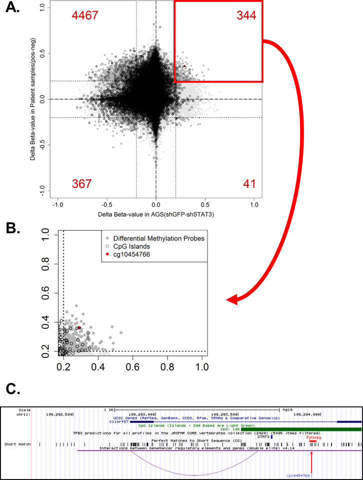
We have previously compared the methylation profile between constitutive STAT3-activated AGS gastric cancer cells, and the subline depleted of STAT3 using Illumina 850K methylation microarray [8, 9]. Additionally, in this study, we compared the methylation profile of three pairs of gastric cancer patients, based on the STAT3 activation status. Integrative computational analyses identified several putative STAT3 targets showing promoter hypomethylation in STAT3-depleted AGS cells and gastric cancer patients showing lower STAT3 activation (as determined by STAT3 nuclear translocation). We further selected probes located within promoter CpG island, showing lower methylation in normal samples but higher methylation in tumor samples in the TCGA gastric cancer dataset. Probes with more than 20% methylation changes in both cell line and patient samples would be defined as significant differential methylation (Fig 1A). One of the probes, cg10454766, showed a significant hypomethylation in both STAT3-depleted AGS cells and patients with lower STAT3 activation (Fig 1B). Further analysis identified a putative STAT3 binding which is in close proximity to cg10454766 (242bp, Fig 1C, blue arrow), and may link to the transcription start site of C11orf87, as demonstrated by GeneHancer (Fig 1C, purple dash line). This probe, cg10454766, was then selected for further analysis.

Fig 1. Identification of cg10454766 as a STAT3-mediated hypermethylation loci.

Fig 1

(A) Scatter plot showing differential methylation differences in gastric cancer cell lines and patient samples. AGS gastric cancer cell lines (shGFP vs shSTAT3) and gastric cancer patients (STAT3 positive vs STAT3 negative) were subjected to Illumina 850K methylation microarray. The significant differences were defined by 20% methylation changes (0.2 delta beta-value). The amount of significant differential probes showed in red numbers for each quadrat. (B) The selected quadrat denoted STAT3-related hypermethylation. Differential methylation probes showed in dark gray. The probes which reside within CpG island were showed in black circle. The selected probe, cg10454766, with most significant difference was shown in red dot. (C) Genomic landscape of cg10454766 (C11orf87) obtained from UCSC Genome browser. The predictive STAT3 binding site were performed by JASPAR2020, as shown in blue bar. The corresponding CG dinucleotides are shown in short match. The region for bisulfite pyrosequencing validation is showed in red bar, including the selected probe, cg10454766 (red arrow). Genomic interactions were performed by GeneHancer, as shown in purple curve.

The expression and methylation of C11orf87

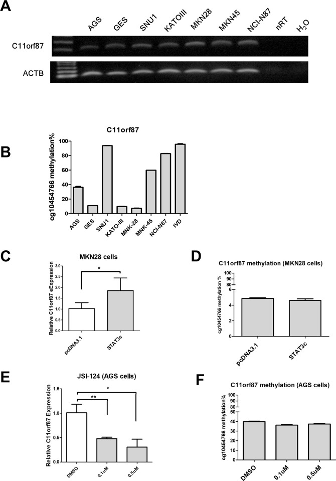
To examine the relationship between expression and methylation on C11orf87, we performed bisulfite pyrosequencing and RT-PCR in GC cell lines and an immortalized normal cell, GES. The results showed that those cell lines exhibited various C11orf87 expression (Fig 2A). Unexpectedly, further bisulfite pyrosequencing also showed various C11orf87 methylation without correlation with its expression (Fig 2B). These results suggested that C11orf87 expression is independent of its methylation.

Fig 2. The expression and methylation of C11orf87 in gastric cancer cell lines.

Fig 2

(A) Expression and (B) methylation of C11orf87 was determined by RT-PCR and bisulfite pyrosequencing, respectively in GC cell lines and an immortalized gastric epithelial cell lines, GES. MKN28 cells were transiently transfected with empty vector (pcDNA3.1) or vector expressing constitutive activated STAT3 mutant (STAT3c). C11orf87 expression and methylation were determined by (C) qRT-PCR and (D) bisulfite pyrosequencing, respectively. AGS gastric cancer cells were treated with DMSO or STAT3 inhibitor (JSI-124) for two days. Expression and methylation of C11orf87 were determined by (E) qRT-PCR and (F) bisulfite pyrosequencing. Significant differences between groups were indicated by *P<0.05, **P<0.01, as determined by unpaired t-test. The original gel images can be found in S1 Fig.

To investigate whether STAT3 control C11orf87 expression, we ectopically expressed a constitutive active STAT3 mutant (STAT3c) in MKN28, a STAT3 inactive cell line [8, 9]. However, ectopic expression of STAT3c promoted C11orf87 expression in MKN28 cells (Fig 2C), without affecting its methylation (Fig 2D). In this regard, we hypothesized that STAT3 may contribute to methylation maintenances rather than de novo methylation. Indeed, treatment of STAT3 inhibitor, JSI-124 [17], resulted in a downregulation of C11orf87 expression in AGS gastric cancer cells (Fig 2E). Surprisingly, treatment of JSI-124 did not affect C11orf87 methylation (Fig 2F). This results suggested that long term STAT3 depletion may be required to disrupt C11orf87 methylation, as previously observed in NR4A3, a STAT3-downregulated target [9]. Taken together, these results suggested that the C11orf87 methylation may be a passenger effect under the STAT3-mediated C11orf87 expression.

The C11orf87 methylation is related to early gastric cancer initiation

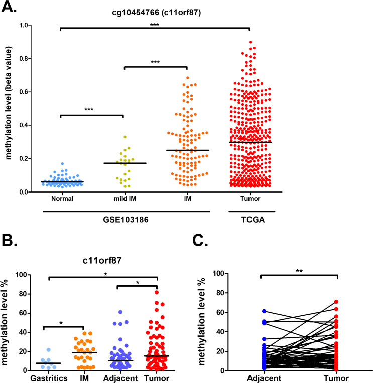
As the methylation wasn’t crucial for controlling C11orf87 expression, we then investigated whether the methylation of C11orf87 could serve as a biomarker for gastric cancer. We first analyzed the DNA methylation of cg10454766 from publicly available dataset obtained from TCGA and a previous study (GSE103186) [18], both of them were performed by Infinium HumanMethylation450 BeadChip. Overall, there was a progressive increase in DNA methylation of cg10454766 from normal gastric epithelium, mild intestinal metaplasia (IM), IM to gastric cancer (Fig 3A). Then, we performed bisulfite pyrosequencing of the CpG sites (chr11:109,293,939–109,293,966, Fig 1C, red bar) in the promoter region of C11orf87 covering cg10454766 in our in-house samples. In agreement with TCGA and the previous study, C11orf87 promoter showed a progressive increase in methylation upon disease progression (Fig 3B). Pairwise analysis of C11orf87 methylation also showed higher methylation in cancer as compared to corresponding adjacent normal (Fig 3C). These results suggested that the methylation of C11orf87 could serve as a diagnostic marker for gastric cancer.

Fig 3. Progressive hypermethylation of cg10454766 in gastric carcinogenesis.

Fig 3

(A) Methylation profile of cg10454766 from two datasets, GSE103186 and TCGA gastric cancer, including normal samples (n = 61), mild IM (n = 22), IM (n = 108) and cancer (n = 395). (B) Methylation of C11orf87 from our in-house samples including gastritis (n = 8), intestinal metaplasia (n = 27), tumor-adjacent normal (n = 47) to gastric tumor patients. (n = 62), as determined by bisulfite pyrosequencing. (C) Pairwise analysis of C11orf87 methylation in tumor-adjacent normal and tumor tissues (n = 47). Black lines denoted median values. Significant differences between groups and pairwise samples were indicated by *P<0.05, **P<0.01, ***P<0.005, as determined by Mann-Whitney U-test or paired t-test, wherever appropriate.

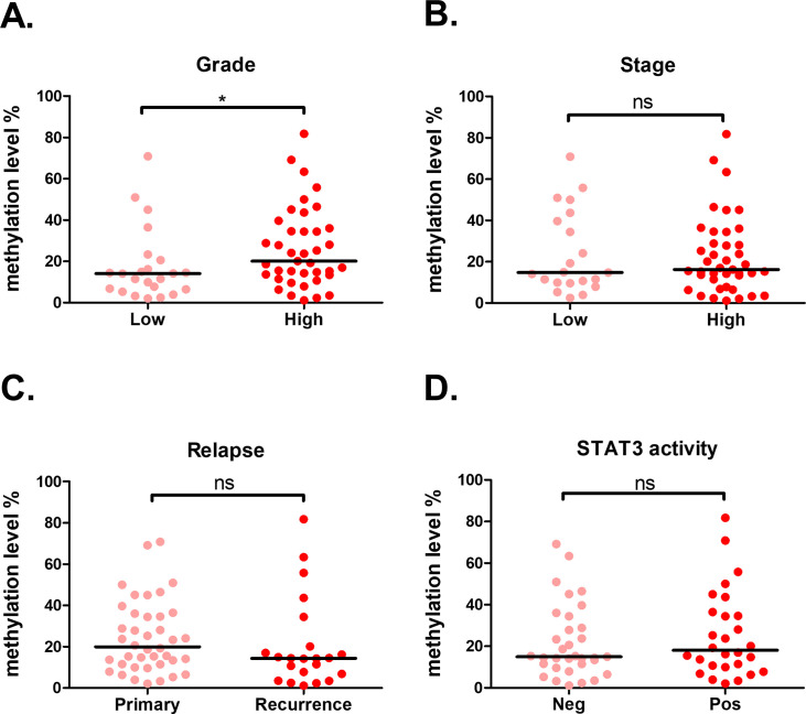
For disease progression analysis, comparison of tumor grade showed that a significant higher C11orf87 methylation was observed in patient samples with higher grade, as compared to low grade samples (Fig 4A). However, C11orf87 methylation was not associated with tumor stage, relapse or STAT3 activity (Fig 4B–4D). As activation of STAT3 was related to poor prognosis in gastric cancer [19], these results suggested that methylation of C11orf87 might independent to cancer progression.

Fig 4. Relationship between C11orf87 methylation of cg10454766 and patients’ clinical parameters.

Fig 4

Comparisons of C11orf87 methylation in gastric cancer patients with different tumor (A) grade (Low: n = 23; High: n = 39), (B) stage (Low: n = 21; High: n = 41), (C) relapse (Primary: n = 40; Recurrence: n = 22), and (D) STAT3 activity (Neg: n = 32; Pos: n = 28, as determined by IHC staining). The black lines denoted median values. Significant differences between groups are indicated by *P<0.05, **P<0.01, ***P<0.005, as determined by Mann-Whitney U-test.

Biomarker evaluation in methylation of C11orf87 from gastric cancer patients and non-cancerous patients

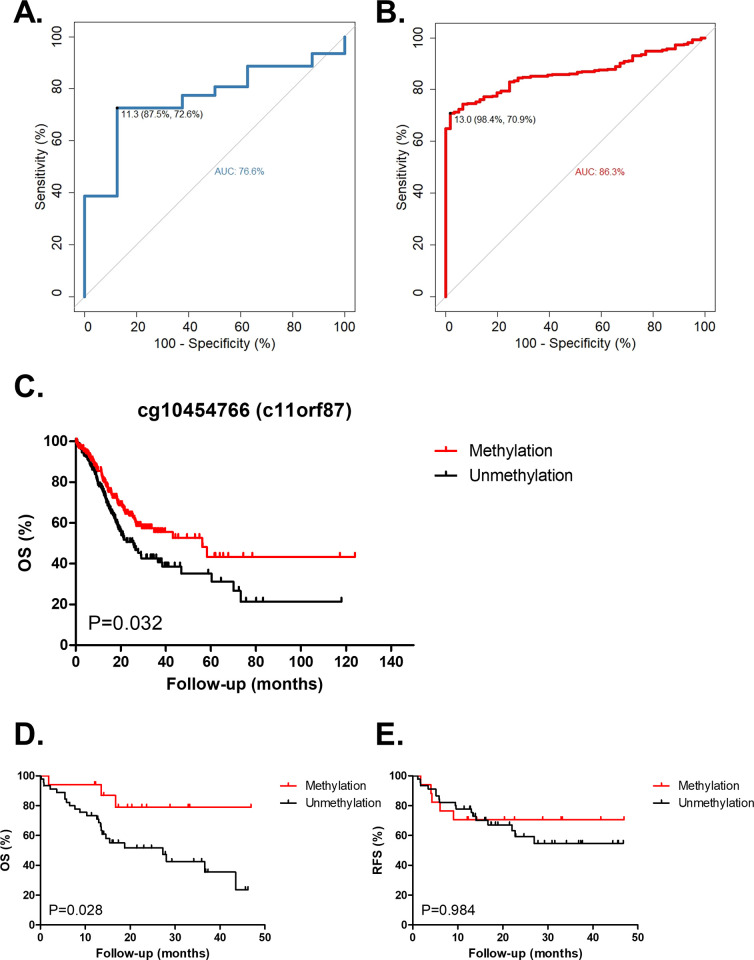
To assess if C11orf87 methylation can be a biomarker for discriminating gastric cancer vs non-cancer, receiver operating characteristics (ROC) curve was performed using the bisulfite pyrosequencing results from our in-house samples (8 gastritis and 62 gastric cancer, Fig 5A), showing an area under curve (AUC) of 76.6%. Using a cut-off of 11.3%, the sensitivity and specificity of cancer detection is 72.6% and 87.5%, respectively.

Fig 5. The methylation of C11orf87 served as a biomarker for diagnosis and diseases outcome.

Fig 5

(A) ROC curve of C11orf87 methylation in our in-house samples, including 8 gastritis and 62 gastric cancer samples. (B) ROC curve of cg10454766 methylation in combined datasets (GSE103186 and TCGA), including 61 normal samples from GSE103186 and 385 tumor samples from TCGA. The best cut-off methylation value for specificity and sensitivity is shown (black dot). (C) Kaplan-Meier analysis of patients with differential C11orf87 methylation status for overall survival (OS) in gastric cancer TCGA dataset (n = 385). Patients were divided into two groups according to the median value of methylation (median = 29.77%). Kaplan-Meier analysis of (D) OS and (E) recurrence-free survival (RFS) in gastric cancer patients from Taiwan. Patients were divided according to same median value from TCGA cohort. P-value is shown from the Log-rank (Mantel-Cox) test.

Besides, we also combined the results from GSE103186 and TCGA gastric cancer dataset, which utilized the same microarray platform, for ROC curve analysis (Fig 5B). ROC curve using 61 normal samples from GSE103186 and 385 cancer samples from the TCGA gastric cancer dataset, showed an area under curve of 86.3%. Using a cut-off of 13.0%, the sensitivity and specificity of cancer detection as positive, the sensitivity and specificity is 70.9% and 98.4%, respectively. These results suggested that C11orf87 methylation could be considered as a novel biomarker with well specificity for gastric cancer diagnosis.

Hypermethylation of C11orf87 is unexpectedly associated with better survival in gastric cancer

To further investigate the methylation of C11orf87 for clinical outcome, we first examined the overall survival (OS) and recurrence free survival (RFS) in our in-house samples (Fig 5C and 5D). Unexpectedly, patients with higher cg10454766 methylation showed a significant better OS, but not RFS in gastric cancer. Additionally, we also performed overall survival (OS) from TCGA gastric cancer cohort (Fig 5E). In agreement with our in-house samples, patients with higher cg10454766 methylation were also associated with better OS in TCGA cohort.

Hypermethylation of C11orf87 is frequently observed in GI-tract cancers

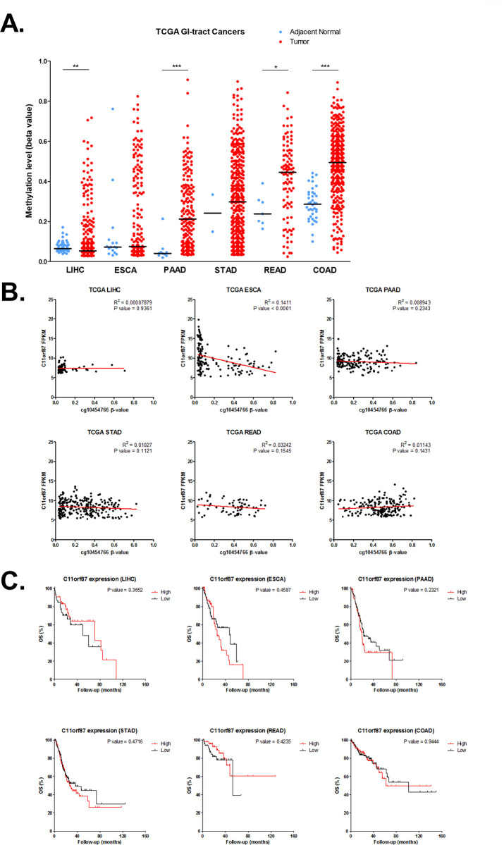
We also analyzed C11orf87 methylation in all of the gastrointestinal tract (GI) cancer from TCGA, including liver hepatocellular carcinoma (LIHC), esophageal carcinoma (ESCA), pancreatic adenocarcinoma (PAAD), stomach adenocarcinoma (STAD), Rectum Adenocarcinoma (READ) and colon adenocarcinoma (COAD). Surprisingly, C11orf87 methylation demonstrated a hypermethylation in most of the GI-tract cancer, as compared to adjacent normal (Fig 6A). This result suggested that C11orf87 methylation may be potentially useful for the detection of all GI cancers. In agreement with our cell lines result, there was no relationship between C11orf87 expression and methylation in most of GI-tract cancers, except for esophageal carcinoma (ESCA) (Fig 6B). Additionally, the expression of C11orf87 in OS from all of GI-tract cancers didn’t show any correlation, which further suggested the biological role of C11orf87 might not relate to clinical outcome (Fig 6C).

Fig 6. The methylation and expression profiles of C11orf87 in GI-tract cancers.

Fig 6

(A) Methylation of C11orf87 in all of GI-tract cancers, including liver hepatocellular carcinoma (LIHC), esophageal carcinoma (ESCA), pancreatic adenocarcinoma (PAAD), stomach adenocarcinoma (STAD), rectum adenocarcinoma (READ), and colon adenocarcinoma (COAD). Blue dots denote adjacent normal while red dots denote cancer. Black lines denote median values. Significant differences between groups are indicated by *P<0.05, **P<0.01, ***P<0.005, as determined by Mann-Whitney U-test. (B) The relationship between C11orf87 expression and methylation in all GI-tract cancers (LIHC: n = 84; ESCA: n = 146; PAAD: n = 160; STAD: n = 247; READ: n = 64; COAD: n = 189). (C) Kaplan-Meier analysis of patients with differential C11orf87 expression for overall survival (OS) in GI-tract cancers from TCGA dataset (LIHC: n = 82; ESCA: n = 145; PAAD: n = 159; STAD: n = 261; READ: n = 98; COAD: n = 248). Patients were divided into two groups according to the median value of expression, while the expression value shown as 0 or missing, were excluded from the analysis.

Discussions

Activation of JAK/STAT signaling plays an important role in gastric carcinogenesis. In particular, STAT3 can serve as a transcriptional activator for oncogene expression [2022]. However, the role of STAT3 as a transcriptional repressor and epigenetic regulator was less explored. By integrated experimental and bioinformatic analyses, we have previously demonstrated that depletion of STAT3 resulted in hypomethylation of STAT3 targets and tumor suppressors, NR4A3 [9], and SPG20 [8], in a gastric cancer cell line with constitutive activation of JAK/STAT signaling. Higher NR4A3 methylation could be observed in gastric cancer patients with STAT3 activation. Patients with higher NR4A3 methylation were associated with poor survival. These results may be due to the recruitment of DNMT, via STAT3, to the STAT3 targets [23]. Previous studies indicated that acetylation on lysine 685 of STAT3 was crucial for the interaction with DNMT1 [24]. Targeted inhibition of STAT3 acetylation may be considered as a novel epigenetic strategy for reactivation of tumor suppressor genes in human cancer [25].

In the current study, by DNA methylation microarray, we further identified a potential STAT3 target, C11orf87, showing hypomethylation in AGS gastric cancer cells depleted with STAT3 and patients with lower STAT3 activation. Although STAT3 can promote the expression of C11orf87, there is no relationship between its expression and methylation. These results, interestingly, contradict with our previous finding that STAT3 acts as an oncogenic protein in the epigenetic silencing of tumor suppressors in gastric cancer [8, 9]. Recently, a study demonstrated that SIRT1, a histone deacetylase that participated in STAT3 deacetylation, was found to be upregulated in advanced gastric cancer [26]. The authors suggested that SIRT1 upregulation may compensate for the damaging effect induced by constitutive activation of STAT3 in gastric cancer. In this regard, SIRT1 may disrupt the interaction between STAT3 and DNMT1 by deacetylation on Lys685, which further limited methylation maintenances. Herein, we postulated that hypermethylation of C11orf87 may serve as a “vestigial marker” for constitutive activation of STAT3 in gastric cancer. This hypothesis may further explain our clinical observation that C11orf87 hypermethylation was related to better survival.

C11orf87, known as neuronal integral membrane protein 1, was found to be predominantly expressed in the brain tissue [27]. However, the involvement of C11orf87 in human cancer has not been characterized. A recent study in head and neck cancer found that p53 mutated tumors could promote differentiation of nerve fibers, which then promoted tumor growth in this tumor microenvironment [28]. As the expression of C11orf87 was controlled by STAT3, we, therefore, postulate that aberrant STAT3 activation may involve in promoting differentiation of nerve fibers via upregulation of C11orf87. Although p53 mutation is frequent in gastric cancer [29], how neuronal-related gene control gastric cancer progression still requires further investigation.

In conclusion, we are the first to demonstrate the progressive increase in C11orf87 methylation in IM and gastric cancer. Hypermethylation of C11orf87 was associated with better prognosis. Importantly, methylation of C11orf87 may act as a novel diagnostic biomarker for gastric cancer. Additionally, C11orf87 methylation is frequently observed in GI cancers, suggesting that it may also be useful for the detection of GI cancers. Besides, methylation of this potential STAT3 target may serve as a marker for the response and efficacy of targeting STAT3 in gastric cancer, as it may respect to STAT3 activity in early carcinogenesis. However, the functional and clinical role of C11orf87 in gastric cancer warrants further investigation.

Supporting information

S1 Fig. The original uncropped and unadjusted gel image for Fig 2A.

(PDF)

Data Availability

The methylation array data has been deposited into in the Gene Expression Omnibus database (accession number: GSE109541).

Funding Statement

This study was supported by grants from the Ministry of Science and Technology, Taiwan (MOST 106-2314-B-194-001-MY3; 107-2314-B-194-001; 108-2314-B-194-001; 108-2314-B-194-003- MY2) to MWYC, Changhua Christian Hospital, Taiwan (106-CCH-IRP-064) to SHL, and the Center for Innovative Research on Aging Society (CIRAS) from The Featured Areas Research Center Program within the framework of the Higher Education Sprout Project by Ministry of Education (MOE) in Taiwan.

References

  • 1.Bray F, Ferlay J, Soerjomataram I, Siegel RL, Torre LA, Jemal A. Global cancer statistics 2018: GLOBOCAN estimates of incidence and mortality worldwide for 36 cancers in 185 countries. CA Cancer J Clin. 2018;68(6):394–424. 10.3322/caac.21492 . [DOI] [PubMed] [Google Scholar]
  • 2.Polk DB, Peek RM Jr. Helicobacter pylori: gastric cancer and beyond. Nat Rev Cancer. 2010;10(6):403–14. 10.1038/nrc2857 . [DOI] [PMC free article] [PubMed] [Google Scholar]
  • 3.Piao JY, Lee HG, Kim SJ, Kim DH, Han HJ, Ngo HK, et al. Helicobacter pylori Activates IL-6-STAT3 Signaling in Human Gastric Cancer Cells: Potential Roles for Reactive Oxygen Species. Helicobacter. 2016;21(5):405–16. 10.1111/hel.12298 . [DOI] [PubMed] [Google Scholar]
  • 4.Lu R, Zhang YG, Sun J. STAT3 activation in infection and infection-associated cancer. Mol Cell Endocrinol. 2017;451:80–7. 10.1016/j.mce.2017.02.023 . [DOI] [PMC free article] [PubMed] [Google Scholar]
  • 5.Jackson CB, Judd LM, Menheniott TR, Kronborg I, Dow C, Yeomans ND, et al. Augmented gp130-mediated cytokine signalling accompanies human gastric cancer progression. J Pathol. 2007;213(2):140–51. 10.1002/path.2218 . [DOI] [PubMed] [Google Scholar]
  • 6.Rocha GA, Rocha AM, Gomes AD, Faria CL Jr., Melo FF, Batista SA, et al. STAT3 polymorphism and Helicobacter pylori CagA strains with higher number of EPIYA-C segments independently increase the risk of gastric cancer. BMC Cancer. 2015;15:528. 10.1186/s12885-015-1533-1 . [DOI] [PMC free article] [PubMed] [Google Scholar]
  • 7.Bronte-Tinkew DM, Terebiznik M, Franco A, Ang M, Ahn D, Mimuro H, et al. Helicobacter pylori cytotoxin-associated gene A activates the signal transducer and activator of transcription 3 pathway in vitro and in vivo. Cancer Res. 2009;69(2):632–9. 10.1158/0008-5472.CAN-08-1191 . [DOI] [PMC free article] [PubMed] [Google Scholar]
  • 8.Wei KL, Chou JL, Chen YC, Jin H, Chuang YM, Wu CS, et al. Methylomics analysis identifies a putative STAT3 target, SPG20, as a noninvasive epigenetic biomarker for early detection of gastric cancer. PLoS One. 2019;14(6):e0218338. 10.1371/journal.pone.0218338 . [DOI] [PMC free article] [PubMed] [Google Scholar]
  • 9.Yeh CM, Chang LY, Lin SH, Chou JL, Hsieh HY, Zeng LH, et al. Epigenetic silencing of the NR4A3 tumor suppressor, by aberrant JAK/STAT signaling, predicts prognosis in gastric cancer. Sci Rep. 2016;6:31690. 10.1038/srep31690 . [DOI] [PMC free article] [PubMed] [Google Scholar]
  • 10.Wu CS, Wei KL, Chou JL, Lu CK, Hsieh CC, Lin JM, et al. Aberrant JAK/STAT Signaling Suppresses TFF1 and TFF2 through Epigenetic Silencing of GATA6 in Gastric Cancer. Int J Mol Sci. 2016;17(9). 10.3390/ijms17091467 . [DOI] [PMC free article] [PubMed] [Google Scholar]
  • 11.Qi H, Yang Z, Dai C, Wang R, Ke X, Zhang S, et al. STAT3 activates MSK1-mediated histone H3 phosphorylation to promote NFAT signaling in gastric carcinogenesis. Oncogenesis. 2020;9(2):15. 10.1038/s41389-020-0195-2 . [DOI] [PMC free article] [PubMed] [Google Scholar]
  • 12.Pan YM, Wang CG, Zhu M, Xing R, Cui JT, Li WM, et al. STAT3 signaling drives EZH2 transcriptional activation and mediates poor prognosis in gastric cancer. Mol Cancer. 2016;15(1):79. 10.1186/s12943-016-0561-z . [DOI] [PMC free article] [PubMed] [Google Scholar]
  • 13.Flavahan WA, Gaskell E, Bernstein BE. Epigenetic plasticity and the hallmarks of cancer. Science. 2017;357(6348). 10.1126/science.aal2380 . [DOI] [PMC free article] [PubMed] [Google Scholar]
  • 14.Biswas S, Rao CM. Epigenetics in cancer: Fundamentals and Beyond. Pharmacol Ther. 2017;173:118–34. 10.1016/j.pharmthera.2017.02.011 . [DOI] [PubMed] [Google Scholar]
  • 15.Jones PA. Functions of DNA methylation: islands, start sites, gene bodies and beyond. Nature reviews Genetics. 2012;13(7):484–92. 10.1038/nrg3230 . [DOI] [PubMed] [Google Scholar]
  • 16.Luczak MW, Jagodzinski PP. The role of DNA methylation in cancer development. Folia histochemica et cytobiologica. 2006;44(3):143–54. . [PubMed] [Google Scholar]
  • 17.Blaskovich MA, Sun J, Cantor A, Turkson J, Jove R, Sebti SM. Discovery of JSI-124 (cucurbitacin I), a selective Janus kinase/signal transducer and activator of transcription 3 signaling pathway inhibitor with potent antitumor activity against human and murine cancer cells in mice. Cancer Res. 2003;63(6):1270–9. . [PubMed] [Google Scholar]
  • 18.Huang KK, Ramnarayanan K, Zhu F, Srivastava S, Xu C, Tan ALK, et al. Genomic and Epigenomic Profiling of High-Risk Intestinal Metaplasia Reveals Molecular Determinants of Progression to Gastric Cancer. Cancer cell. 2018;33(1):137–50 e5. 10.1016/j.ccell.2017.11.018 . [DOI] [PubMed] [Google Scholar]
  • 19.Kim DY, Cha ST, Ahn DH, Kang HY, Kwon CI, Ko KH, et al. STAT3 expression in gastric cancer indicates a poor prognosis. Journal of gastroenterology and hepatology. 2009;24(4):646–51. 10.1111/j.1440-1746.2008.05671.x . [DOI] [PubMed] [Google Scholar]
  • 20.Levy DE, Darnell JE Jr, Stats: transcriptional control and biological impact. Nat Rev Mol Cell Biol. 2002;3(9):651–62. 10.1038/nrm909 . [DOI] [PubMed] [Google Scholar]
  • 21.Polak KL, Chernosky NM, Smigiel JM, Tamagno I, Jackson MW. Balancing STAT Activity as a Therapeutic Strategy. Cancers (Basel). 2019;11(11). 10.3390/cancers11111716 . [DOI] [PMC free article] [PubMed] [Google Scholar]
  • 22.Lim CP, Cao X. Structure, function, and regulation of STAT proteins. Mol Biosyst. 2006;2(11):536–50. 10.1039/b606246f . [DOI] [PubMed] [Google Scholar]
  • 23.Zhang Q, Wang HY, Marzec M, Raghunath PN, Nagasawa T, Wasik MA. STAT3- and DNA methyltransferase 1-mediated epigenetic silencing of SHP-1 tyrosine phosphatase tumor suppressor gene in malignant T lymphocytes. Proc Natl Acad Sci U S A. 2005;102(19):6948–53. 10.1073/pnas.0501959102 . [DOI] [PMC free article] [PubMed] [Google Scholar]
  • 24.Lee H, Zhang P, Herrmann A, Yang C, Xin H, Wang Z, et al. Acetylated STAT3 is crucial for methylation of tumor-suppressor gene promoters and inhibition by resveratrol results in demethylation. Proc Natl Acad Sci U S A. 2012;109(20):7765–9. 10.1073/pnas.1205132109 . [DOI] [PMC free article] [PubMed] [Google Scholar]
  • 25.Kang HJ, Yi YW, Hou SJ, Kim HJ, Kong Y, Bae I, et al. Disruption of STAT3-DNMT1 interaction by SH-I-14 induces re-expression of tumor suppressor genes and inhibits growth of triple-negative breast tumor. Oncotarget. 2017;8(48):83457–68. 10.18632/oncotarget.4054 . [DOI] [PMC free article] [PubMed] [Google Scholar]
  • 26.Zhang S, Yang Y, Huang S, Deng C, Zhou S, Yang J, et al. SIRT1 inhibits gastric cancer proliferation and metastasis via STAT3/MMP-13 signaling. J Cell Physiol. 2019;234(9):15395–406. 10.1002/jcp.28186 . [DOI] [PubMed] [Google Scholar]
  • 27.Etemadikhah M, Niazi A, Wetterberg L, Feuk L. Transcriptome analysis of fibroblasts from schizophrenia patients reveals differential expression of schizophrenia-related genes. Sci Rep. 2020;10(1):630. 10.1038/s41598-020-57467-z . [DOI] [PMC free article] [PubMed] [Google Scholar]
  • 28.Amit M, Takahashi H, Dragomir MP, Lindemann A, Gleber-Netto FO, Pickering CR, et al. Loss of p53 drives neuron reprogramming in head and neck cancer. Nature. 2020;578(7795):449–54. 10.1038/s41586-020-1996-3 . [DOI] [PMC free article] [PubMed] [Google Scholar]
  • 29.Rhyu MG, Park WS, Jung YJ, Choi SW, Meltzer SJ. Allelic deletions of MCC/APC and p53 are frequent late events in human gastric carcinogenesis. Gastroenterology. 1994;106(6):1584–8. 10.1016/0016-5085(94)90414-6 . [DOI] [PubMed] [Google Scholar]

Decision Letter 0

Qian Tao

29 Jul 2020

PONE-D-20-20115

Methylomic analysis identifies C11orf87 as a novel epigenetic biomarker for GI cancers

PLOS ONE

Dear Dr. Chan,

Thank you for submitting your manuscript to PLOS ONE. After careful consideration, we feel that it has merit but does not fully meet PLOS ONE’s publication criteria as it currently stands. Therefore, we invite you to submit a revised version of the manuscript that addresses all the points raised by the two reviewers.

Please ensure that your decision is justified on PLOS ONE’s publication criteria and not, for example, on novelty or perceived impact.

==============================

Please submit your revised manuscript by Sep 12 2020 11:59PM. If you will need more time than this to complete your revisions, please reply to this message or contact the journal office at plosone@plos.org. When you're ready to submit your revision, log on to https://www.editorialmanager.com/pone/ and select the 'Submissions Needing Revision' folder to locate your manuscript file.

Please include the following items when submitting your revised manuscript:

  • A rebuttal letter that responds to each point raised by the academic editor and reviewer(s). You should upload this letter as a separate file labeled 'Response to Reviewers'.

  • A marked-up copy of your manuscript that highlights changes made to the original version. You should upload this as a separate file labeled 'Revised Manuscript with Track Changes'.

  • An unmarked version of your revised paper without tracked changes. You should upload this as a separate file labeled 'Manuscript'.

If you would like to make changes to your financial disclosure, please include your updated statement in your cover letter. Guidelines for resubmitting your figure files are available below the reviewer comments at the end of this letter.

If applicable, we recommend that you deposit your laboratory protocols in protocols.io to enhance the reproducibility of your results. Protocols.io assigns your protocol its own identifier (DOI) so that it can be cited independently in the future. For instructions see: http://journals.plos.org/plosone/s/submission-guidelines#loc-laboratory-protocols

We look forward to receiving your revised manuscript.

Kind regards,

Qian Tao

Academic Editor

PLOS ONE

Journal Requirements:

When submitting your revision, we need you to address these additional requirements.

1. Please ensure that your manuscript meets PLOS ONE's style requirements, including those for file naming. The PLOS ONE style templates can be found at

https://journals.plos.org/plosone/s/file?id=wjVg/PLOSOne_formatting_sample_main_body.pdf and

https://journals.plos.org/plosone/s/file?id=ba62/PLOSOne_formatting_sample_title_authors_affiliations.pdf

2. In your Methods section, please provide additional information about the participant recruitment method and the demographic details of your participants. Please ensure you have provided sufficient details to replicate the analyses such as: a) the recruitment date range (month and year),  b) a description of how participants were recruited, and c) descriptions of where participants were recruited and where the research took place.

3.We note that you have indicated that data from this study are available upon request. PLOS only allows data to be available upon request if there are legal or ethical restrictions on sharing data publicly. For information on unacceptable data access restrictions, please see http://journals.plos.org/plosone/s/data-availability#loc-unacceptable-data-access-restrictions.

In your revised cover letter, please address the following prompts:

a) If there are ethical or legal restrictions on sharing a de-identified data set, please explain them in detail (e.g., data contain potentially identifying or sensitive patient information) and who has imposed them (e.g., an ethics committee). Please also provide contact information for a data access committee, ethics committee, or other institutional body to which data requests may be sent.

b) If there are no restrictions, please upload the minimal anonymized data set necessary to replicate your study findings as either Supporting Information files or to a stable, public repository and provide us with the relevant URLs, DOIs, or accession numbers. Please see http://www.bmj.com/content/340/bmj.c181.long for guidelines on how to de-identify and prepare clinical data for publication. For a list of acceptable repositories, please see http://journals.plos.org/plosone/s/data-availability#loc-recommended-repositories.

We will update your Data Availability statement on your behalf to reflect the information you provide.

4.Thank you for stating the following in the Acknowledgments Section of your manuscript:

[This study was supported by grants from the Ministry of Science and Technology, Taiwan (MOST

106-2314-B-194-001-MY3; 107-2314-B-194-001; 108-2314-B-194-001; 108-2314-B-194-003-

MY2), Changhua Christian Hospital, Taiwan (106-CCH-IRP-064) and the Center for Innovative

Research on Aging Society (CIRAS) from The Featured Areas Research Center Program within

the framework of the Higher Education Sprout Project by Ministry of Education (MOE) in Taiwan.]

We note that you have provided funding information that is not currently declared in your Funding Statement. However, funding information should not appear in the Acknowledgments section or other areas of your manuscript. We will only publish funding information present in the Funding Statement section of the online submission form.

Please remove any funding-related text from the manuscript and let us know how you would like to update your Funding Statement. Currently, your Funding Statement reads as follows:

 [This study was supported by grants from the Ministry of Science and Technology, Taiwan (MOST 106-2314-B-194-001-MY3; 107-2314-B-194-001; 108-2314-B-194-001; 108-2314-B-194-003-MY2) to MWYC, and Changhua Christian Hospital, Taiwan (106-CCH-IRP-064) to SHL.

The funders had no role in study design, data collection and analysis, decision to publish, or preparation of the manuscript.]

[Note: HTML markup is below. Please do not edit.]

Reviewers' comments:

Reviewer's Responses to Questions

Comments to the Author

1. Is the manuscript technically sound, and do the data support the conclusions?

The manuscript must describe a technically sound piece of scientific research with data that supports the conclusions. Experiments must have been conducted rigorously, with appropriate controls, replication, and sample sizes. The conclusions must be drawn appropriately based on the data presented.

Reviewer #1: Partly

Reviewer #2: Partly

**********

2. Has the statistical analysis been performed appropriately and rigorously?

Reviewer #1: Yes

Reviewer #2: I Don't Know

**********

3. Have the authors made all data underlying the findings in their manuscript fully available?

The PLOS Data policy requires authors to make all data underlying the findings described in their manuscript fully available without restriction, with rare exception (please refer to the Data Availability Statement in the manuscript PDF file). The data should be provided as part of the manuscript or its supporting information, or deposited to a public repository. For example, in addition to summary statistics, the data points behind means, medians and variance measures should be available. If there are restrictions on publicly sharing data—e.g. participant privacy or use of data from a third party—those must be specified.

Reviewer #1: Yes

Reviewer #2: Yes

**********

4. Is the manuscript presented in an intelligible fashion and written in standard English?

PLOS ONE does not copyedit accepted manuscripts, so the language in submitted articles must be clear, correct, and unambiguous. Any typographical or grammatical errors should be corrected at revision, so please note any specific errors here.

Reviewer #1: Yes

Reviewer #2: No

**********

5. Review Comments to the Author

Please use the space provided to explain your answers to the questions above. You may also include additional comments for the author, including concerns about dual publication, research ethics, or publication ethics. (Please upload your review as an attachment if it exceeds 20,000 characters)

Reviewer #1: Tran et al reported C11orf87 as a novel epigenetic biomarker for GI cancers. The authors found that C11orf87, a potential STAT3 target, was hypomethylated in gastric cancer patients and a cell line with STAT3 lower activation using DNA methylation microarray. C11orf87 was hypermethylated in GI cancers compared to their adjacent normal tissues, which was associated with better survival, thus may serve as a biomarker for GI cancers. There are a few specific issues the authors should address before publication.

Special comments:

1. The authors postulated the relationship of STAT3 activation with epigenetically silenced C11orf87 in GI cancers. However, some key experiments should be performed to identify this conclusion, such as the effect of C11orf87 methylation and expression in gastric cell lines with STAT3 reactivation.

2. The C11orf87 expression and methylation levels in GI cell lines with different STAT3 status should be examined.

3. What is the expression pattern of C11orf87 in GI tumor tissues? Is there any correlation with its methylation level?

4. As a novel biomarker of GI cancers, what are the biological functions of C11orf87 in GI cells?

Reviewer #2: 1.The detection results of different methylation sequencing methods are very different. Are the methylation detection methods used in the TCGA and GSE103186 database the same? Is the combined analysis reasonable?

2.Because the authors screened the target genes of STAT3 through DNA methylation microarray, please analyze and verify the relationship between STAT3 and C11orf87.

3.What is the expression of C11orf87 in TGCA database and in-house samples? The correlation analysis between its methylation and expression ? And, the correlation analysis between its expression and prognosis?

4.In a variety of gastrointestinal tumors, including gastric cancer, the methylation level of C11orf87 in cancer tissues is higher than that in adjacent tissues and/or normal tissues. Therefore, the author proposes that C11orf87 may be used as a biomarker for gastric cancer. However, gastric cancer patients with high C11orf87 methylation have a better prognosis, which seems to be contrary to previous results. The authors think that it may be caused by the role of STAT3 in gastric cancer, but lack of relevant experimental results.

**********

6. PLOS authors have the option to publish the peer review history of their article (what does this mean?). If published, this will include your full peer review and any attached files.

If you choose “no”, your identity will remain anonymous but your review may still be made public.

Do you want your identity to be public for this peer review? For information about this choice, including consent withdrawal, please see our Privacy Policy.

Reviewer #1: No

Reviewer #2: No

[NOTE: If reviewer comments were submitted as an attachment file, they will be attached to this email and accessible via the submission site. Please log into your account, locate the manuscript record, and check for the action link "View Attachments". If this link does not appear, there are no attachment files.]

While revising your submission, please upload your figure files to the Preflight Analysis and Conversion Engine (PACE) digital diagnostic tool, https://pacev2.apexcovantage.com/. PACE helps ensure that figures meet PLOS requirements. To use PACE, you must first register as a user. Registration is free. Then, login and navigate to the UPLOAD tab, where you will find detailed instructions on how to use the tool. If you encounter any issues or have any questions when using PACE, please email PLOS at figures@plos.org. Please note that Supporting Information files do not need this step.

PLoS One. 2021 Apr 22;16(4):e0250499. doi: 10.1371/journal.pone.0250499.r002

Author response to Decision Letter 0


23 Nov 2020

Point-to-point responses

Comments from Reviewer #1:

We thank the reviewer for the insightful review and interest in our manuscript. We have now carefully read each comment, and incorporated the recommendations and suggestions in the following manner:

1. The authors postulated the relationship of STAT3 activation with epigenetically silenced C11orf87 in GI cancers. However, some key experiments should be performed to identify this conclusion, such as the effect of C11orf87 methylation and expression in gastric cell lines with STAT3 reactivation.

Response: We thank the review for this important question. We now performed additional experiments to investigate the relationship between C11orf87 methylation and expression in a panel of gastric cancer cell lines. The results showed that those cell lines exhibited various C11orf87 expression (Fig. 2A). Unexpectedly, further bisulfite pyrosequencing also showed various C11orf87 methylation without correlation with its expression (Fig. 2B). These results suggested that C11orf87 expression is independent of its methylation.

These new results can be found in Figure 2A, B and Page 7 of the result section.

2. The C11orf87 expression and methylation levels in GI cell lines with different STAT3 status should be examined.

Response: Again, we thank the reviewer for this important question. We now performed additional experiments to examine the effect of STAT3 on C11orf87 expression. We ectopically expressed a constitutive active STAT3 mutant (STAT3c) in MKN28, a STAT3 inactive cell line. However, ectopic expression of STAT3c promoted C11orf87 expression in MKN28 cells (Fig. 2C), without affecting its methylation (Fig. 2D). In this regard, we hypothesized that STAT3 may contribute to methylation maintenances rather than de novo methylation. Indeed, treatment of STAT3 inhibitor, JSI-124, resulted in a downregulation of C11orf87 expression (Fig. 2E). Taken together, these results suggested that the C11orf87 methylation may be a passenger effect under the STAT3-mediated C11orf87 expression.

These new results can be found in Figure 2C-E and Page 7 of the Result sections.

3. What is the expression pattern of C11orf87 in GI tumor tissues? Is there any correlation with its methylation level?

Response: We thank the reviewer for this question. Unfortunately, we don’t have any RNA of the in-house tumor tissue samples to perform such experiments. We therefore performed additional experiments to investigate the relationship between C11orf87 methylation and expression in a panel of gastric cancer cell lines. The results showed that those cell lines exhibited various C11orf87 expression (Fig. 2A). Unexpectedly, further bisulfite pyrosequencing also showed various C11orf87 methylation without correlation with their expression (Fig. 2B). These results suggested that C11orf87 expression is independent of its methylation. As the methylation wasn’t crucial for controlling C11orf87 expression, we then investigated whether the methylation of C11orf87 could serve as a biomarker for gastric cancer. Interestingly, we found that C11orf87 methylation can be an epigenetic biomarker for GI cancers.

4. As a novel biomarker of GI cancers, what are the biological functions of C11orf87 in GI cells?

Response: We thank the reviewer for this important question. C11orf87, known as neuronal integral membrane protein 1, was found to be predominantly expressed in the brain tissue. However, the involvement of C11orf87 in human cancer has not been characterized. A recent study in head and neck cancer found that p53 mutated tumors could promote differentiation of nerve fibers, which then promoted tumor growth in this tumor microenvironment [1]. As the expression of C11orf87 was controlled by STAT3, we, therefore, postulate that aberrant STAT3 activation may involve in promoting differentiation of nerve fibers via upregulation of C11orf87. Although p53 mutation is frequent in gastric cancer [2], how neuronal-related gene control gastric cancer progression still requires further investigation.

These statements have been added in Page 11 of the Discussion section.

Comments from Reviewer #2:

We thank the reviewer for the insightful review and interest in our manuscript. We have now carefully read each comment, and incorporated the recommendations and suggestions in the following manner:

1.The detection results of different methylation sequencing methods are very different. Are the methylation detection methods used in the TCGA and GSE103186 database the same? Is the combined analysis reasonable?

Response: We thank the reviewer for this important question. We apologize that we didn’t state clearly the methodology of these two dataset. Methylation analysis of TCGA and GSE103186 dataset are indeed from the same microarray platform (Infinium HumanMethylation450 Beadchip). Therefore, we combined these two dataset for the analyses. We have added a statement to clarify they are indeed from the same microarray platform (Page 8 of the Result section).

2.Because the authors screened the target genes of STAT3 through DNA methylation microarray, please analyze and verify the relationship between STAT3 and C11orf87.

Response: Again, we thank the reviewer for this important question. We now performed additional experiments to examine the effect of STAT3 on C11orf87 expression. We ectopically expressed a constitutive active STAT3 mutant (STAT3c) in MKN28, a STAT3 inactive cell line. However, ectopic expression of STAT3c promoted C11orf87 expression in MKN28 cells (Fig. 2C), without affecting its methylation (Fig. 2D). In this regard, we hypothesized that STAT3 may contribute to methylation maintenances rather than de novo methylation. Indeed, treatment of STAT3 inhibitor, JSI-124, resulted in a downregulation of C11orf87 expression (Fig. 2E). Taken together, these results suggested that the C11orf87 methylation may be a passenger effect under the STAT3-mediated C11orf87 expression.

3.What is the expression of C11orf87 in TGCA database and in-house samples? The correlation analysis between its methylation and expression? And, the correlation analysis between its expression and prognosis?

Response: We thank the reviewer for this question. Unfortunately, we don’t have any RNA of our in-house tumor tissues to perform such experiments. As the methylation wasn’t crucial for controlling C11orf87 expression, we then investigated whether the methylation of C11orf87 could serve as a biomarker for gastric cancer. Interestingly, we found that C11orf87 methylation can be an epigenetic biomarker for GI cancers.

These new results can be found in Figure 2 and Page 7 of the result section.

4.In a variety of gastrointestinal tumors, including gastric cancer, the methylation level of C11orf87 in cancer tissues is higher than that in adjacent tissues and/or normal tissues. Therefore, the author proposes that C11orf87 may be used as a biomarker for gastric cancer. However, gastric cancer patients with high C11orf87 methylation have a better prognosis, which seems to be contrary to previous results. The authors think that it may be caused by the role of STAT3 in gastric cancer, but lack of relevant experimental results.

Response: We thank the reviewer for this important question. As mentioned in point #2 above, we found that ectopic expression of STAT3c promoted C11orf87 expression in MKN28 cells, without affecting its methylation. While treatment of STAT3 inhibitor, JSI-124, resulted in a downregulation of C11orf87 expression. Taken together, these results suggested that the C11orf87 methylation may be a passenger effect under the STAT3-mediated C11orf87 expression.

Recently, a study demonstrated that SIRT1, a histone deacetylase that participated in STAT3 deacetylation, was found to be upregulated in advanced gastric cancer [3]. The authors suggested that SIRT1 upregulation may compensate for the damaging effect induced by constitutive activation of STAT3 in gastric cancer. In this regard, SIRT1 may disrupt the interaction between STAT3 and DNMT1 by deacetylation on Lys685, which further limited methylation maintenances. Herein, we postulated that hypermethylation of C11orf87 may serve as a “vestigial marker” for constitutive activation of STAT3 in gastric cancer. This hypothesis may further explain our clinical observation that C11orf87 hypermethylation was related to better survival.

We have added those statement in page 10 of the Discussion section.

References

1. Amit, M., et al., Loss of p53 drives neuron reprogramming in head and neck cancer. Nature, 2020. 578(7795): p. 449-454.

2. Rhyu, M.G., et al., Allelic deletions of MCC/APC and p53 are frequent late events in human gastric carcinogenesis. Gastroenterology, 1994. 106(6): p. 1584-8.

3. Zhang, S., et al., SIRT1 inhibits gastric cancer proliferation and metastasis via STAT3/MMP-13 signaling. J Cell Physiol, 2019. 234(9): p. 15395-15406.

Attachment

Submitted filename: Tran et al_C11orf87_response_rev.pdf

Decision Letter 1

Qian Tao

23 Dec 2020

PONE-D-20-20115R1

Methylomic analysis identifies C11orf87 as a novel epigenetic biomarker for GI cancers

PLOS ONE

Dear Dr. Chan,

Thank you for submitting your manuscript to PLOS ONE. After careful consideration, we feel that it has merit but does not fully meet PLOS ONE’s publication criteria as it currently stands. Therefore, we invite you to submit a revised version of the manuscript that addresses all the points raised by the two reviewers.

Please submit your revised manuscript by Feb 06 2021 11:59PM. If you will need more time than this to complete your revisions, please reply to this message or contact the journal office at plosone@plos.org. When you're ready to submit your revision, log on to https://www.editorialmanager.com/pone/ and select the 'Submissions Needing Revision' folder to locate your manuscript file.

Please include the following items when submitting your revised manuscript:

  • A rebuttal letter that responds to each point raised by the academic editor and reviewer(s). You should upload this letter as a separate file labeled 'Response to Reviewers'.

  • A marked-up copy of your manuscript that highlights changes made to the original version. You should upload this as a separate file labeled 'Revised Manuscript with Track Changes'.

  • An unmarked version of your revised paper without tracked changes. You should upload this as a separate file labeled 'Manuscript'.

If you would like to make changes to your financial disclosure, please include your updated statement in your cover letter. Guidelines for resubmitting your figure files are available below the reviewer comments at the end of this letter.

If applicable, we recommend that you deposit your laboratory protocols in protocols.io to enhance the reproducibility of your results. Protocols.io assigns your protocol its own identifier (DOI) so that it can be cited independently in the future. For instructions see: http://journals.plos.org/plosone/s/submission-guidelines#loc-laboratory-protocols

We look forward to receiving your revised manuscript.

Kind regards,

Qian Tao

Academic Editor

PLOS ONE

[Note: HTML markup is below. Please do not edit.]

Reviewers' comments:

Reviewer's Responses to Questions

Comments to the Author

1. If the authors have adequately addressed your comments raised in a previous round of review and you feel that this manuscript is now acceptable for publication, you may indicate that here to bypass the “Comments to the Author” section, enter your conflict of interest statement in the “Confidential to Editor” section, and submit your "Accept" recommendation.

Reviewer #1: (No Response)

Reviewer #2: (No Response)

**********

2. Is the manuscript technically sound, and do the data support the conclusions?

The manuscript must describe a technically sound piece of scientific research with data that supports the conclusions. Experiments must have been conducted rigorously, with appropriate controls, replication, and sample sizes. The conclusions must be drawn appropriately based on the data presented.

Reviewer #1: Partly

Reviewer #2: (No Response)

**********

3. Has the statistical analysis been performed appropriately and rigorously?

Reviewer #1: Yes

Reviewer #2: (No Response)

**********

4. Have the authors made all data underlying the findings in their manuscript fully available?

The PLOS Data policy requires authors to make all data underlying the findings described in their manuscript fully available without restriction, with rare exception (please refer to the Data Availability Statement in the manuscript PDF file). The data should be provided as part of the manuscript or its supporting information, or deposited to a public repository. For example, in addition to summary statistics, the data points behind means, medians and variance measures should be available. If there are restrictions on publicly sharing data—e.g. participant privacy or use of data from a third party—those must be specified.

Reviewer #1: Yes

Reviewer #2: (No Response)

**********

5. Is the manuscript presented in an intelligible fashion and written in standard English?

PLOS ONE does not copyedit accepted manuscripts, so the language in submitted articles must be clear, correct, and unambiguous. Any typographical or grammatical errors should be corrected at revision, so please note any specific errors here.

Reviewer #1: Yes

Reviewer #2: (No Response)

**********

6. Review Comments to the Author

Please use the space provided to explain your answers to the questions above. You may also include additional comments for the author, including concerns about dual publication, research ethics, or publication ethics. (Please upload your review as an attachment if it exceeds 20,000 characters)

Reviewer #1: In the revised manuscript, the authors demonstrated that C11orf87 methylation could be an epigenetic biomarker for GI cancers, and its expression is independent of its methylation. There are only a few comments for the authors which they should take into account before publication.

Comments:

1. The labeling is misplaced in Figure 2A.

2. The authors identified a potential STAT3 target, C11orf87, through using DNA methylation microarray, which is hypomethylated in gastric cancer patients with lower STAT3 activation and AGS cells with STAT3 inactivation. The authors thus inferred a possible correlation between C11orf87 methylation/expression and STAT3 activation. However, after ectopic expression of STAT3c, no methylation changes of C11orf87 was found in MKN28 with STAT3 inactivation (Figure 2D), and also no bars were shown. The authors should repeat the experiment and do statistical analysis (Figure 2B, 2D). The methylation analysis of C11orf87 in cells with the treatment of STAT3 inhibitor JSI-124 should also be examined.

Reviewer #2: 1.Fig2a. there are 9 samples,but only 8 bands for C11orf87. 7 binds for Actin.

2.Fig2c,2d. please detect expression of C11orf87 by qRT-PCR.

**********

7. PLOS authors have the option to publish the peer review history of their article (what does this mean?). If published, this will include your full peer review and any attached files.

If you choose “no”, your identity will remain anonymous but your review may still be made public.

Do you want your identity to be public for this peer review? For information about this choice, including consent withdrawal, please see our Privacy Policy.

Reviewer #1: No

Reviewer #2: No

[NOTE: If reviewer comments were submitted as an attachment file, they will be attached to this email and accessible via the submission site. Please log into your account, locate the manuscript record, and check for the action link "View Attachments". If this link does not appear, there are no attachment files.]

While revising your submission, please upload your figure files to the Preflight Analysis and Conversion Engine (PACE) digital diagnostic tool, https://pacev2.apexcovantage.com/. PACE helps ensure that figures meet PLOS requirements. To use PACE, you must first register as a user. Registration is free. Then, login and navigate to the UPLOAD tab, where you will find detailed instructions on how to use the tool. If you encounter any issues or have any questions when using PACE, please email PLOS at figures@plos.org. Please note that Supporting Information files do not need this step.

PLoS One. 2021 Apr 22;16(4):e0250499. doi: 10.1371/journal.pone.0250499.r004

Author response to Decision Letter 1


26 Feb 2021

Point-to-point responses

Comments from Reviewer #1:

We thank the reviewer for the insightful review and interest in our manuscript. We have now carefully read each comment, and incorporated the recommendations and suggestions in the following manner:

1. The labeling is misplaced in Figure 2A.

Response: We apologize for this mistake, we have now aligned the labeling in the correct place.

2. The authors identified a potential STAT3 target, C11orf87, through using DNA methylation microarray, which is hypomethylated in gastric cancer patients with lower STAT3 activation and AGS cells with STAT3 inactivation. The authors thus inferred a possible correlation between C11orf87 methylation/expression and STAT3 activation. However, after ectopic expression of STAT3c, no methylation changes of C11orf87 was found in MKN28 with STAT3 inactivation (Figure 2D), and also no bars were shown. The authors should repeat the experiment and do statistical analysis (Figure 2B, 2D). The methylation analysis of C11orf87 in cells with the treatment of STAT3 inhibitor JSI-124 should also be examined.

Response: We thank the reviewer for this important question. We have repeated the bisulfite pyrosequencing in Fig 2B and D, and the results are very consistent, showing low SD. We have replaced Fig 2B and D with error bars. Regarding AGS cells treated with STAT3 inhibitor (JSI-124), we have now performed bisulfite pyrosequencing to examine the changes of C11orf87 methylation. Surprisingly, treatment of JSI-124 did not affect C11orf87 methylation in AGS cells (Fig. 2F). This results suggested that long term STAT3 depletion may be required to disrupt C11orf87 methylation, as previously observed in NR4A3, a STAT3-downregulated target [1].

This new result can be found in Fig 2F and Page 7 of the Result section.

References

1. Yeh CM, Chang LY, Lin SH, Chou JL, Hsieh HY, et al. (2016) Epigenetic silencing of the NR4A3 tumor suppressor, by aberrant JAK/STAT signaling, predicts prognosis in gastric cancer. Sci Rep 6: 31690.

Comments from Reviewer #2:

We thank the reviewer for the insightful review and interest in our manuscript. We have now carefully read each comment, and incorporated the recommendations and suggestions in the following manner:

1. Fig2a. there are 9 samples, but only 8 bands for C11orf87. 7 binds for Actin.

Response: We apologize for this mistake, we have now aligned the labeling in the correct place.

2.Fig2c,2d. please detect expression of C11orf87 by qRT-PCR.

Response: Expression of C11orf87 in Fig 2C and E is now replaced by qRT-PCR. Thank you for this comment.

Attachment

Submitted filename: response_rev.pdf

Decision Letter 2

Qian Tao

8 Apr 2021

Methylomic analysis identifies C11orf87 as a novel epigenetic biomarker for GI cancers

PONE-D-20-20115R2

Dear Dr. Chan,

We’re pleased to inform you that your manuscript has been judged scientifically suitable for publication and will be formally accepted for publication once it meets all outstanding technical requirements.

Within one week, you’ll receive an e-mail detailing the required amendments. When these have been addressed, you’ll receive a formal acceptance letter and your manuscript will be scheduled for publication.

An invoice for payment will follow shortly after the formal acceptance. To ensure an efficient process, please log into Editorial Manager at http://www.editorialmanager.com/pone/, click the 'Update My Information' link at the top of the page, and double check that your user information is up-to-date. If you have any billing related questions, please contact our Author Billing department directly at authorbilling@plos.org.

If your institution or institutions have a press office, please notify them about your upcoming paper to help maximize its impact. If they’ll be preparing press materials, please inform our press team as soon as possible -- no later than 48 hours after receiving the formal acceptance. Your manuscript will remain under strict press embargo until 2 pm Eastern Time on the date of publication. For more information, please contact onepress@plos.org.

Kind regards,

Qian Tao

Academic Editor

PLOS ONE

Additional Editor Comments (optional):

Reviewers' comments:

Acceptance letter

Qian Tao

13 Apr 2021

PONE-D-20-20115R2

Methylomic analysis identifies C11orf87 as a novel epigenetic biomarker for GI cancers

Dear Dr. Chan:

I'm pleased to inform you that your manuscript has been deemed suitable for publication in PLOS ONE. Congratulations! Your manuscript is now with our production department.

If your institution or institutions have a press office, please let them know about your upcoming paper now to help maximize its impact. If they'll be preparing press materials, please inform our press team within the next 48 hours. Your manuscript will remain under strict press embargo until 2 pm Eastern Time on the date of publication. For more information please contact onepress@plos.org.

If we can help with anything else, please email us at plosone@plos.org.

Thank you for submitting your work to PLOS ONE and supporting open access.

Kind regards,

PLOS ONE Editorial Office Staff

on behalf of

Dr. Qian Tao

Academic Editor

PLOS ONE

Associated Data

    This section collects any data citations, data availability statements, or supplementary materials included in this article.

    Supplementary Materials

    S1 Fig. The original uncropped and unadjusted gel image for Fig 2A.

    (PDF)

    Attachment

    Submitted filename: Tran et al_C11orf87_response_rev.pdf

    Attachment

    Submitted filename: response_rev.pdf

    Data Availability Statement

    The methylation array data has been deposited into in the Gene Expression Omnibus database (accession number: GSE109541).


    Articles from PLoS ONE are provided here courtesy of PLOS

    RESOURCES