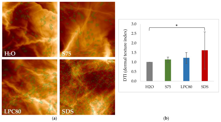
Figure 7.
(a). Representative images of corneocyte surface morphology as assessed by AFM after exposure to S75, LPC80, SDS and water as control (H2O). Images were taken on corneocytes removed by adhesive tapes. Circular Nano Objects (CNOs) are depicted in green. (b) Dermal Texture Index (DTI) is defined as the average count of CNOs in a field of observation (20 μm)2. Values are means of n = 8 ± SD. Statistically significant differences are marked with asterisks (* p < 0.05) and were tested with a one-way ANOVA + post-hoc Tukey test with p < 0.05 as minimum level of significance.

