Abstract
Background
Among the challenges for personalizing the management of mechanically ventilated patients with coronavirus disease (COVID-19)-associated acute respiratory distress syndrome (ARDS) are the effects of different positive end-expiratory pressure (PEEP) levels and body positions in regional lung mechanics. Right-left lung aeration asymmetry and poorly recruitable lungs with increased recruitability with alternating body position between supine and prone have been reported. However, real-time effects of changing body position and PEEP on regional overdistension and collapse, in individual patients, remain largely unknown and not timely monitored. The aim of this study was to individualize PEEP and body positioning in order to reduce the mechanisms of ventilator-induced lung injury: collapse and overdistension.
Methods
We here report a series of five consecutive mechanically ventilated patients with COVID-19-associated ARDS in which sixteen decremental PEEP titrations were performed in the first days of mechanical ventilation (8 titration pairs: supine position immediately followed by 30° targeted lateral position). The choice of lateral tilt was based on X-Ray. This targeted lateral position strategy was defined by selecting the less aerated lung to be positioned up and the more aerated lung to be positioned down. For each PEEP level, global and regional collapse and overdistension maps and percentages were measured by electrical impedance tomography. Additionally, we present the incidence of lateral asymmetry in a cohort of forty-four patients.
Results
The targeted lateral position strategy resulted in significantly smaller amounts of overdistension and collapse when compared with the supine one: less collapse along the PEEP titration was found within the left lung in targeted lateral (P = 0.014); and less overdistension along the PEEP titration was found within the right lung in targeted lateral (P = 0.005). Regarding collapse within the right lung and overdistension within the left lung: no differences were found for position. In the cohort of forty-four patients, ventilation inequality of > 65/35% was observed in 15% of cases.
Conclusions
Targeted lateral positioning with bedside personalized PEEP provided a selective attenuation of overdistension and collapse in mechanically ventilated patients with COVID-19-associated ARDS and right-left lung aeration/ventilation asymmetry.
Trial registration
Trial registration number: NCT04460859
Keywords: Acute respiratory distress syndrome, Coronavirus disease, Mechanical ventilation, Ventilator-induced lung injury, Positive end-expiratory pressure, Body position
Background
Most critically ill patients with coronavirus disease (COVID-19) [1] develops acute respiratory distress syndrome (ARDS), needs mechanical ventilation for prolonged time, and exhibits high mortality [2]. In a large cohort study with critically ill patients with COVID-19 referred for intensive care unit (ICU) admission [2], positive end-expiratory pressure (PEEP) levels were higher than those reported for the management of moderate-to-severe ARDS in the pre–COVID-19 era; and, along with high fraction of inspired oxygen (FIO2) and low partial pressure of arterial oxygen ratio (PaO2/FIO2) at ICU admission, were an independent factor associated with high mortality. Lung heterogeneity, hypoxemia disproportional to mechanics, right-left lung aeration/ventilation asymmetry [3], and poorly recruitable lungs with increased recruitability with alternating body position between supine and prone [4] have been reported. However, real-time effects of changing body position and PEEP on regional overdistension and collapse, in individual patients, remain largely unknown and not timely monitored.
Lung collapse usually predominates within the most dependent units where the transpulmonary pressure (PL = airways pressure − pleural pressure) is the lowest, while lung overdistension predominates within the most nondependent ones where the PL is the highest. When there is right-left lung heterogeneity of collapse and overdistension, as in many patients with COVID-19-associated ARDS, a targeted lateral positioning strategy is conceivable: by one-sided lateral position, the lung with more collapsed units in supine position can be positioned gravity-nondependent (mostly) and, conversely, the lung with more overdistended units in supine position can be positioned gravity-dependent. Such targeted lateral position, by which PL becomes larger in the nondependent units and smaller in the dependent ones, may afford simultaneous regional/selective recruitment and relief of overdistension effects.
The aim of this study was to individualize PEEP and body positioning in order to reduce lung collapse and overdistension. We hypothesized that targeted lateral positioning with bedside personalized PEEP would provide a selective attenuation of overdistension and collapse in mechanically ventilated patients with COVID-19-associated ARDS and right-left lung aeration/ventilation asymmetry.
Methods
Patients with COVID-19-associated ARDS in the first days of mechanical ventilation were included. ARDS was defined according to the Berlin definition [5]. COVID-19 was confirmed by positive nasopharyngeal polymerase chain reaction for SARS-CoV-2. Patients were excluded in case of a contraindication to electrical impedance tomography (EIT): pacemaker, implantable defibrillator, skin lesion.
The design was a prospective observational study. The settings were the ICU of the Department of Anaesthesiology, Resuscitation and Intensive Medicine, First Faculty of Medicine, Charles University, General University Hospital in Prague, Czech Republic; and the ICU of the Pulmonology Division, Cardiopulmonary Department, Heart Institute, University of São Paulo, São Paulo, Brazil. All the experiment protocol for involving humans was in accordance to guidelines of national/international/institutional or Declaration of Helsinki. The study was approved by the Ethics Committee of the General University Hospital, Prague, and by the Ethics Committee of the Heart Institute, University of São Paulo, São Paulo.
We used EIT [6] with decremental PEEP titration algorithm (PEEPEIT-titration), which provides information on regional overdistension and collapse [7, 8], to individualize PEEP and body position aiming to minimize ventilator-induced lung injury (VILI) mechanisms, namely collapse and overdistension. Electrical impedance tomography (Enlight 1800; Timpel SA, Brazil) is a noninvasive, radiation-free, real-time imaging method that measures global and regional changes in lung volumes [9]. Pulmonary EIT data were recorded at 50 Hz with 32 electrodes equidistantly placed around the circumference of the thorax just below the level of the axilla. Lung collapse and overdistension percentages were determined by comparing each electrical impedance tomography pixel-compliance during PEEPEIT-titration [7, 8]. Each pixel-compliance was determined by dividing tidal impedance change by the variation in pressure during the respiratory cycle. Therefore, overdistension was identified when, for a given pixel, aeration increased and compliance worsened. On the other hand, reversal of collapse was identified if aeration increased and compliance improved.
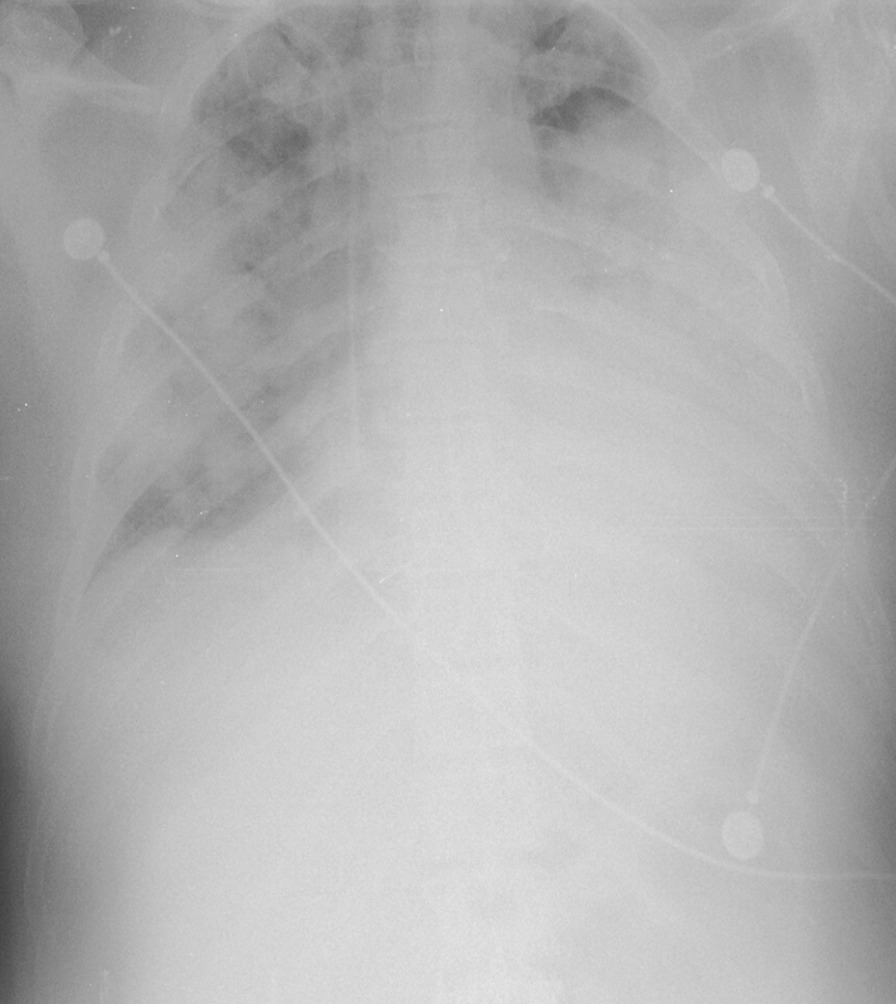
Sixteen PEEPEIT-titration were performed during the first days of mechanical ventilation in five consecutive patients with COVID-19-associated ARDS in supine immediately followed by targeted lateral position (30°). Thus 8 PEEPEIT-titration pairs were obtained. The choice of lateral tilt was based on X-Ray. This targeted lateral position strategy was defined by selecting the less aerated lung to be positioned up and the more aerated lung to be positioned down. During all the procedures, the patients were deeply sedated and under muscle paralysis.
Immediately before all PEEPEIT-titration, the same lung recruitment maneuver was performed both in supine position and the corresponding targeted lateral position: 2 min of PEEP 24 cmH2O and driving pressure of 15 cmH2O. The recruitment maneuver allows the measurement of ventilation distribution in all the recruitable pixels. The PEEPEIT-titration, which started at a PEEP level of 26 cmH2O, were performed with decremental PEEP steps of 2 cmH2O until reaching a lower PEEP level set by the clinician.
The EIT data of all PEEPEIT-titration was analyzed in order to quantify the amounts of lung collapse and overdistension at each PEEP step. The lung images were divided in two regions, right and left lung.
It was unclear what percentage of patients with COVID-19-associated ARDS could be potentially suitable for the approach of our study by presenting sufficient right-left lung inequality. To address this issue, we measured the distribution of ventilation to right and left lung from additional 44 patients with COVID-19-associated ARDS. This distribution of ventilation was measured by EIT in supine position. All these patients were ventilated with PEEP set according to the PEEP–FIO2 table used in the original ARDS Network trial.
Statistical analysis
The Shapiro–Wilk test was used to test data for normality. The two-way repeated measures ANOVA was used to determine if there was a statistically significant interaction effect between our two within-subjects factors on our continuous dependent variable (i.e., whether a two-way interaction exists). Our continuous dependent variable was % collapse or % overdistension. Our two independent variables were position (supine, lateral) and PEEP [two within-subjects factors]. Simple and main effects were also tested where appropriated. The Bonferroni adjustment for multiple tests was applied for post hoc comparisons. The statistical analyses were conducted with SPSS (version 25; IBM Corp, IBM SPSS Statistics for Windows, Armonk, NY). Individual P values to indicate statistical tests’ significance are reported were relevant. Values presented are mean and SEM unless otherwise stated.
Results
The five patients (Table 1) that were studied with pairs of PEEPEIT-titration (supine immediately followed by targeted lateral position) exhibited less aeration and ventilation in the left lung (Fig. 1). Thus the right (down) lateral tilt was decided. Below the results of the analyzes of their sixteen PEEPEIT-titration (eight pairs):
Table 1.
Patients’ characteristics
| Sex | Age (yr) | BMI (kg/m2) | PaO2/FIO2 ratio (mm Hg)*—supine | PaO2/FIO2 ratio (mm Hg)†—lateral | DP (cm HO)‡ | Respiratory system compliance (ml/cm H2O)§ | Duration of mechanical ventilation at recruitment (days)¶ |
|---|---|---|---|---|---|---|---|
| M | 44 | 30,0 | 396,0 | 438,0 | 8,4 | 35,0 | 1 |
| F | 85 | 26,8 | 71,0 | N/A | 7,2 | 36,3 | 2 |
| F | 70 | 24,2 | 224,0 | 308,0 | 6,8 | 35,0 | 2 |
| F | 82 | 33,3 | 521,0 | 464,0 | 3,0 | 60,6 | 1 |
| M | 53 | 32,1 | 367,0 | 441,0 | 3 7 | 88,2 | 9 |
| Mean | 66,8 | 29,3 | 315,8 | 412,8 | 5,8 | 51,0 | 3,0 |
| SD | 17,9 | 3,8 | 172,9 | 70,8 | 2,3 | 23,5 | 3,4 |
Definition of abbreviations: BMI = body mass index; PaO2/FIO2 Ratio = ratio of arterial oxygen partial pressure (PaO2 in mmHg) to fractional inspired oxygen (FIO2 expressed as a fraction, not a percentage); DP = driving pressure; N/A = not available
*At the end of the lung recruitment maneuver in supine position
†At the end of the lung recruitment maneuver in lateral position
‡At PEEP of 12 cm H2O
§At PEEP of 12 cm H2O
¶Unavailable because of loss of data
Fig. 1.

Initial X-Ray in supine body position. The choice of lateral tilt was based on the initial X-Ray that was taken in supine body position. The targeted lateral position strategy was defined by selecting the less aerated lung to be positioned up and the more aerated lung to be positioned down. Please note the left-to-right lung asymmetry present on this initial X-Ray: unequivocally more opacities within the left lung
Collapse-left lung
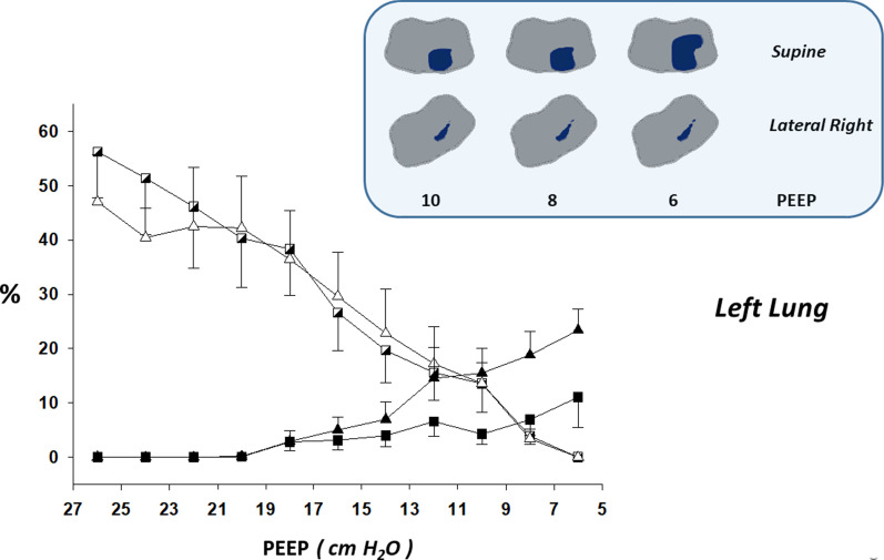
There was a statistically significant two-way interaction between position (supine vs. targeted lateral) and PEEP (P = 0.014; two-way repeated measures ANOVA) in the % of collapse within the left lung: less collapse along the PEEP titration was found within the left lung in targeted lateral (right down) than supine position (Fig. 2). Additionally, when the simple main effects were tested, the following significant differences were found: PEEP 14 (P = 0.034), PEEP 10 (P = 0.028), PEEP 8 (P = 0.019), and PEEP 6 cmH2O (P = 0.007).
Fig. 2.
Lung collapse and overdistension by electrical impedance tomography in supine vs. targeted lateral body position within the left lung. Left-to-right lung asymmetry was present on initial X-Ray taken in supine body position: unequivocally more opacities within the left lung. Thus lateral right positioning (30°) was indicated (“targeted”) and performed with the platform-based rotation bed Multicare® (LINET). Line graphs of electrical impedance tomography (EIT)-based estimations of collapse and overdistension during decremental positive end-expiratory pressure (PEEP) titrations (supine vs. targeted lateral body position) are shown (mean ± SEM). Some illustrative and representative EIT images of collapse are also shown: collapsed pixels in purple. Note that the amount of collapsed units within the left lung present in the supine body position was minimized in the lateral right one. X axis: Decremental PEEP levels of the EIT-PEEP titrations. Y axis: Percent of overdistended and collapsed lung units out of the total lung imaged by EIT. Triangle: Supine body position. Square: Targeted lateral body position (lateral right). Black triangle and black square: Percent of collapsed lung units out of the total lung imaged by EIT. White triangle and white semi-filled square: Percent of overdistended lung units out of the total lung imaged by EIT
Overdistension-right lung
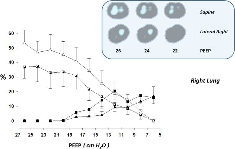
There was a marginal two-way interaction between position and PEEP (P = 0.073; two-way repeated measures ANOVA). The main effect of position showed a statistically significant difference in the % of overdistension within the right lung: less overdistension along the PEEP titration in targeted lateral (right down) than supine position (P = 0.005; Fig. 3). The main effect of PEEP on right lung overdistension showed a statistically significant difference (P < 0.0005). Additionally, for many PEEP levels significant P values were found in the pairwise comparisons with adjustment for multiple comparisons (Bonferroni).
Fig. 3.
Lung collapse and overdistension by electrical impedance tomography in supine vs. targeted lateral body position within the right lung. Left-to-right lung asymmetry was present on initial X-Ray taken in supine body position: unequivocally more opacities within the left lung. Thus lateral right positioning (30°) was indicated (“targeted”) and performed with the platform-based rotation bed Multicare® (LINET). Line graphs of electrical impedance tomography (EIT)-based estimations of collapse and overdistension during decremental positive end-expiratory pressure (PEEP) titrations (supine vs. targeted lateral body position) are shown (mean ± SEM). Some illustrative and representative EIT images of overdistension are also shown: overdistended pixels in white. Note the asymmetric distribution of overdistension between the right and left lungs (concentration and predominance of overdistension within the right lung); and that the amount of overdistended units within the right lung in the supine body position was minimized in the lateral right one. Also note that the regional distribution of overdistension in the supine body position was much less gravitational-dependent than it is usually present in “typical” acute respiratory distress syndrome. X axis: Decremental PEEP levels of the EIT-PEEP titrations. Y axis: Percent of overdistended and collapsed lung units out of the total lung imaged by EIT. Triangle: Supine body position. Square: Targeted lateral body position (lateral right). Black triangle and black square: Percent of collapsed lung units out of the total lung imaged by EIT. White triangle and white semi-filled square: Percent of overdistended lung units out of the total lung imaged by EIT
Collapse-right lung and overdistension-left lung
No statistically significant differences were found for position.
Regarding the percentage of patients with COVID-19-associated ARDS presenting sufficient right-left lung inequality: the mean of the % of ventilation of the right lung was 54.9 ± 8.2 and the mean of the % of ventilation of the left lung was 45.3 ± 8.1. A right-left lung asymmetrical ventilation of > 65/35% was observed in 15% of these patients. This distribution of ventilation was measured by EIT in supine position and under a mean PEEP level of 11.4 ± 3.1 cmH2O (set according to the PEEP–FIO2 table used in the original ARDS Network trial).
The optimum PEEP of PEEPEIT-titration was defined by the best compromise between pulmonary overdistension and collapse, i.e. the smallest sum of overdistension and collapse. The optimum PEEP was 10.8 ± 5.3 cmH2O in supine position and was 11.2 ± 5.9 cmH2O in targeted lateral position.
Hemodynamic compromise was not detected for any patients during all procedures of the study.
Discussion
A major focus of mechanical ventilation for COVID-19 is the avoidance of VILI while facilitating gas exchange via lung-protective ventilation. This is the first description of using EIT with targeted lateral positioning to personalize PEEP in adult patients with COVID-19-associated ARDS. A randomized and controlled trial demonstrated the feasibility and efficacy of a postural recruitment maneuver in children with anesthesia-induced atelectasis [10]. Besides being applied in children with healthy lungs, another difference between the Acosta et al. and our study is the lack of PEEP titrations. Very recently, Zhao et al. reported the use of EIT for individualized ventilation strategy in one patient with COVID-19 [11]. Similarly to the study in children with anesthesia-induced atelectasis [10], a key difference between the Zhao et al. case report and our case series is the lack of a strategy to personalize PEEP during the lateral positioning.
The vertical gradient of PL, which is mainly due to gravity, changes with body mass and posture [12]. Agostoni and D’Angelo showed that the PL gradient increased when body position was changed from supine to lateral position [13]. They demonstrated that lateral position leads to higher PL in the most nondependent units and lower PL in the most dependent ones. That is mainly because the thoracic right-to-left distance is longer than the anterior–posterior. Thus, generally, lateral positioning increases heterogeneity of PL across the parenchyma, but this depends on PEEP and baseline lung conditions: recent reports in children have shown that an optimized PEEP after lateralization can minimize hyperdistension (maximizing ventilation) in a nondependent, sicker lung, while reasonably keeping functional residual capacity in dependent, healthier lung [14]. Our PEEPEIT-titration seems to be a promising tool to find such personalized PEEP at the bedside.
Another potential beneficial effect of the targeted lateral positioning is an improved ventilation/perfusion matching due to: 1) attenuation of regional overdistension within the more aerated lung and, consequently, less diversion of pulmonary blood flow away from these units; 2) diminution of regional collapse within the less aerated lung. The consequent improvement of oxygenation may be important in these patients to manage their disproportional hypoxemia and “buy time” with minimum additional damage.
Our findings reinforce the importance of timely PEEP titrations [15] tackling the dynamically changing phases of this disease. They suggest the relevance of personalized PEEP adjustments every time body positions are changed. The recommendation of applying nonpersonalized low or high PEEP may lead to insufficient and/or excessive PEEP in terms of protection of VILI [16].
Conclusions
Targeted lateral positioning with bedside personalized PEEP provided a selective attenuation of overdistension and collapse in mechanically ventilated patients with COVID-19-associated ARDS and right-left lung aeration/ventilation asymmetry.
Acknowledgements
Not applicable.
Abbreviations
- COVID-19
Coronavirus disease
- ARDS
Acute respiratory distress syndrome
- ICU
Intensive care unit
- PEEP
Positive end-expiratory pressure
- FIO2
Fraction of inspired oxygen
- PaO2
Arterial oxygen partial pressure
- PaO2/FIO2
Partial pressure of arterial oxygen ratio
- PL
Transpulmonary pressure (PL = airways pressure − pleural pressure)
- EIT
Electrical impedance tomography
- PEEPEIT-titration
PEEP titration algorithm using EIT
- VILI
Ventilator-induced lung injury
Authors' contributions
MM, MO, JBB, GCA, DH, LT and MA collected the data. JBB and GCA analyzed the patient data and drafted the manuscript. MM, MO, GCA, EK, MA and OK developed the concept. MM, MO, JBB, GCA, EK and MA interpreted the data. All authors revised the manuscript critically. All authors read and approved the final manuscript.
Funding
This work was financially supported by Technology Agency of the Czech Republic (FW 01010679) and Progres 206–041. The funding has no influence in study design, data analysis or publication.
Availability of data and materials
The data are with the authors and will be available upon reasonable request. The data is available from the corresponding author: João Batista Borges.
Declarations
Ethics approval and consent to participate
The study was approved by the Ethics Committee of the General University Hospital, Prague (833/20 S-IV), and by the Ethics Committee of the Heart Institute, University of São Paulo, São Paulo (CAAE: 30938720.8.0000.0068). Informed consent was obtained from all patients included in the study for PEEPEIT-titration. We confirm that all the experiment protocol for involving humans was in accordance to guidelines of national/international/institutional or Declaration of Helsinki.
Consent for publication
Not applicable. Our manuscript does not contain any individual person’s data in any form (including individual details, images or videos).
Competing interests
The authors report no conflict of interests.
Footnotes
Publisher's Note
Springer Nature remains neutral with regard to jurisdictional claims in published maps and institutional affiliations.
Mikuláš Mlček and Michal Otáhal contributed equally to this work
References
- 1.Coronaviridae Study Group of the International Committee on Taxonomy of V: The species Severe acute respiratory syndrome-related coronavirus: classifying 2019-nCoV and naming it SARS-CoV-2. Nature Microbiol 2020, 5(4):536–544. [DOI] [PMC free article] [PubMed]
- 2.Grasselli G, Greco M, Zanella A, Albano G, Antonelli M, Bellani G, Bonanomi E, Cabrini L, Carlesso E, Castelli G, et al. Risk factors associated with mortality among patients with COVID-19 in intensive care units in lombardy Italy. JAMA Internal Med. 2020;180(10):1345–1355. doi: 10.1001/jamainternmed.2020.3539. [DOI] [PMC free article] [PubMed] [Google Scholar]
- 3.Bhatraju PK, Ghassemieh BJ, Nichols M, Kim R, Jerome KR, Nalla AK, Greninger AL, Pipavath S, Wurfel MM, Evans L, et al. Covid-19 in critically Ill patients in the seattle region - case series. N Engl J Med. 2020;382(21):2012–2022. doi: 10.1056/NEJMoa2004500. [DOI] [PMC free article] [PubMed] [Google Scholar]
- 4.Pan C, Chen L, Lu C, Zhang W, Xia JA, Sklar MC, Du B, Brochard L, Qiu H. Lung recruitability in COVID-19-associated acute respiratory distress syndrome: a single-center observational study. Am J Respir Crit Care Med. 2020;201(10):1294–1297. doi: 10.1164/rccm.202003-0527LE. [DOI] [PMC free article] [PubMed] [Google Scholar]
- 5.Force ADT, Ranieri VM, Rubenfeld GD, Thompson BT, Ferguson ND, Caldwell E, Fan E, Camporota L, Slutsky AS. Acute respiratory distress syndrome: the Berlin definition. JAMA. 2012;307(23):2526–2533. doi: 10.1001/jama.2012.5669. [DOI] [PubMed] [Google Scholar]
- 6.Borges JB, Cronin JN, Crockett DC, Hedenstierna G, Larsson A, Formenti F. Real-time effects of PEEP and tidal volume on regional ventilation and perfusion in experimental lung injury. Intensive Care Med Exp. 2020;8(1):10. doi: 10.1186/s40635-020-0298-2. [DOI] [PMC free article] [PubMed] [Google Scholar]
- 7.Costa EL, Borges JB, Melo A, Suarez-Sipmann F, Toufen C, Jr, Bohm SH, Amato MB. Bedside estimation of recruitable alveolar collapse and hyperdistension by electrical impedance tomography. Intensive Care Med. 2009;35(6):1132–1137. doi: 10.1007/s00134-009-1447-y. [DOI] [PubMed] [Google Scholar]
- 8.Fumagalli J, Santiago RRS, Teggia Droghi M, Zhang C, Fintelmann FJ, Troschel FM, Morais CCA, Amato MBP, Kacmarek RM, Berra L, et al. Lung recruitment in obese patients with acute respiratory distress syndrome. Anesthesiology. 2019;130(5):791–803. doi: 10.1097/ALN.0000000000002638. [DOI] [PubMed] [Google Scholar]
- 9.Victorino JA, Borges JB, Okamoto VN, Matos GF, Tucci MR, Caramez MP, Tanaka H, Sipmann FS, Santos DC, Barbas CS, et al. Imbalances in regional lung ventilation: a validation study on electrical impedance tomography. Am J Respir Crit Care Med. 2004;169(7):791–800. doi: 10.1164/rccm.200301-133OC. [DOI] [PubMed] [Google Scholar]
- 10.Acosta CM, Volpicelli G, Rudzik N, Venturin N, Gerez S, Ricci L, Natal M, Tusman G. Feasibility of postural lung recruitment maneuver in children: a randomized, controlled study. Ultrasound J. 2020;12(1):34. doi: 10.1186/s13089-020-00181-8. [DOI] [PMC free article] [PubMed] [Google Scholar]
- 11.Zhao Z, Zhang JS, Chen YT, Chang HT, Hsu YL, Frerichs I, Adler A. The use of electrical impedance tomography for individualized ventilation strategy in COVID-19: a case report. BMC Pulm Med. 2021;21(1):38. doi: 10.1186/s12890-021-01411-y. [DOI] [PMC free article] [PubMed] [Google Scholar]
- 12.D'Angelo E, Bonanni MV, Michelini S, Agostoni E. Topography of the pleural pressure in rabbits and dogs. Respir Physiol. 1970;8(2):204–229. doi: 10.1016/0034-5687(70)90016-2. [DOI] [PubMed] [Google Scholar]
- 13.Agostoni E, D'Angelo E, Bonanni MV. The effect of the abdomen on the vertical gradient of pleural surface pressure. Respir Physiol. 1970;8(3):332–346. doi: 10.1016/0034-5687(70)90040-X. [DOI] [PubMed] [Google Scholar]
- 14.Schibler A, Henning R. Positive end-expiratory pressure and ventilation inhomogeneity in mechanically ventilated children. Pediat Crit Care Med. 2002;3(2):124–128. doi: 10.1097/00130478-200204000-00006. [DOI] [PubMed] [Google Scholar]
- 15.van der Zee P, Somhorst P, Endeman H, Gommers D. Electrical impedance tomography for positive end-expiratory pressure titration in COVID-19-related acute respiratory distress syndrome. Am J Respir Crit Care Med. 2020;202(2):280–284. doi: 10.1164/rccm.202003-0816LE. [DOI] [PMC free article] [PubMed] [Google Scholar]
- 16.Fan E, Beitler JR, Brochard L, Calfee CS, Ferguson ND, Slutsky AS, Brodie D. COVID-19-associated acute respiratory distress syndrome: is a different approach to management warranted? Lancet Respir Med. 2020;8(8):816–821. doi: 10.1016/S2213-2600(20)30304-0. [DOI] [PMC free article] [PubMed] [Google Scholar]
Associated Data
This section collects any data citations, data availability statements, or supplementary materials included in this article.
Data Availability Statement
The data are with the authors and will be available upon reasonable request. The data is available from the corresponding author: João Batista Borges.


