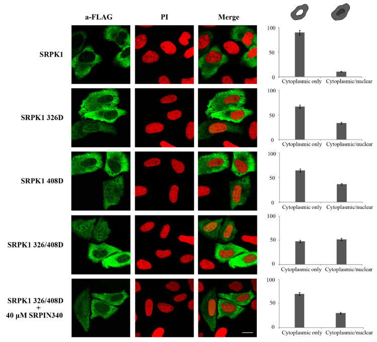
Figure 8.
The phosphorylation-mimicking mutants of SRPK1 were partially localized in the nucleus. Representative confocal images of wild-type FLAG-SRPK1, FLAG-SRPK1326D, FLAG-SRPK1408D, FLAG-SRPK1326/408D, and FLAG-SRPK1326/408D plus SRPIN340. SRPK1 was detected using the M5 anti-FLAG monoclonal antibody, while the nuclei were stained with PI. Scale bar: 10 µM. A diagrammatic representation of wild-type FLAG-SRPK1 and mutant FLAG-SRPK1 staining patterns is shown in the upper part of the right panel. The percentage of SRPK1 staining patterns relative to the indicated staining pattern was determined for ≈150 cells in two different experiments, where the means ± standard errors of the measurements are shown.

