Figure 3.
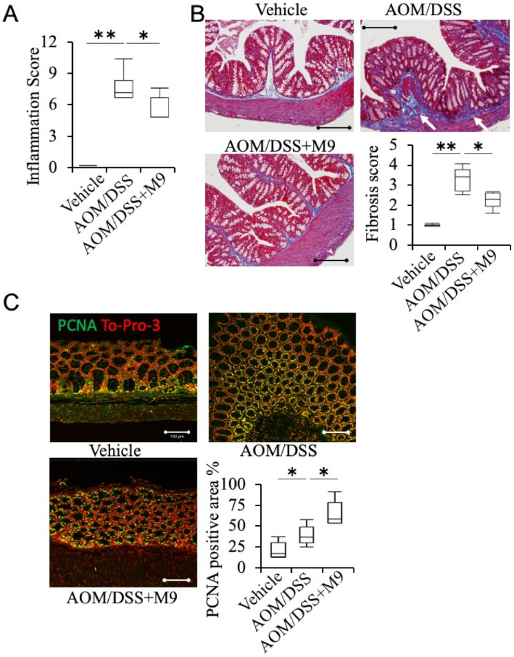
Treatment of Probio-M9 ameliorated AOM/DSS-induced fibrosis and accelerated tissue repair in non-tumor areas. (A) Inflammation score in non-tumor areas was evaluated from hematoxylin-eosin (HE) histological sections of the colons. n = 10. (B) Representative images of the non-tumor areas of the colon evaluated with Masson’s trichrome (MT) staining and the corresponding fibrosis scores. Averaged fibrosis score was grouped as 0%, 1–25%, 26–50%, 51–75%, or 76–100% of the submucosa affected corresponding to a fibrosis score of 0, 1, 2, 3, and 4, respectively. n = 10. Scale bar: 200 μm. (C) Representative image of immunohistochemistry staining of PCNA in non-tumor areas. To-Pro-3 was used to label the nucleus. The ratio of the expression area in the mucosal layer is shown in the graph. n = 5. * p < 0.05; ** p < 0.01. Scale bar: 100 μm.

