Abstract
Plants belonging to the Asteraceae family are widely used as traditional medicinal herbs around the world for the treatment of numerous diseases. In this work, the chemical profiles of essential oils (EOs) of the above-ground parts of Pluchea dioscoridis (L.) DC. and Erigeron bonariensis (L.) were studied in addition to their cytotoxic and anti-aging activities. The extracted EOs from the two plants via hydrodistillation were analyzed by gas chromatography-mass spectroscopy (GC-MS). GC-MS of EO of P. dioscoridis revealed the identification of 29 compounds representing 96.91% of the total oil. While 35 compounds were characterized from EO of E. bonariensis representing 98.21%. The terpenoids were found the main constituents of both plants with a relative concentration of 93.59% and 97.66%, respectively, including mainly sesquiterpenes (93.40% and 81.06%). α-Maaliene (18.84%), berkheyaradulen (13.99%), dehydro-cyclolongifolene oxide (10.35%), aromadendrene oxide-2 (8.81%), β-muurolene (8.09%), and α-eudesmol (6.79%), represented the preponderance compounds of EO of P. dioscoridis. While, trans-α-farnesene (25.03%), O-ocimene (12.58%), isolongifolene-5-ol (5.53%), α-maaliene (6.64%), berkheyaradulen (4.82%), and α-muurolene (3.99%), represented the major compounds EO of E. bonariensis. A comparative study of our results with the previously described data was constructed based upon principal component analysis (PCA) and agglomerative hierarchical clustering (AHC), where the results revealed a substantial variation of the present studied species than other reported ecospecies. EO of P. dioscoridis exhibited significant cytotoxicity against the two cancer cells, MCF-7 and A-549 with IC50 of 37.3 and 22.3 μM, respectively. While the EO of the E. bonariensis showed strong cytotoxicity against HepG2 with IC50 of 25.6 μM. The EOs of P. dioscoridis, E. bonariensis, and their mixture (1:1) exhibited significant inhibitory activity of the collagenase, elastase, hyaluronidase, and tyrosinase comparing with epigallocatechin gallate (EGCG) as a reference. The results of anti-aging showed that the activity of mixture (1:1) > P. dioscoridis > E. bonariensis against the four enzymes.
Keywords: horseweed, wavy-leaf fleabane, sesquiterpenes, cytotoxicity, anti-senility
1. Introduction
Natural products derived from the plant kingdom represented potent resources for foods, cosmetics, and traditional medicines [1,2]. Many scientists focused on the study of the chemical characterization of essential oils (EOs) along with their pharmaceutical effects for many decades [3,4,5,6]. Due to the complicated composition from different isoprenoids based compounds [7], EOs exhibited several significant biological effects including anti-inflammatory, antipyretic [8], antioxidant [9,10], allelopathy [8,11,12,13,14,15,16,17], antiulcer [18], antimicrobial [8,19], and hepatoprotective [20]. EOs have been reported as potent agents against degenerative diseases via inhibition of oxidative stress due to the strong free radical scavenging activity [21].
Plants belonging to Conyza genus (Family Asteraceae), including around 150 plant species [22], were described as important traditional medicinal plants in the treatment of toothache, skin diseases, rheumatism, haemorrhoidal, diarrhoeal, and injuries bleeding [23,24]. Some members of Asteraceae were named Conyza formerly; however, some taxa names have been changed later based on taxonomic criteria. From these taxa, Conyza dioscoridis (L.) Desf. that its accepted name now is Pluchea dioscoridis (L.) DC., and Conyza linifolia (Willd.) Täckh. that now have been accepted as Erigeron bonariensis L. [25,26]. Studying various criteria of the plant species such as morphological, anatomical, molecular, and chemical properties, providing valuable information for taxonomists, thereby, some taxa names have been changed [27,28]. EOs analysis has been reported to provide profitable information for chemotaxonomy [27,29].
Pluchea dioscoridis (L.) DC. (syn. Conyza dioscoridis (L.) Desf.) is a widely distributed wild plant in the Nile delta, Mediterranean coast, Sinai Peninsula, Western Desert, and Eastern Desert [25]. This plant was described in folk medicines for the treatment of some diseases as ulcer, colic, carminative, epilepsy in children, rheumatic pains, and cold [30]. Many documents described that the different extracts of this plant have several potent biological activities comprising anti-inflammation, antiulcer, antidiabetic, antinociceptive, antipyretic, ant-diarrheal, antibacterial, antifungal, and free radical scavenging activities, along with diuretic effect [30,31,32,33]. Many metabolites were isolated and characterized from P. dioscoridis including steroids, triterpenes [30], flavonoids, and phenolic acids [30,32].
The chemical constituents of EO of E. bonariensis collected from Alexandria, Egypt has been reported in addition to its antimicrobial and insecticidal activities [34]. In this study, 25 compounds were identified from EO of E. bonariensis including sesqui- and monoterpenes. From the total of this oil, α-bergamotene, and D-limonene represented the mains with concentrations of 27.4 and 22.5%. In the same report, EO of C. linifolia was documented to exhibit antibacterial potentiality against B. subtilis with MIC of 125 mg/mL [34,35]. Little reports concerning the chemical profiles as well as biological activities of Erigeron bonariensis L. (syn. Conyza linifolia (Willd.) Täckh.) have been recorded.
We hypothesized that these two plant species were formerly named Conyza, and their names were changed. Therefore, the chemical characterization of their EOs could be valuable in their chemotaxonomy. Herein, this work aimed to (i) identify the chemical profiles of EOs from P. dioscoridis and E. bonariensis, collected from Egypt, (ii) establish comparative profiles of the two plants based upon chemometric analysis with other reported ecospecies, (iii) study the cytotoxic activity of the EOs of the two plants against several human cancer cell lines, and (iv) assess in vitro anti-aging potentialities of the EOs of the two plants.
2. Results and Discussion
2.1. Chemical Compositions of EO of P. dioscoridis
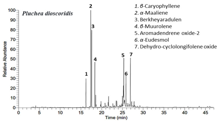
The chemical characterization of P. dioscoridis EO was extracted via hydro-distillation afforded golden yellow (0.037%). The chemical profiles of the extracted EO were assigned depending upon the GC-MS analysis. The GC-MS chromatogram of the EO is presented in Figure 1 exhibiting the main peaks from all identified components. Twenty-nine compounds were identified from the EO of P. dioscoridis represented 96.91% of the total oil. All the identified compounds along with their chemical and physical properties were summarized in Table 1.
Figure 1.
Gas chromatography-mass spectroscopy (GC-MS) chromatograme of the essential oils (EO) of Pluchea dioscoridis. The main peaks were numbered (1–7).
Table 1.
Components of essential oils of Pluchea dioscoridis and Erigeron bonariensis.
| No | Rt [a] | Compound Name | MF | KILit [b] | KIExp [c] | Relative Concentration % | |
|---|---|---|---|---|---|---|---|
| P. discodirdis | E. bonariensis | ||||||
| Monoterpenes | 0.19% | 14.16% | |||||
| 1 | 4.13 | α-Pinene | C10H16 | 933 | 934 | 0.19 ± 0.01 | 0.18 ± 0.01 |
| 2 | 5.43 | α-Myrcene | C10H16 | 991 | 990 | ------ | 0.20 ± 0.01 |
| 3 | 6.41 | O-Ocimene | C10H16 | 1012 | 1012 | ------ | 12.58 ± 0.09 |
| 4 | 6.91 | D-Limonene | C10H16 | 1035 | 1036 | ------ | 1.20 ± 0.04 |
| Sesquiterpenes | 93.4% | 81.06% | |||||
| 5 | 16.22 | β-Caryophyllene | C15H24 | 1418 | 1420 | 4.95 ± 0.07 | 2.17 ± 0.04 |
| 6 | 16.96 | Aromandendrene | C15H24 | 1439 | 1448 | ------ | 0.12 ± 0.03 |
| 7 | 17.17 | α-Guaiene | C15H24 | 1439 | 1439 | 0.21 ± 0.02 | 0.11 ± 0.02 |
| 8 | 17.39 | α-Maaliene | C15H24 | 1480 | 1479 | 18.84 ± 0.08 | 6.64 ± 0.09 |
| 9 | 17.60 | Berkheyaradulen | C15H24 | 1492 | 1493 | 13.99 ± 0.09 | 4.82 ± 0.06 |
| 10 | 18.36 | β-Muurolene | C15H24 | 1493 | 1493 | 8.09 ± 0.05 | 2.37 ± 0.04 |
| 11 | 18.57 | α-Muurolene | C15H24 | 1498 | 1499 | 2.20 ± 0.04 | 3.99 ± 0.07 |
| 12 | 18.98 | Bicyclogermacrene | C15H24 | 1500 | 1501 | ------ | 0.66 ± 0.01 |
| 13 | 19.67 | trans-α-Farnesene | C15H24 | 1508 | 1507 | ------ | 25.03 ± 0.13 |
| 14 | 19.73 | α-Bisabolene | C15H24 | 1509 | 1511 | 0.58 ± 0.03 | ------ |
| 15 | 19.84 | γ-Cadinene | C15H24 | 1514 | 1515 | 1.36 ± 0.04 | ------ |
| 16 | 20.35 | α-Sesquiphellandrene | C15H24 | 1516 | 1517 | 0.44 ± 0.01 | ------ |
| 17 | 20.45 | cis-Lanceol | C15H24O | 1525 | 1527 | 0.16 ± 0.02 | 0.09 ± 0.01 |
| 18 | 20.54 | Isolongifolene-5-ol | C15H24O | 1534 | 1535 | 0.19 ± 0.01 | 5.53 ± 0.07 |
| 19 | 20.64 | Germacrene D-4-ol | C15H24 | 1574 | 1576 | ------ | 2.35 ± 0.04 |
| 20 | 20.81 | Spathulenol | C15H24O | 1576 | 1577 | 0.81 ± 0.03 | 0.10 ± 0.01 |
| 21 | 20.98 | Isoaromadendrene epoxide | C15H24O | 1580 | 1579 | ------ | 1.50 ± 0.03 |
| 22 | 21.41 | Calarenepoxide | C15H24O | 1592 | 1592 | ------ | 1.07 ± 0.02 |
| 23 | 21.56 | Caryophyllene oxide | C15H24O | 1594 | 1593 | 0.86 ± 0.02 | 0.08 ± 0.01 |
| 24 | 21.66 | Salvial-4(14)-en-1-one | C15H24O | 1595 | 1595 | 2.20 ± 0.04 | 0.38 ± 0.01 |
| 25 | 21.93 | Ledene alcohol | C15H24O | 1729 | 1731 | ------ | 0.97 ± 0.03 |
| 26 | 22.1 | Carotol | C15H26O | 1597 | 1598 | 0.78 ± 0.02 | ------ |
| 27 | 22.42 | Humuladienone | C15H24O | 1607 | 1605 | ------ | 0.32 ± 0.01 |
| 28 | 23.09 | Neoclovenoxid | C15H24O | 1608 | 1610 | 0.90 ± 0.02 | 0.51 ± 0.03 |
| 29 | 23.33 | Cubenol | C15H26O | 1642 | 1642 | ------ | 0.74 ± 0.02 |
| 30 | 23.49 | Farnesol | C15H26O | 1722 | 1720 | 1.35 ± 0.05 | ------ |
| 31 | 23.55 | Ledene oxide-(i) | C15H24O | 1668 | 1667 | ------ | 10.93 ± 0.10 |
| 32 | 23.65 | Dendrolasin | C15H22O | 1574 | 1575 | 2.85 ± 0.06 | 8.37 ± 0.09 |
| 33 | 23.81 | Torreyol | C15H26O | 1645 | 1644 | ------ | 0.55 ± 0.02 |
| 34 | 25 | Isospathulenol | C15H24O | 1625 | 1627 | 1.90 ± 0.05 | ------ |
| 35 | 25.22 | tau-Muurolol | C15H26O | 1646 | 1646 | 3.88 ± 0.08 | 0.51 ± 0.03 |
| 36 | 25.32 | Aromadendrene oxide-2 | C15H24O | 1650 | 1649 | 8.81 ± 0.11 | 0.38 ± 0.02 |
| 37 | 25.85 | α-Eudesmol | C15H26O | 1652 | 1653 | 6.79 ± 0.08 | 0.77 ± 0.01 |
| 38 | 26.69 | γ-Cadinol | C15H26O | 1654 | 1655 | 0.91 ± 0.04 | ------ |
| 39 | 26.92 | Dehydro-cyclolongifolene oxide | C15H24O | 1657 | 1658 | 10.35 ± 0.12 | ------ |
| Diterpenes | ------ | 2.44% | |||||
| 40 | 30.47 | Neophytadiene | C20H38 | 1840 | 1840 | ------ | 2.44 |
| Carotenoids derived compounds | 0.28% | ------ | |||||
| 41 | 27.36 | α-Ionone | C13H20O | 1426 | 1426 | 0.28 ± 0.01 | ------ |
| Others | 1.33% | 0.55% | |||||
| 42 | 18.66 | 1-Butanone, 1-(2,3,4,5-tetramethylphenyl)- | C14H20O | 1660 | 1661 | 0.46 ± 0.03 | 0.32 ± 0.02 |
| 43 | 26.32 | Methyl 2,5-octadecadiynoate | C19H30O2 | 1980 | 1980 | 0.64 ± 0.03 | 0.13 ± 0.01 |
| 44 | 42.28 | n-Pentacosane | C25H52 | 2500 | 2500 | ------ | 0.10 ± 0.01 |
| 45 | 45.48 | n-Heptacosane | C27H56 | 2700 | 2700 | 0.23±0.01 | ------ |
| Total | 96.91% | 98.21% | |||||
[a] Rt: retention time, [b] Literature Kovats retention index on DB-5 column with reference to n-alkanes [36], [c] experimental Kovats retention index; values of each compound are average ± SD from duplicates. The identification of essential oil (EO) components was performed based on the (a) mass spectral data of compounds (MS) and (b) Kovats indices with those of Wiley spectral library collection and NIST (National Institute of Standards and Technology) library database.
The constituents of EO of P. dioscoridis were characterized by the presence of four classes of compounds including sesqui- (93.40%), and monoterpenes (0.19%), carotenoid derived compounds (0.28%) in addition to other acyclic compounds (1.33%). The terpenoids were found as abundant compounds with a relative concentration of 93.59% in addition to traces of carotenoids and acyclic compounds with a complete absence of diterpenoids. GC-MS analysis of EO derived from E. bonariensis, revealed the presence of four categories of compounds comprising sesqui- (81.06%), and monoterpenes (14.16%), diterpenes (2.44%) in addition to other acyclic compounds (0.55%). Furthermore, the terpenoids were characterized as the main components by a relative concentration of 95.22% with traces of diterpenoids and other compounds. These results deduced the fact of the preponderance of the terpenoids in the different species of Conyza genus [32,34,37].
Sesquiterpenoids were found as the main compounds of the EO of P. dioscoridis with mixtures of oxygenated and non-oxygenated compounds. The abundance of sesquiterpenes was found in full agreement with previous data of EOs of this plant [32,38]. From all identified sesquiterpenes, α-maaliene (18.84%), berkheyaradulen (13.99%), dehydro-cyclolongifolene oxide (10.35%), aromadendrene oxide-2 (8.81%), β-muurolene (8.09%), α-eudesmol (6.79%), β-caryophyllene (4.95%), t-muurolol (3.88%), represented the major compounds.
Berkheyaradulen, muurolene, eudesmol, tau-muurolol, and caryophyllene, were found as marker compounds for this plant in the previous study [32] and this data is in the same line with our results. While the reported data of EO of the leaves of this plant [38] exhibited variations in chemical constituents than those data described previously by our team [32] and also than our results herein. Elshamy, et al. [32] documented that α-cadinol is the main sesquiterpene and this data is different than our results in which γ-cadinol is present as a minor compound. Additionally, eudesmol and tau-muurolol were reported as major sesquiterpenes in EO of the leaves of this plant, and this data agreed with our results.
The results of GC-MS of EO of P. dioscoridis revealed that the monoterpenes are traces with only one compound, α-pinene (0.19%). The scarcity of monoterpenes is consistent with the results of Elshamy, et al. [32] and El-Seedi, et al. [38].
In EO derived from P. dioscoridis, diterpenes were completely absent and this result is inconsistent with the published data [32,37], while El-Seedi, et al. [38] characterized only one diterpene, phytol, from the leaves of this plant. α-ionone was the only identified carotenoid-derived compound from EO of P. dioscoridis that was not reported before from this plant [32].
The other compounds (1.33%) including hydrocarbons were characterized as traces in EO of P. dioscoridis that was in agreement with the previous data [32,39]. In contrast, El-Seedi, et al. [38] documented that the monoterpenoid compounds represented a high concentration (26.6%) of the total mass of EO of the leaves of P. dioscoridis.
2.2. Chemical Compositions of EO of E. bonariensis
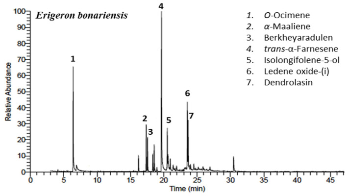
The hydro-distillation of the above-ground parts of E. bonariensis afforded golden yellow EO (0.049%). The chemical characterization of the extracted EO was performed based on the GC-MS analysis. Figure 2 represented the GC-MS chromatogram including the major peaks. Thirty-five components were assigned representing 98.21% of the total oil mass. The characterized constituents as well as retentions times (RIs), molecular formulas (MFs), and literature and calculated Kovats indexes (KIs) were compacted in Table 1.
Figure 2.
GC-MS chromatogrames of the EO of Erigeron bonariensis. The main peaks were numbered (1–7).
In EO of E. bonariensis, sesquiterpenes represented also the main constituents including several oxygenated and non-oxygenated metabolites. With a relative concentration of 81.06% of sesquiterpenes, our results are completely agreed with the previous data by Harraz, et al. [34] that reported a relative concentration of 92.50%; trans-α-Farnesene (25.03%), isolongifolene-5-ol (5.53%), α-maaliene (6.64%), berkheyaradulen (4.82%), and α-muurolene (3.99%) were found the main sesquiterpenoid contents. The main sesquiterpene, trans-α-farnesene, was widely distributed in the EOs of Conyza species such as C. bonariensis (≈E. bonariensis) collected from Venezuela and Vietnam [40], C. canadensis [39], and C. sumatrensis [41]. However, in the only stated study of EO of E. bonariensis [34], α-bergamotene was described as the main compound in addition to some farnesene derivatives such as, β-farnesene, and (E)-farnesene epoxide. The abundance of α-maaliene (6.64%), berkheyaradulen (4.82%), and α-muurolene (3.99%) were found in perfect harmony with our results of EO of P. dioscoridis. The variations of secondary metabolites comprising EOs might be attributed to the plant age and development, plant organs, as well as the environmental factors including such as altitude, seasonality, atmospheric composition and temperature, and water availability [11,42,43].
Monoterpenes represented a remarkable concentration of the EO of E. bonariensis with a wealth of O-ocimene (12.58%). O-Ocimene was reported here for the first time in EO of this plant, contrariwise, Harraz, et al. [34] reported the complete absence of it from EO of the aerial parts of this plant collected from Alexandria, Egypt.
The diterpenoids were represented by a relative concentration of 2.44% from over all mass of the oil of E. bonariensis. The total relative concentration of diterpenes was determined in the EO of E. bonariensis with only one compound, neophytadiene, which is not reported before from the EO of this plant [34].
Carotenoid derived compounds were not identified from the EO of E. bonariensis and this result is in harmony with Mabrouk, et al. [37]; also, hydrocarbons and the other components were represented by traces in EO of E. bonariensis (0.55%) that agreed the previous described studies [32,39].
2.3. Chemometric Analysis
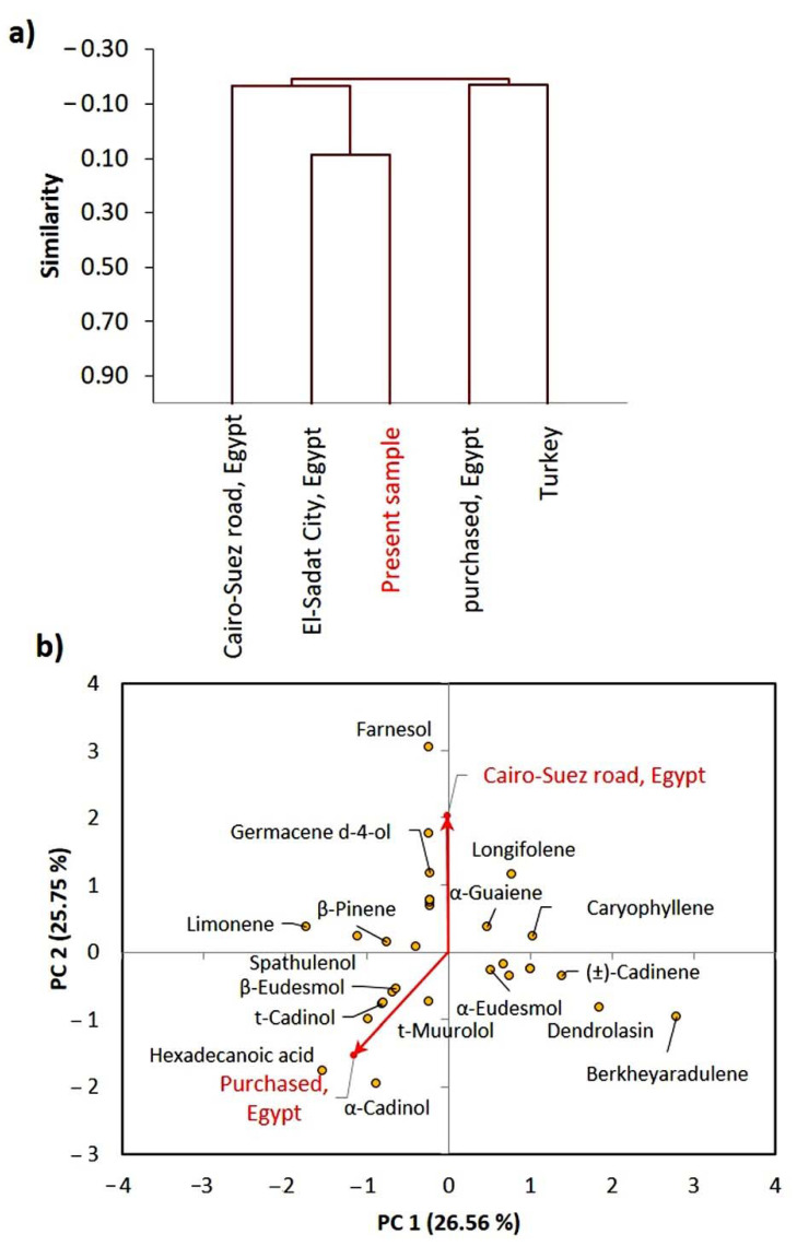
The EOs chemical compositions of the major compounds (>3%), reported from different ecospecies of P. dioscoridis and E. bonariensis were constructed in a matrix. These collected data were subjected to agglomerative hierarchical clustering (AHC) and principal component analysis (PCA). The cluster analysis of P. dioscoridis EOs showed that the present studied sample of P. dioscoridis is closely correlated to the Egyptian ecospecies collected from El-Sadat City, and little correlated to that collected from Cairo–Suez desert road, Egypt (Figure 3a). However, the present sample was different than those purchased from a commercial source in Cairo, Egypt. This means that the commercial samples are not in pure form or may be mixed with other plants.
Figure 3.
Chemometric analysis of the EOs from the present studied Pluchea dioscoridis ecospecies and other reported ecospecies. (a) agglomerative hierarchical clustering (AHC) and (b) principal component analysis (PCA).
The PCA of the P. dioscoridis ecospecies showed that the sample collected from Cairo-Suez desert road, Egypt is mainly characterized by farnesol, germacene d-4-ol, and longifolene (Figure 3b). However, the purchased sample from a commercial source in Cairo, Egypt is characterized by hexadecanoic acid and α-cadinol.
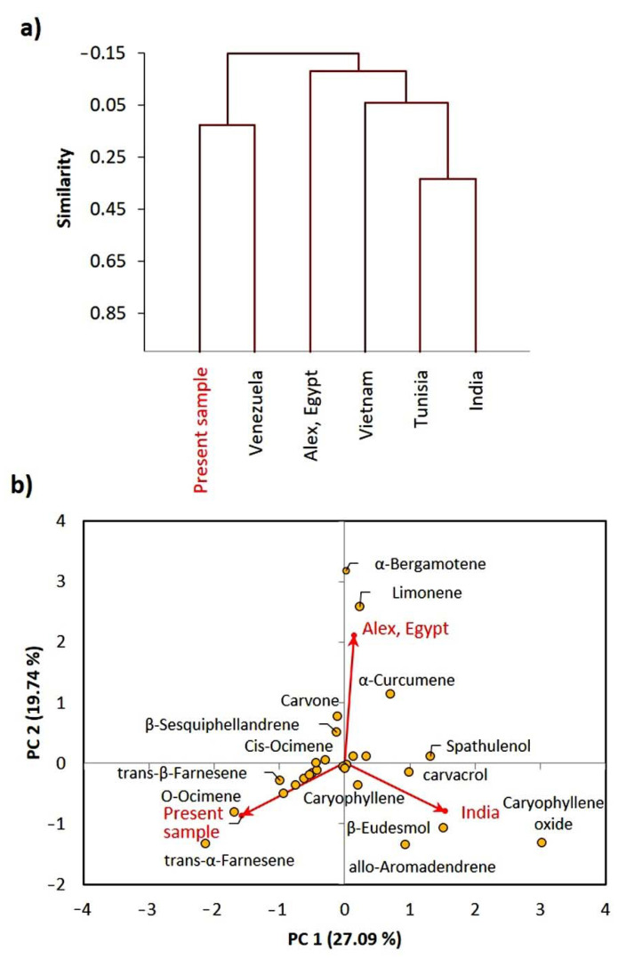
On the other side, the cluster analysis of E. bonariensis EOs revealed that the present Egyptian sample is closely related to the Venezuelan ecospecies, while it was different than other ecospecies (Figure 4a).
Figure 4.
Chemometric analysis of the EOs from the present studied Erigeron bonariensis ecospecies and other reported ecospecies. (a) agglomerative hierarchical clustering (AHC) and (b) principal component analysis (PCA).
While the Indian and Tunisian ecospecies showed a close relation in the composition of the EO. The PCA showed that the present sample of E. bonariensis is characterized by trans-α-Farnesene, O-ocimene, and trans-β-Farnesene (Figure 4b). The sample collected from Alexandria, Egypt, showed a close correlation with α-bergamotene, limonene, and α-curcumene, while the Indian ecospecies is characterized by β-eudesmol, caryophyllene oxide, allo-aromadendrene, and carvacrol.
The observed variation among the present samples and other reported ones revealed the profitable information derived from the EOs analysis, which could be a useful tool in chemotaxonomy [27].
2.4. Anti-Aging Activity
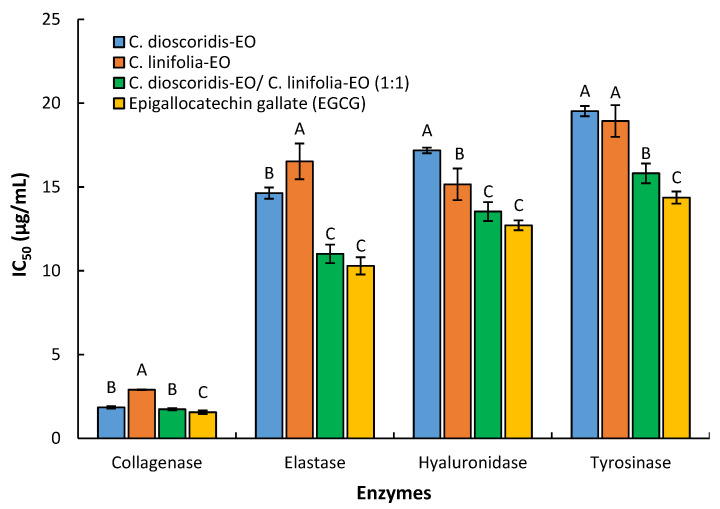
The EOs from P. dioscoridis, E. bonariensis, and the mixture of the two EOs (1:1) have a strong inhibitory activity of the collagenase, elastase, hyaluronidase, and tyrosinase (Figure 5). All the EO treatments exhibited potent inhibition of collagenase enzyme with IC50 of 1.85, 2.90, and 1.73 µg/mL for P. dioscoridis, E. bonariensis, and the mixture, respectively. Furthermore, the three EO treatments strongly inhibit the elastase enzyme with respective values of IC50 of 14.63, 16.52, and 11.01 µg/mL. Furthermore, strong suppression of hyaluronidase was demonstrated via the three EO treatments based upon the respective observed values of IC50 of 17.18, 15.16, and 13.54 µg/mL. By the same, the three tested displayed strong tyrosinase enzyme inhibition with IC50 values at 19.52, 18.93, and 15.81 µg/mL, respectively. All the results were constructed based upon comparing with the polyphenolic compound, epigallocatechin gallate (EGCG), as a standard ant-aging reference [44] that exhibit inhibition of collagenase, elastase, hyaluronidase, and tyrosinase with IC50 of 1.56, 10.29, 12.71, and 14.37 µg/mL.
Figure 5.
Anti-aging activities of the EOs extracted from Pluchea dioscoridis and Erigeron bonariensis against the four enzymes: collagenase, elastase, hyaluronidase, and tyrosinase. Values are IC50 (µg/mL) as an average of three replicates and the bars representing the standard deviation. Different letters (A, B, and C) within each enzyme mean values significant at 0.05 probability level after Duncan’s test.
In the matrix of extracellular, the elastin and hyaluronan degradation were principally correlated with the two respective proteolytic enzymes, elastase, and hyaluronidase that cause the main reasons for aging of the skin such as wrinkles, sagging. Moreover, tyrosinase caused the regulation of the synthesis of melanin in human melanocytes that lead to skin ailments.
In the present study, the anti-aging of the 1:1 mixture of the two EOs was evaluated to study the synergetic effects of the combination of the two EOs. Results revealed that the EO of P. dioscoridis, E. bonariensis, and the mixture of the two EOs (1:1) have strong anti-aging activity. These results might be attributed to the chemical components of these oils. The anti-aging activity was directly correlated with antioxidant potentiality [45]. The main constituents in both EOs, sesquiterpenes, were described to play a significant role as antioxidants, anti-inflammatory agents, and thus anti-aging [45]. Tu and Tawata [45] reported that EO of the leaves of Alpinia zerumbet exhibit antioxidant and anti-aging activities due to the high concentration of terpenoids, especially sesquiterpenes. Furthermore, the monoterpenes were documented as active anti-aging agents in EO of Juniperus communis [46] and Origanum vulgare [47]. These reports concluded that the increase of free radical scavenging constituents in EOs lead to an increase in their anti-aging activity. Based upon this fact, the high concentrations of terpenes especially the oxygenated sesqui- and monoterpenes caused increasing in the anti-aging activity of EOs of these two plants. All these reported data deduced the role of synergetic effects between the components of the EOs. This fact of the role of synergetic effect was very clear in our results in which the mixture of the two EOs (1:1) exhibited better activity than the individual EO of each plant. In the mixture of the two EOs, the raising of concentration of oxygenated terpenes as well the synergetic effects between the components caused increasing of the inhibition potentiality.
2.5. Cytotoxic Activity of EOs of P. dioscoridis and E. bonariensis
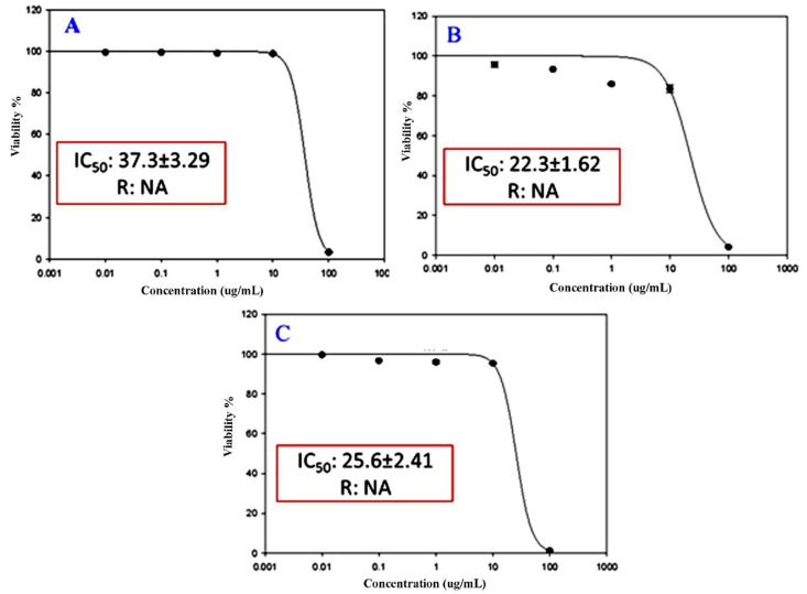
The cytotoxicity of EOs of the above-ground parts of the two plants, P. dioscoridis and E. bonariensis, as well as a mixture of the two EOs (1:1) against the three cancer cell lines, breast adenocarcinoma cells (MCF-7), lung cancer cells (A-549), and hepatocellular carcinoma cells (HepG2) are shown in Figure 6. The results exhibited that the EO of P. dioscoridis have a significant inhibition of the two cancer cells, MCF-7 and A-549, with IC50 of 37.3 and 22.3 μM, respectively (Figure 6A,B), without any activity against HepG2. While, the EO of the E. bonariensis showed inhibitory potentiality only against HepG2 with IC50 of 25.6 μM (Figure 6C), with negative results against MCF-7 and A-549. The 1:1 mixture of the two EOs did not exhibit any activity against the three cancer cells.
Figure 6.
Cytotoxicity of EOs of (A) Pluchea dioscoridis against MCF-7 cells, (B) P. dioscoridis against A-549 cells, and (C) Erigeron bonariensis against HepG2.
The significant activities of the two EOs might be attributed to the chemical composition in which the synergetic effect of the compounds contributes to this activity [48]. The sesquiterpenes in both forms, oxygenated and hydrocarbons, represented very effective compounds as anticancer leaders [49,50]. Several reports deduced that the increasing of sesquiterpene contents in EOs caused increasing in anticancer activity [51,52]. For example, caryophyllene with high concentration in EOs was reported as a known potential cytotoxic agent especially against the growth of breast adenocarcinoma cells (MCF-7) [53,54].
The present data revealed that these two EOs are selective against the tested cancer cells. This selectivity was in full agreement with several documented results of EOs derived from other plants. For example, EO derived from Sideritis perfoliata, Satureia thymbra, Salvia officinalis, Laurus nobilis, and Pistacia palestina were found to have selective inhibitory effects against, amelanotic melanoma (C32), renal celladenocarcinoma (ACHN), hormone-dependent prostatecarcinoma (LNCaP), and breast cancer (MCF-7) [55]. Moreover, EOs extracted from the three plants, Satureja montana, Coriandrum sativum, and Ocimum basilicum, were found to have selective cytotoxic activity against HeLa, MDA-MB-453, K562, and MRC-5 [56]. The disappearance of the mixtures of the two EOs (1:1) might be ascribed to the negative synergetic effects of each EO upon the other and this phenomenon was reported in some reports. Haroun and Al-Kayali [57] found that the different extracts of Thymbra spicata showed positive synergetic effects via combination with some references antibiotics against some strains of bacteria and a while negative synergetic effects against other strains.
3. Materials and Methods
3.1. Plant Materials Collection and Preparation
The above-ground parts of P. dioscoridis and E. bonariensis, were collected from two populations along the Cairo–Alexandria desert road, Egypt in November 2019. From each population, the healthy and fresh plant samples were clipped from three individuals and pooled as composite samples (two per each plant; P. dioscoridis and E. bonariensis). The two plants were authenticated according to Tackholm [58] and Boulos [25]. Voucher specimens (CZ-D-x908-019 & CZ-L-x909-019) have been deposited in the herbarium of the National Research Center, Egypt. The above-ground parts were dried in the shade, ground into a fine powder, and packed in paper bags till further analysis [13].
3.2. Extraction of EOs
The air-dried powder of the above-ground parts of the P. dioscoridis, and E. bonariensis, (200 gm, each) were subjected separately to Clevenger-type apparatuses using round flask (2.5 L) comtaining water (1.5 L) for hydro-distillation for 3 h. The oily layer of each plant was isolated separately by n-hexane, then dried using anhydrous Na2SO4 (0.5 g), and finally stored in glass vials in the freezer till further analysis via GC-MS. This extraction of the EO of each plant was repeated as duplicates.
3.3. Gas Chromatography-Mass Spectroscopy (GC-MS) Analysis and Chemical Components Investigations
The four EOs samples (two samples for each plant) were analyzed via Gas Chromatography-Mass Spectroscopy (GC-MS) at National Research Center, Egypt [8]. The adjustment of the GC/MS instrument specifications has occurred as the following conditions: TRACE GC Ultra Gas Chromatographs (THERMO Scientific™ Corporate, Waltham, MA, USA), lined with a Thermo Scientific ISQ™ EC single quadrupole mass spectrometer. The GC-MS system was equipped with a TR-5 MS column with a dimension of 30 m × 0.32 mm i.d., 0.25 μm film thickness. Helium as carrier gas at a flow rate of 1.0 mL/min with a split ratio of 1:10 using the following temperature program: 60 °C for 1 min; rising at 4.0 °C/min to 240 °C and held for 1 min was used for the analyses. Both injector and detector were held at 210 °C. An aliquot of 1 μL of diluted samples in hexane (1:10, v/v) was always injected. Mass spectra were recorded by electron ionization (EI) at 70 eV, using a spectral range of m/z 40–450.
Chemical constituent of the EOs under investigations was characterized by Automated Mass spectral Deconvolution and Identification (AMDIS) software (www.amdis.net, accessed on 2 January 2020), retention indexes (relative to n-alkanes C8-C22), comparison of the mass spectrum with authentics (if available), and Wiley spectral library collection and NSIT library database (Gaithersburg, MD, USA; Wiley, Hoboken, NJ, USA).
3.4. Anti-Aging Activity of the EOs
3.4.1. Anti-Collagenase Assay
The anti-collagenase assay of the two studied plants EOs as well as the 1:1 mixture were performed according to Thring, et al. [59] with minor modifications for use in a microplate reader. The assay was performed in 50 mM tricine buffer (pH 7.5) with 400 mM NaCl and 10 mM CaCl2. Collagenase from Clostridium histolyticum (ChC–EC.3.4.23.3) was dissolved in a buffer for use at an initial concentration of 0.8 units/mL according to the supplier’s activity data. The synthetic substrate N-[3-(2-furyl) acryloyl]-Leu-Gly-Pro-Ala (FALGPA) was dissolved in tricine buffer to 2 mM. Two studied EOs and the mixture of EOs of the two plants (1:1, w/w), separately, were incubated with the enzyme in a buffer for 15 min before adding substrate to start the reaction. Absorbance at 490 nm was measured using a Microplate reader (TECAN, Group Ltd., Männedorf, Switzerland). Epigallocatechin gallate (EGCG) was used as a positive control.
3.4.2. Anti-Elastase Assay
For anti-elastase inhibitory assay the two studied plants EOs as well as the 1:1 mixture, this assay was performed according to Kim, et al. [60] with minor modifications. Briefly; Porcine pancreatic elastase, was dissolved to make a 3.33 mg/mL stock solution in sterile water. The substrate, N-succinyl-Ala-Ala-Ala-p-nitroanilide (AAAPVN) was dissolved in buffer at 1.6 mM. The test EOs were incubated with the enzyme for 15 min before adding substrate to begin the reaction. The final reaction mixture (250 μL total volume) contained buffer, 0.8 mM AAAPVN, 1 μg/mL PE and 25 μg test sample. The studied EOs and a mixture of EOs of the two plants (1:1, w/w), separately, were incubated. EGCG was used as a positive control. Absorbance values at 400 nm were measured in 96 well microtitre plates using a Microplate reader (TECAN, Inc.). The percentage inhibition for this assay is calculated.
3.4.3. Anti-Tyrosinase Assay
Assays of tyrosinase inhibition of the two plants EOs, as well as the 1:1 mixture, were carried out via measuring of L-DOPA chrome formation according to the described protocol of Batubara, et al. [61]. Briefly, the two EOs and a mixture of them (1:1, w/w), separately, were dissolved in a solvent with three certain concentrations (10, 100, and 250 μg/mL). The assays were performed by insertion of the following components: (a) phosphate buffer (120 μL, 20 mM, pH 6.8), (b) 20 μL sample, and (c) 20 μL mushroom tyrosinase (500 U/mL in 20 mM phosphate buffer) in 96-well plates. After 15 min of incubation at 25 °C, the intiation of reaction was occurred by insertion of 20 μL L-tyrosine solution (0.85 mM) for every well and followed by incubation for 10 min at room temperature. The activity of the enzyme was monitored at 475 nm using a Microplate reader (TECAN, Inc.). EGCG was used as a positive control. The calculation of the tyrosinase inhibition % was performed via the following equation:
| Tyrosinase inhibition (%) = [(A − B) − (C − D)]/(A−B) × 100 | (1) |
where A is the absorbance of the control with the enzyme, B is the absorbance of the control without the enzyme, C is the absorbance of the test sample with the enzyme, and D is the absorbance of the test sample without the enzyme.
3.4.4. Anti-Hyaluronidase Assay
The fluorimetric Morgan–Elson assay method was performed according to Reissig, et al. [62] that modified by Takahashi, et al. [63]. In a brief description, a 5 μL of tested EOs and a mixture of EOs of the two plants (1:1, w/w), separately, were incubated for 10 min at 37 °C with bovine hyaluronidase (1.50 U) in 100 μL of 20 mM sodium phosphate buffer solution (pH 7.0), sodium chloride (77 mM), in addition to 0.01% bovine serum albumin (BSA). The assay reaction was initiated via adding the hyaluronic acid sodium salt (100 μL) from rooster comb (0.03% in 300 mM sodium phosphate, pH 5.35) to the incubation mixture, then the mixture was incubated at 37 °C for 45 min. The precipitation of undigested hyaluronic acid was carried out by 1 mL acidic solution of albumin, involving 0.1% BSA in sodium acetate (24 mM) and acetic acid (79 mM, pH 3.75). The mixture was stoped by allowing it for 10 min at room temperature, and fluorescence was detected using a Tecan Infinite microplate reader at 545 nm excitation and 612 nm emission EGCG was used as a positive control.
The percentage of the collagenase, elastase, and hyaluronidase inhibition was calculated via the following equation:
| Enzyme inhibition (%) = [1 − (S/C) × 100] | (2) |
where S: the corrected absorbance of the samples containing elastase inhibitor (the enzyme activity in the presence of the samples); and C: the corrected absorbance of controls (the enzyme activity in the absence of the samples).
The IC50, the concentration required to inhibit 50% of the enzyme under the assay conditions, was estimated from graphic plots of the dose-response curve for each concentration using Graphpad Prism software (San Diego, CA, USA).
3.5. Cytotoxicity of the Two EOs
Cytotoxic activity of the P. dioscoridis and E. bonariensis EOs and a mixture of them (1:1, w/w), separately were carried out against the three human cancer cells, breast adenocarcinoma cells (MCF-7), lung cancer cells (A-549), and hepatocellular carcinoma cells (HepG2), using sulforhodamine B (SRB) protocol.
3.5.1. Cell Culture
The three cancer cell lines, breast adenocarcinoma cells (MCF-7), lung cancer cells (A-549), and hepatocellular carcinoma cells (HepG2) were obtained from VACCERA, Mokatam, Giza, Egypt. Cells were maintained in DMEM media supplemented with 100 mg/mL of streptomycin, 100 units/mL of penicillin, and 10% of heat-inactivated fetal bovine serum in humidified, 5% (v/v) CO2 atmosphere at 37 °C.
3.5.2. Cytotoxicity Assay
Cell viability was assessed by SRB assay. Aliquots of 100 μL cell suspension (5 × 10³ cells) were in 96-well plates and incubated in complete media for 24 h. Cells were treated with another aliquot of 100 μL media containing EOs and a mixture of them (1:1, w/w), separately, at various concentrations ranging from (0.01, 0.1, 1, 10, and 100 ug/mL). After 72 h of drug exposure, cells were fixed by replacing media with 150 μL of 10% TCA and incubated at 4 °C for 1 h. The TCA solution was removed, and the cells were washed 5 times with distilled water. Aliquots of 70 μL SRB solution (0.4% w/v) were added and incubated in a dark place at room temperature for 10 min. Plates were washed 3 times with 1% acetic acid and allowed to air-dry overnight. Then, 150 μL of TRIS (10 mM) was added to dissolve protein-bound SRB stain; the absorbance was measured at 540 nm using a BMG LABTECH®- FLUOstar Omega microplate reader (Ortenberg, Germany) [64,65].
3.6. Data Treatment
The data of the anti-aging activity of various enzymes were presented in three replications and subjected to one-way ANOVA followed by Duncan’s test using CoStat version 6.311 (CoHort, Monterey, CA, USA, http://www.cohort.com).
A matrix of the concentration of a total of 30 major chemical compounds (>3%) identified in the EO of five P. dioscoridis ecospecies was constructed, these samples were (1) present sample; (2) purchased from a market in Cairo, Egypt; (3) collected from El-Sadat City, Egypt; (4) collected from Cairo-Suez desert road, Egypt dring April; and (5) collected from Turkey. While for E. bonariensis, a matrix of 27 major chemical compounds (>3%) represented six samples was designed, these samples were (1) present sample; (2) collected from Alexandria, Egypt; (3) collected from Monastir, Tunisia; (4) collected from Venezuela; (5) collected from Vietnam; and (6) collected from India. The matrices were subjected to principal component analysis (PCA) and agglomerative hierarchical clustering (AHC) via XLSTAT statistical computer software package (version 2018, Addinsoft Inc., New York, NY, USA).
4. Conclusions
Herein, the GC-MS analysis of EOs of the above-ground parts of P. dioscoridis and E. bonariensis, revealed the identification of 29 and 35 compounds, respectively. Sesquiterpenes were characterized as the main components of EOs derived from the two plants. The major components of EO of P. dioscoridis were α-maaliene, berkheyaradulen, dehydro-cyclolongifolene oxide, aromadendrene oxide-2, and β-muurolene. While, trans-α-farnesene, O-ocimene, and α-maaliene represented the abundant constituents of E. bonariensis EO. The observed variation in the EOs composition among the studied ecospecies and that reported support the changing of the taxa names. EO of P. dioscoridis exhibited cytotoxicity against the two cancer cells, MCF-7 and A-549, while the EO of the E. bonariensis showed activity only against HepG2. The EOs of P. dioscoridis and E. bonariensis as well as the mixture of them (1:1), exhibited significant anti-aging activity in which the mixture (1:1) > P. dioscoridis > E. bonariensis. All these data deduced the studied EOs of these two plants may be used as antiaging and anticancer leading agents.
Acknowledgments
The authors gratefully acknowledge the National Research Centre, Cairo, Egypt for the support.
Author Contributions
Conceptualization, A.M.A.-E., A.M.E. and A.I.E.; methodology, A.M.E., R.F.A., A.M.A.-E., A.E.-N.G.E.-G., A.I.E., and M.I.N.; data curation, A.M.A.-E. A.I.E.; software, A.M.A.-E., A.E.-N.G.E.-G. and A.M.E.; validation, A.M.E., R.F.A., A.M.A.-E., A.E.-N.G.E.-G., A.I.E. and M.I.N.; formal analysis, A.M.E., R.F.A., A.M.A.-E., A.E.-N.G.E.G., A.I.E. and M.I.N.; investigation, A.M.E., R.F.A., A.M.A.-E., A.E.-N.G.E.-G., A.I.E. and M.I.N.; funding acquisition, A.M.E.; writing–original draft preparation, A.M.A.-E. and A.I.E.; writing–review and editing, A.M.A.-E., R.F.A., A.M.A.-E., A.E.-N.G.E.-G., M.I.N. and A.I.E. All authors have read and agreed to the published version of the manuscript.
Funding
This research received no external funding.
Institutional Review Board Statement
Not applicable.
Informed Consent Statement
Not applicable.
Data Availability Statement
The data presented in this study are available in the article.
Conflicts of Interest
The authors declare no conflict of interest.
Sample Availability
Samples of the compounds are not available from the authors.
Footnotes
Publisher’s Note: MDPI stays neutral with regard to jurisdictional claims in published maps and institutional affiliations.
References
- 1.Cseke L.J., Kirakosyan A., Kaufman P.B., Warber S., Duke J.A., Brielmann H.L. Natural Products from Plants. CRC Press; Boca Raton, FL, USA: 2016. [Google Scholar]
- 2.Abdallah H.M.I., Elshamy A.I., El Gendy A.E.-N.G., Abd El-Gawad A.M., Omer E.A., De Leo M., Pistelli L. Anti-inflammatory, antipyretic, and antinociceptive effects of a Cressa cretica aqueous extract. Planta Med. 2017;83:1313–1320. doi: 10.1055/s-0043-108650. [DOI] [PubMed] [Google Scholar]
- 3.Edris A.E. Pharmaceutical and therapeutic potentials of essential oils and their individual volatile constituents: A review. Phytother. Res. 2007;21:308–323. doi: 10.1002/ptr.2072. [DOI] [PubMed] [Google Scholar]
- 4.Della Pepa T., Elshafie H.S., Capasso R., De Feo V., Camele I., Nazzaro F., Scognamiglio M.R., Caputo L. Antimicrobial and phytotoxic activity of Origanum heracleoticum and O. majorana essential oils growing in Cilento (Southern Italy) Molecules. 2019;24:2576. doi: 10.3390/molecules24142576. [DOI] [PMC free article] [PubMed] [Google Scholar]
- 5.Gruľová D., Caputo L., Elshafie H.S., Baranová B., De Martino L., Sedlák V., Gogaľová Z., Poráčová J., Camele I., De Feo V. Thymol chemotype Origanum vulgare L. essential oil as a potential selective bio-based herbicide on monocot plant species. Molecules. 2020;25:595. doi: 10.3390/molecules25030595. [DOI] [PMC free article] [PubMed] [Google Scholar]
- 6.Al-Rowaily S.L., Abd-ElGawad A.M., Assaeed A.M., Elgamal A.M., Gendy A.E.-N.G.E., Mohamed T.A., Dar B.A., Mohamed T.K., Elshamy A.I. Essential oil of Calotropis procera: Comparative chemical profiles, antimicrobial activity, and allelopathic potential on weeds. Molecules. 2020;25:5203. doi: 10.3390/molecules25215203. [DOI] [PMC free article] [PubMed] [Google Scholar]
- 7.Abd El-Gawad A., El Gendy A., Elshamy A., Omer E. Chemical composition of the essential oil of Trianthema portulacastrum L. Aerial parts and potential antimicrobial and phytotoxic activities of its extract. J. Essent. Oil Bear. Plants. 2016;19:1684–1692. doi: 10.1080/0972060X.2016.1205523. [DOI] [Google Scholar]
- 8.Elshamy A.I., Ammar N.M., Hassan H.A., Al-Rowaily S.L., Ragab T.I., El Gendy A.E.-N.G., Abd-ElGawad A.M. Essential oil and its nanoemulsion of Araucaria heterophylla resin: Chemical characterization, anti-inflammatory, and antipyretic activities. Ind. Crops Prod. 2020;148:112272. doi: 10.1016/j.indcrop.2020.112272. [DOI] [Google Scholar]
- 9.Abd-ElGawad A.M., Elshamy A.I., El-Nasser El Gendy A., Al-Rowaily S.L., Assaeed A.M. Preponderance of oxygenated sesquiterpenes and diterpenes in the volatile oil constituents of Lactuca serriola L. revealed antioxidant and allelopathic activity. Chem. Biodivers. 2019;16:e1900278. doi: 10.1002/cbdv.201900278. [DOI] [PubMed] [Google Scholar]
- 10.Assaeed A., Elshamy A., El Gendy A.E.-N., Dar B., Al-Rowaily S., Abd-ElGawad A. Sesquiterpenes-rich essential oil from above ground parts of Pulicaria somalensis exhibited antioxidant activity and allelopathic effect on weeds. Agronomy. 2020;10:399. doi: 10.3390/agronomy10030399. [DOI] [Google Scholar]
- 11.Elshamy A.I., Abd-ElGawad A.M., El-Amier Y.A., El Gendy A.E.N.G., Al-Rowaily S.L. Interspecific variation, antioxidant and allelopathic activity of the essential oil from three Launaea species growing naturally in heterogeneous habitats in Egypt. Flavour Fragr. J. 2019;34:316–328. doi: 10.1002/ffj.3512. [DOI] [Google Scholar]
- 12.Abd El-Gawad A.M., El-Amier Y.A., Bonanomi G. Essential oil composition, antioxidant and allelopathic activities of Cleome droserifolia (Forssk.) Delile. Chem. Biodivers. 2018;15:e1800392. doi: 10.1002/cbdv.201800392. [DOI] [PubMed] [Google Scholar]
- 13.Abd El-Gawad A.M. Chemical constituents, antioxidant and potential allelopathic effect of the essential oil from the aerial parts of Cullen plicata. Ind. Crops Prod. 2016;80:36–41. doi: 10.1016/j.indcrop.2015.10.054. [DOI] [Google Scholar]
- 14.Abd-ElGawad A.M., El Gendy A.E.-N.G., Assaeed A.M., Al-Rowaily S.L., Alharthi A.S., Mohamed T.A., Nassar M.I., Dewir Y.H., Elshamy A.I. Phytotoxic effects of plant essential oils: A systematic review and structure-activity relationship based on chemometric analyses. Plants. 2021;10:36. doi: 10.3390/plants10010036. [DOI] [PMC free article] [PubMed] [Google Scholar]
- 15.Abd-ElGawad A.M., El Gendy A.E.-N.G., Assaeed A.M., Al-Rowaily S.L., Omer E.A., Dar B.A., Al-Taisan W.a.A., Elshamy A.I. Essential oil enriched with oxygenated constituents from invasive plant Argemone ochroleuca exhibited potent phytotoxic effects. Plants. 2020;9:998. doi: 10.3390/plants9080998. [DOI] [PMC free article] [PubMed] [Google Scholar]
- 16.Abd El-Gawad A.M., El-Amier Y.A., Bonanomi G. Allelopathic activity and chemical composition of Rhynchosia minima (L.) DC. essential oil from Egypt. Chem. Biodivers. 2018;15:e1700438. doi: 10.1002/cbdv.201700438. [DOI] [PubMed] [Google Scholar]
- 17.Elshamy A.I., Abd-ElGawad A.M., El Gendy A.E.N.G., Assaeed A.M. Chemical characterization of Euphorbia heterophylla L. essential oils and their antioxidant activity and allelopathic potential on Cenchrus echinatus L. Chem. Biodivers. 2019;16:e1900051. doi: 10.1002/cbdv.201900051. [DOI] [PubMed] [Google Scholar]
- 18.Arunachalam K., Balogun S.O., Pavan E., de Almeida G.V.B., de Oliveira R.G., Wagner T., Cechinel Filho V., de Oliveira Martins D.T. Chemical characterization, toxicology and mechanism of gastric antiulcer action of essential oil from Gallesia integrifolia (Spreng.) Harms in the in vitro and in vivo experimental models. Biomed. Pharmacother. 2017;94:292–306. doi: 10.1016/j.biopha.2017.07.064. [DOI] [PubMed] [Google Scholar]
- 19.Saleh I., Abd-ElGawad A., El Gendy A.E.-N., Abd El Aty A., Mohamed T., Kassem H., Aldosri F., Elshamy A., Hegazy M.-E.F. Phytotoxic and antimicrobial activities of Teucrium polium and Thymus decussatus essential oils extracted using hydrodistillation and microwave-assisted techniques. Plants. 2020;9:716. doi: 10.3390/plants9060716. [DOI] [PMC free article] [PubMed] [Google Scholar]
- 20.Damtie D., Braunberger C., Conrad J., Mekonnen Y., Beifuss U. Composition and hepatoprotective activity of essential oils from Ethiopian thyme species (Thymus serrulatus and Thymus schimperi) J. Essent. Oil Res. 2019;31:120–128. doi: 10.1080/10412905.2018.1512907. [DOI] [Google Scholar]
- 21.Tomaino A., Cimino F., Zimbalatti V., Venuti V., Sulfaro V., De Pasquale A., Saija A. Influence of heating on antioxidant activity and the chemical composition of some spice essential oils. Food Chem. 2005;89:549–554. doi: 10.1016/j.foodchem.2004.03.011. [DOI] [Google Scholar]
- 22.Wang A., Wu H., Zhu X., Lin J. Species identification of Conyza bonariensis assisted by chloroplast genome sequencing. Front. Genet. 2018;9:374. doi: 10.3389/fgene.2018.00374. [DOI] [PMC free article] [PubMed] [Google Scholar]
- 23.Peng L., Hu C., Zhang C., Lu Y., Man S., Ma L. Anti-cancer activity of Conyza blinii saponin against cervical carcinoma through MAPK/TGF-β/Nrf2 signaling pathways. J. Ethnopharmacol. 2020;251:112503. doi: 10.1016/j.jep.2019.112503. [DOI] [PubMed] [Google Scholar]
- 24.Ayaz F., Küçükboyacı N., Demirci B. Chemical composition and antimicrobial activity of the essential oil of Conyza canadensis (L.) Cronquist from Turkey. J. Essent. Oil Res. 2017;29:336–343. doi: 10.1080/10412905.2017.1279989. [DOI] [Google Scholar]
- 25.Boulos L. Flora of Egypt. Volume 3 Al Hadara Publishing; Cairo, Egypt: 2002. [Google Scholar]
- 26.List T.P. Version 1.1. [(accessed on 1 January 2013)]; Available online: http://www.theplantlist.org/
- 27.Abd El-Gawad A., Elshamy A., El Gendy A.E.-N., Gaara A., Assaeed A. Volatiles profiling, allelopathic activity, and antioxidant potentiality of Xanthium strumarium leaves essential oil from Egypt: Evidence from chemometrics analysis. Molecules. 2019;24:584. doi: 10.3390/molecules24030584. [DOI] [PMC free article] [PubMed] [Google Scholar]
- 28.El-Taher A.M., Gendy A.E.-N.G.E., Alkahtani J., Elshamy A.I., Abd-ElGawad A.M. Taxonomic implication of integrated chemical, morphological, and anatomical attributes of leaves of eight Apocynaceae taxa. Diversity. 2020;12:334. doi: 10.3390/d12090334. [DOI] [Google Scholar]
- 29.Mohamed T.A., Elshamy A.I., Abd-ElGawad A.M., Hussien T.A., El-Toumy S.A., Efferth T., Hegazy M.E.F. Cytotoxic and chemotaxonomic study of isolated metabolites from Centaurea aegyptiaca. J. Chin. Chem. Soc. 2021;68:159–168. doi: 10.1002/jccs.202000156. [DOI] [Google Scholar]
- 30.El Zalabani S., Hetta M., Ross S., Abo Youssef A., Zakiand M., Ismail A. Antihyperglycemic and antioxidant activities and chemical composition of Conyza dioscoridis (L.) Desf. DC. growing in Egypt. Aust. J. Basic Appl. Sci. 2012;6:257–265. [Google Scholar]
- 31.Atta A.H., Nasr S.M., Mouneir S., Alwabel N., Essawy S. Evaluation of the diuretic effect of Conyza dioscorides and Alhagi maurorum. Int. J. Pharm. Pharm. Sci. 2010;2:162–165. [Google Scholar]
- 32.Elshamy A.I., El Gendy A., Farrag A., Nassar M.I. Antidiabetic and antioxidant activities of phenolic extracts of Conyza dioscoridis L. shoots. Int. J. Pharm. Pharm. Sci. 2015;7:65–72. [Google Scholar]
- 33.Awaad A.S., El-Meligy R., Qenawy S., Atta A., Soliman G.A. Anti-inflammatory, antinociceptive and antipyretic effects of some desert plants. J. Saudi Chem. Soc. 2011;15:367–373. doi: 10.1016/j.jscs.2011.02.004. [DOI] [Google Scholar]
- 34.Harraz F.M., Hammoda H.M., El Ghazouly M.G., Farag M.A., El-Aswad A.F., Bassam S.M. Chemical composition, antimicrobial and insecticidal activities of the essential oils of Conyza linifolia and Chenopodium ambrosioides. Nat. Prod. Res. 2015;29:879–882. doi: 10.1080/14786419.2014.988714. [DOI] [PubMed] [Google Scholar]
- 35.Atta A.H., Mouneir S.M. Antidiarrhoeal activity of some Egyptian medicinal plant extracts. J. Ethnopharmacol. 2004;92:303–309. doi: 10.1016/j.jep.2004.03.017. [DOI] [PubMed] [Google Scholar]
- 36.Adams R.P. Identification of Essential Oil Components By Gas Chromatography/Mass Spectrometry. Volume 456 Allured Publishing Corporation; Carol Stream, IL, USA: 2007. [Google Scholar]
- 37.Mabrouk S., Elaissi A., Ben Jannet H., Harzallah-Skhiri F. Chemical composition of essential oils from leaves, stems, flower heads and roots of Conyza bonariensis L. from Tunisia. Nat. Prod. Res. 2011;25:77–84. doi: 10.1080/14786419.2010.513685. [DOI] [PubMed] [Google Scholar]
- 38.El-Seedi H.R., Azeem M., Khalil N.S., Sakr H.H., Khalifa S.A., Awang K., Saeed A., Farag M.A., AlAjmi M.F., Pålsson K. Essential oils of aromatic Egyptian plants repel nymphs of the tick Ixodes ricinus (Acari: Ixodidae) Exp. Appl. Acarol. 2017;73:139–157. doi: 10.1007/s10493-017-0165-3. [DOI] [PMC free article] [PubMed] [Google Scholar]
- 39.Lis A., Góra J. Essential oil of Conyza canadensis (L.) Cronq. J. Essent. Oil Res. 2000;12:781–783. doi: 10.1080/10412905.2000.9712214. [DOI] [Google Scholar]
- 40.Araujo L., Moujir L.M., Rojas J., Rojas L., Carmona J., Rondón M. Chemical composition and biological activity of Conyza bonariensis essential oil collected in Mérida, Venezuela. Nat. Prod. Commun. 2013;8:1175–1178. doi: 10.1177/1934578X1300800838. [DOI] [PubMed] [Google Scholar]
- 41.Hoi T.M., Huong L.T., Chinh H.V., Hau D.V., Satyal P., Tai T.A., Dai D.N., Hung N.H., Hien V.T., Setzer W.N. Essential oil compositions of three invasive Conyza species collected in Vietnam and their larvicidal activities against Aedes aegypti, Aedes albopictus, and Culex quinquefasciatus. Molecules. 2020;25:4576. doi: 10.3390/molecules25194576. [DOI] [PMC free article] [PubMed] [Google Scholar]
- 42.Abd-ElGawad A.M., Elshamy A.I., El-Amier Y.A., El Gendy A.E.-N.G., Al-Barati S.A., Dar B.A., Al-Rowaily S.L., Assaeed A.M. Chemical composition variations, allelopathic, and antioxidant activities of Symphyotrichum squamatum (Spreng.) Nesom essential oils growing in heterogeneous habitats. Arab. J. Chem. 2020;13:4237–4245. doi: 10.1016/j.arabjc.2019.07.005. [DOI] [Google Scholar]
- 43.Abd-ElGawad A.M., Elshamy A.I., Al-Rowaily S.L., El-Amier Y.A. Habitat affects the chemical profile, allelopathy, and antioxidant properties of essential oils and phenolic enriched extracts of the invasive plant Heliotropium curassavicum. Plants. 2019;8:482. doi: 10.3390/plants8110482. [DOI] [PMC free article] [PubMed] [Google Scholar]
- 44.Xiong L.-G., Chen Y.-J., Tong J.-W., Gong Y.-S., Huang J.-A., Liu Z.-H. Epigallocatechin-3-gallate promotes healthy lifespan through mitohormesis during early-to-mid adulthood in Caenorhabditis elegans. Redox Biol. 2018;14:305–315. doi: 10.1016/j.redox.2017.09.019. [DOI] [PMC free article] [PubMed] [Google Scholar]
- 45.Tu P.T.B., Tawata S. Anti-oxidant, anti-aging, and anti-melanogenic properties of the essential oils from two varieties of Alpinia zerumbet. Molecules. 2015;20:16723–16740. doi: 10.3390/molecules200916723. [DOI] [PMC free article] [PubMed] [Google Scholar]
- 46.Pandey S., Tiwari S., Kumar A., Niranjan A., Chand J., Lehri A., Chauhan P.S. Antioxidant and anti-aging potential of Juniper berry (Juniperus communis L.) essential oil in Caenorhabditis elegans model system. Ind. Crops Prod. 2018;120:113–122. doi: 10.1016/j.indcrop.2018.04.066. [DOI] [Google Scholar]
- 47.Laothaweerungsawat N., Sirithunyalug J., Chaiyana W. Chemical compositions and anti-skin-ageing activities of Origanum vulgare L. essential oil from tropical and mediterranean region. Molecules. 2020;25:1101. doi: 10.3390/molecules25051101. [DOI] [PMC free article] [PubMed] [Google Scholar]
- 48.Pezzani R., Salehi B., Vitalini S., Iriti M., Zuñiga F.A., Sharifi-Rad J., Martorell M., Martins N. Synergistic effects of plant derivatives and conventional chemotherapeutic agents: An update on the cancer perspective. Medicina. 2019;55:110. doi: 10.3390/medicina55040110. [DOI] [PMC free article] [PubMed] [Google Scholar]
- 49.Mohamed T.A., Albadry H.A., Elshamy A.I., Younes S.H., Shahat A.A., El-wassimy M.T., Moustafa M.F., Hegazy M.E.F. A new Tetrahydrofuran sesquiterpene skeleton from Artemisia sieberi. J. Chin. Chem. Soc. 2020;86:1–5. doi: 10.1002/jccs.202000198. [DOI] [Google Scholar]
- 50.Xie Z.Q., Ding L.F., Wang D.S., Nie W., Liu J.X., Qin J., Song L.D., Wu X.D., Zhao Q.S. Sesquiterpenes from the leaves of Magnolia delavayi Franch. and their cytotoxic activities. Chem. Biodivers. 2019;16:e1900013. doi: 10.1002/cbdv.201900013. [DOI] [PubMed] [Google Scholar]
- 51.Blowman K., Magalhães M., Lemos M.F.L., Cabral C., Pires I.M. Anticancer properties of essential oils and other natural products. J. Evid. Based Compl. Altern. Med. 2018;2018:3149362. doi: 10.1155/2018/3149362. [DOI] [PMC free article] [PubMed] [Google Scholar]
- 52.Danin A. Plants of Desert Dunes. Springer Science & Business Media; Berlin, Germany: 2012. [Google Scholar]
- 53.Wright B.S., Bansal A., Moriarity D.M., Takaku S., Setzer W.N. Cytotoxic leaf essential oils from Neotropical Lauraceae: Synergistic effects of essential oil components. Nat. Prod. Commun. 2007;2:1241–1244. doi: 10.1177/1934578X0700201210. [DOI] [Google Scholar]
- 54.Ali N.A.A., Chhetri B.K., Dosoky N.S., Shari K., Al-Fahad A.J., Wessjohann L., Setzer W.N. Antimicrobial, antioxidant, and cytotoxic activities of Ocimum forskolei and Teucrium yemense (Lamiaceae) essential oils. Medicines. 2017;4:17. doi: 10.3390/medicines4020017. [DOI] [PMC free article] [PubMed] [Google Scholar]
- 55.Loizzo M.R., Tundis R., Menichini F., Saab A.M., Statti G.A., Menichini F. Cytotoxic activity of essential oils from Labiatae and Lauraceae families against in vitro human tumor models. Anticancer Res. 2007;27:3293–3299. [PubMed] [Google Scholar]
- 56.Elgndi M.A., Filip S., Pavlić B., Vladić J., Stanojković T., Žižak Ž., Zeković Z. Antioxidative and cytotoxic activity of essential oils and extracts of Satureja montana L., Coriandrum sativum L. and Ocimum basilicum L. obtained by supercritical fluid extraction. J. Supercrit. Fluids. 2017;128:128–137. doi: 10.1016/j.supflu.2017.05.025. [DOI] [Google Scholar]
- 57.Haroun M.F., Al-Kayali R.S. Synergistic effect of Thymbra spicata L. extracts with antibiotics against multidrug-resistant Staphylococcus aureus and Klebsiella pneumoniae strains. Iran. J. Basic Med. Sci. 2016;19:1193–1200. [PMC free article] [PubMed] [Google Scholar]
- 58.Tackholm V. Students’ Flora of Egypt. 2nd ed. Cairo University Press; Cairo, Egypt: 1974. [Google Scholar]
- 59.Thring T.S.A., Hili P., Naughton D.P. Anti-collagenase, anti-elastase and anti-oxidant activities of extracts from 21 plants. BMC Compl. Altern. Med. 2009;9:27. doi: 10.1186/1472-6882-9-27. [DOI] [PMC free article] [PubMed] [Google Scholar]
- 60.Kim Y.-J., Uyama H., Kobayashi S. Inhibition effects of (+)-catechin–aldehyde polycondensates on proteinases causing proteolytic degradation of extracellular matrix. Biochem. Biophys. Res. Commun. 2004;320:256–261. doi: 10.1016/j.bbrc.2004.05.163. [DOI] [PubMed] [Google Scholar]
- 61.Batubara I., Darusman L., Mitsunaga T., Rahminiwati M., Djauhari E. Potency of Indonesian medicinal plants as tyrosinase inhibitor and antioxidant agent. J. Biol. Sci. 2010;10:138–144. doi: 10.3923/jbs.2010.138.144. [DOI] [Google Scholar]
- 62.Reissig J.L., Strominger J.L., Leloir L.F. A modified colorimetric method for the estimation of N-acetylamino sugars. J. Biol. Chem. 1955;217:959–966. doi: 10.1016/S0021-9258(18)65959-9. [DOI] [PubMed] [Google Scholar]
- 63.Takahashi T., Ikegami-Kawai M., Okuda R., Suzuki K. A fluorimetric Morgan–Elson assay method for hyaluronidase activity. Anal. Biochem. 2003;322:257–263. doi: 10.1016/j.ab.2003.08.005. [DOI] [PubMed] [Google Scholar]
- 64.Skehan P., Storeng R., Scudiero D., Monks A., McMahon J., Vistica D., Warren J.T., Bokesch H., Kenney S., Boyd M.R. New colorimetric cytotoxicity assay for anticancer-drug screening. J. Natl. Cancer Inst. 1990;82:1107–1112. doi: 10.1093/jnci/82.13.1107. [DOI] [PubMed] [Google Scholar]
- 65.Allam R.M., Al-Abd A.M., Khedr A., Sharaf O.A., Nofal S.M., Khalifa A.E., Mosli H.A., Abdel-Naim A.B. Fingolimod interrupts the cross talk between estrogen metabolism and sphingolipid metabolism within prostate cancer cells. Toxicol. Lett. 2018;291:77–85. doi: 10.1016/j.toxlet.2018.04.008. [DOI] [PubMed] [Google Scholar]
Associated Data
This section collects any data citations, data availability statements, or supplementary materials included in this article.
Data Availability Statement
The data presented in this study are available in the article.


