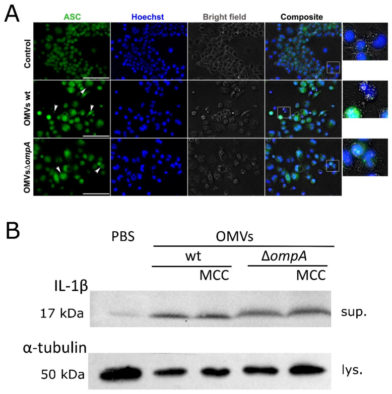
Figure 3.
Inflamasomme induction in J774 macrophages stimulated with OMVs from A. baumannii wt and its ∆ompA mutant. (A) Immunofluorescence analysis of J774 mice macrophages incubated for 24 h with OMVs produced by A. baumannii wt strain and its ompA deletion mutant. Cells were stained with Hoechst and anti-ASC primary antibodies. ASC specks were visible as accumulated dots (indicated with white arrows). In the composite images, rectangles show magnified parts. Scale bar shows 100 µm; (B) detection of activated form of IL-1β (17 kDa) by Western blot. J774 macrophages were incubated with OMVs or PBS for 24 h. MCC—NLRP3 inflammasome inhibitor MCC950. Sup.—supernatants, lys.—cells lysates. Membrane was developed with Pierce ECL Western Blotting Substrate (Thermo Fisher Scientific, Walkersville, MD, USA).

