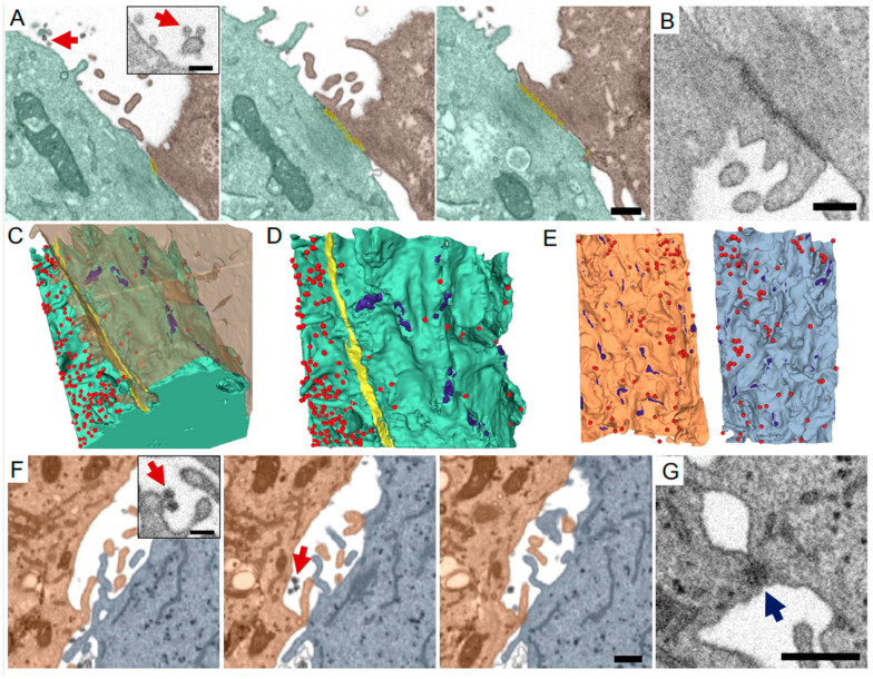
Figure 2.
FIB-SEM imaging and 3-D reconstructions of SARS-CoV-2 viruses and cell-cell contacts. Infected Vero E6 cells (A–D) or Calu-3 human lung epithelial cells (Cortese et al., 2020) (E–G) processed for and imaged by FIB-SEM. (A) Periodic FIB-SEM micrographs of every 10th 8 nm slice showing a tight junction-mediated contact (yellow) between two Vero E6 cells (green and brown) and viruses in the extracellular space (red arrows). Inset shows high magnification view of viruses and cell membranes. (B) Higher magnification micrograph of a tight junction connecting the two cells in A. (C,D) 3-D reconstructions of plasma membranes of both cells in A (C). Virus particles appear as red spheres, small focal adhesions (likely desmosomes) are shown in purple, tight junction is shown in yellow. Viral density is dramatically different on either side of the contiguous tight junction (D). (E) 3-D reconstructions of two neighboring lung epithelial cells in contact (orange, light blue), separated to reveal the surfaces in closest proximity. Desmosome-like junctions were seen between the two cells (purple, and G). Viral particles (red) are evenly distributed on the plasma membranes. (F) FIB-SEM micrographs of every 5th 8 nm slice from lung epithelial cells [15]. Same cells as in E. Red arrows mark viruses. (G) Higher magnification micrograph of a desmosome (blue arrow) between the two cells in E-F. Scale bars (A,F,G): 500 nm. (A,F) insets: 200 nm. (B): 300 nm.

